Hi boog99! Would you be willing to mark the areas in question on my pics I posted? If not, I understand.

The green arrow is pointing to what I was concerned about.
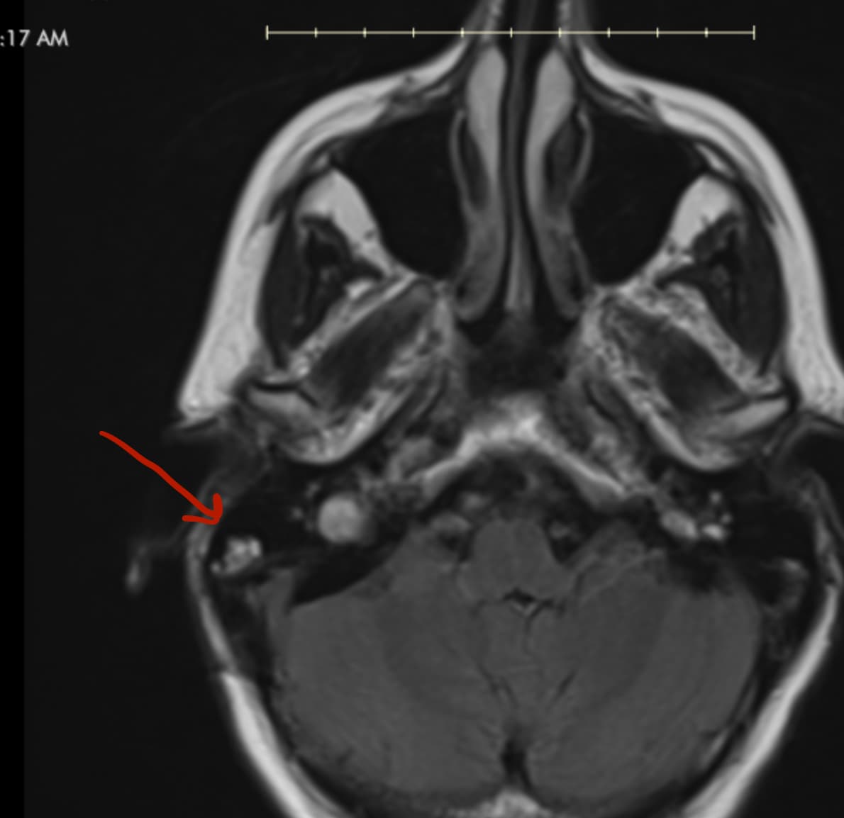
You are pointing to the correct spot. I think we are pointing to the same spot but in different “sequences”. I suspect the green arrow is from a “T2” sequence while the Red arrow is from a “Flair” sequence.
But yes, just ask about this area and make sure it isn’t anything sinister.
1 Like
Thanks so much, boogs99!! I went back to some older MRI imaging. Interesting, what’s there now, doesn’t appear to have been there before the MVD. I’m going to send my ENT a message as I can’t get an appt for 3 weeks. I hate to message him imaging questions without an appt, but if it’s an infection it needs addressed sooner rather than later.

How can you tell if your mri is t1 or t2 weighted? I thought it would be as simple as looking at the MRI report, but it doesn’t indicate which weighting was used.
The DICOM viewer I’m using, HOROS, displays which sequence I’m selecting, so it is easy to tell. I can tell the last image you posted is T2 because the CSF is bright. (i think)
Anywho, perhaps the radiology department responsible for your latest imaging report is readily accessible to answer questions in addition to your ENT? Just a thought…
1 Like
Ha! They are not accessible, hence, why I ask so many questions here lol! Thanks for the info!
Radiologists aren’t accessible in Canada either.
Good luck!
That is correct. T2 shows CFS fluid as bright white where T1 displays the CSF fluid as black.
2 Likes
Would T2 also show other fluid (such as otitis media fluid) as bright white?
This is above my pay grade. But you can look into cholesteatomas and diffusion weighted images, if that was included in your imaging, as a possibility also.
Do you have a fever at all?
No, I don’t. But I can feel a lot of fluid in the right middle ear.
So, I messaged the ENT (and sent our screenshots) and he told me to ask the neurosurgeon?!?!?!? He said he “assumed” it was the thrombus (I circled the white area we talked about), but he said he wasn’t for sure. I don’t think the thrombus would show up as white on a T2 MRI image. @KoolDude and boogs99, what are your thoughts? It almost has to be some sort of fluid.
It seems like an unusual path for the sigmoid sinus…and I don’t think thrombus has a hyperintense signal on T2 imaging… edit: turns out acute thrombus can be hyperintense!
Does your ENT not have access to a radiologist?
What about flipping an email to Dr. P?
That’s my next plan of attack. My ENT has access to the imaging and a radiologist. Of course, he should also be able to read it himself since the area involves the head/neck/ear. He just didn’t want to get involved.
Did Dr. P review the imaging yesterday?
I think I agree with @boogs99 on this one. It appears to be some kind of fluid perhaps mixed with scar tissues in an around Mastoid Air Cells (Not a doctor but my assumption here). Having said that, a clot or slow flow of blood can show up as bright white in the sinuses in fat suppressed T2 MRI so it is not entirely wrong to assume that.
I think you will need to investigate this with neurosurgeon and neurovascular surgeon. I also agree contacting Dr. P as Boogs99 suggested.
An example from internet of clots showing up as hyperintense in T2 weighed MRI
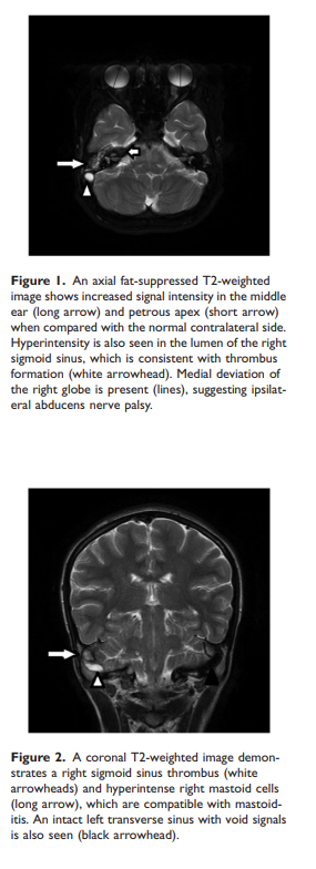
Source : https://journals.sagepub.com/doi/pdf/10.1177/0300060518823404
2 Likes
Thank you both so incredibly much!!! The thing that made me mad is that my ENT wasn’t even willing to take the time to look at the full imaging to help out (and I’m already a patient of his).
I’ve been trying to tell my neurologist for weeks that my head is having high pressure. He blew it off until this morning when I showed him pictures of my imaging with the lack of blood flow. Now all of a sudden he wants to try Diamox.
Yeah I’m infuriated on your behalf. You have CVST following MVD surgery and your ENT can’t be bothered to look into something that you brought concern to!!! I think as a patient you have a right to ask questions and there is an expectation of answers. To ignore your questions seems irresponsible.
Anywho, I hope you get relief and answers soon!
2 Likes
First the good news is, you are now advocating for yourself and you are equipped with some evidence that can’t be ignored. But before they put you on Diamox forever, I think you need to consider the advice of Dr. P and have him look into the sinuses since I happen to think there might be more than just clotting going on. As you have suspected and as @boogs99 has found, there could be a new narrowing of the right side sinuses perhaps induced by excess fluid or scar tissue or surgeon moving them around. Veins in general have thin wall and are easily collapsible so it is not far fetched that the surgery might have induced some narrowing which can cause blood flow stasis which could in turn cause clots. Or it could be some infection in the Mastoid area as Boogs99 might have suspected, although you do not have high fever, that does not rule out a brewing infection in the mastoid area which is linked to clotting in the sinuses after this type of surgery. So regardless of the causal mechanism of the clots, this will eventually need to be treated by Neurovascular surgeon with or without the help of neurosurgeon since your headaches are due to fluid build up in the brain.
Looking in the internet to see how this MVD surgery is done, I am astonished to find how close the incision is to the transverse & Sigmoid sinuses (see below images from the internet).

Source : Microvascular Decompression (MVD) - Facial Pain Association
3 Likes