Hi everyone,
I’m looking for some insight from those who have been through this.
My Background: Six months ago, I saw Mr. Patrick Axon (Cambridge). He reviewed my scans, confirmed I have bilateral IJV compressions, and stated he could offer decompression surgery if my symptoms worsened. Over the last few months, they have gotten significantly worse. My main symptoms are:
Severe fatigue & profound brain fog (including word-swapping)
Positional dizziness and a ‘full, heavy head’ sensation upon waking
The Conflict Today: I wanted to consult a neurosurgeon before making a final decision on surgery, so I saw a neurosurgeon in Oxford today. He also worked in Cambridge previously and knows Dr. Axon. He looked at my CT scans and completely dismissed them. He thinks the compressions are ‘mild’ and insignificant, and claims they cannot be causing my venous outflow symptoms.
The Pivot: However, when I asked about a Catheter Venogram with Manometry, he said I could have one in Cambridge by Dr. Higgins if I wanted it.
My Questions for the Group:
Should I get the Catheter Venogram? Given that he dismissed the static CT, will the manometry (pressure wire) test provide the objective proof needed to show who is right?
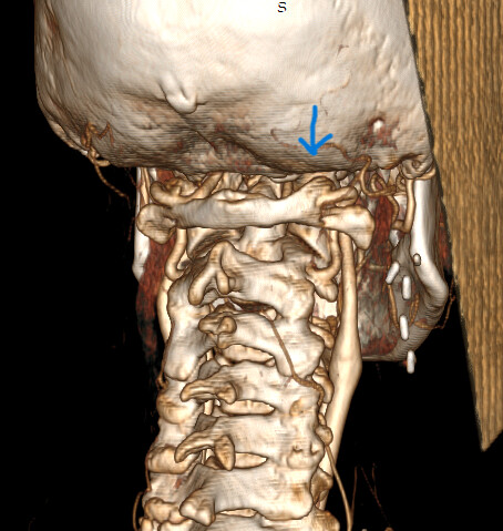
Looking at my attached scans (Bottom to top, Left is Right): Do you agree with the Oxford neurosurgeon that this looks ‘mild,’ or does this look like a classic mechanical blockage?
Should I stop seeing general neurosurgeons and just go back to Mr. Axon and his team now that my symptoms are worse?
Thank you so much for any advice or shared experiences!
@emre - I don’t know what measure the Oxford neurologist uses to determine mildly compressed from moderately or severely compressed, but your IJVs DO NOT appear mildly compressed to me. The fact that you have any compression & coinciding symptoms indicates that your compression is significant enough your body is responding negatively to it.
Catheter venograms carry a reasonable level of risk both during & after the imaging is done. You’ve got imaging that shows your situation adequately. The little bit of extra info you’ll gain may not be worth the risk.
We’ve seen a broad range of compression levels which have caused symptoms for our members. the least I recall is about 45% w/ the greatest being 100%. The neurologist you saw may be speaking “from his experience” but his experience cannot dictate situations that are outside of the parameters he’s set.
Dr. Axon has years of experience in diagnosing & doing surgery for IJV compressions. I would trust his judgment over a doctor who doesn’t specifically do that surgery.
I agree with @Isaiah_40_31 that there is significant compression, & Mr Axon is very experienced with this surgery, so if he agrees and has offered surgery I would trust his judgement- he doesn’t offer the surgery to everyone. I thought Mr Higgins wasn’t practising any more, so that’s interesting, he has worked with Mr Axon quite a bit…but there are risks with the catheter venogram so I would consider carefully whether you want to have that done… I can’t label the images I’m afraid, but there does seem to be some C1 involvement in the compression, and possibly other blood vessels in the sagittal views (the bottom two images), although that possibly doesn’t look the same in the axial plane.
I’m sorry that you were hoping for confirmation & reassurance from the neurologist, but we see on here regularly that many doctors dismiss quite significant compression as not being enough to cause symptoms and that’s wrong. I hope you can feel confident in moving forward with Mr Axon, would you be having surgery or going on the NHS waiting list?
@Isaiah_40_31 can you look at this image @Emre posted ? Do they also have Ponticulus Posticus or is it just the angle of the 3D image? I ask because my bilateral styloidectomy resolved multiple symptoms but not all and my remaining symptoms can be explained by this. Dr. Kamran Aghayev found it within minutes looking at my pre op CTA last week. 6 years of imaging and he is the first to see it and note that my head pressure and occipital/face pain could be coming from this anomaly.
Good evening, have saw Mr Axon a couple of years ago and although it have elongated styloids, he looked at my scans and recommended I see a spinal surgeon.
He does not do surgery unless necessary and in my case it was the correct advice. I had a long wait but I ended up having neurosurgery on C1 C2 and many of my symptoms have been resolved Mr Axon it’s very experienced and knows his stuff - trust him. Good luck. X
@MGORNEAU - I don’t see a PP in this image of @emre’s, but I’m not a professional in diagnosing it. When it’s very obvious, I can see it, but there are more subtle variations that I’m not sure I’d recognize.
I’ve annotated what I see in this image. The area that looks odd to me is the vessel that is surrounding & has 2 smaller vessels protruding from it. I’ve never seen that before & don’t know the implications of that situation.
@Isaiah_40_31 thank you for looking, The yellow “haven’t seen this before” area is actually the exact spot where I wondered if there might be an arcuate foramen/ponticulus posticus hidden behind the vessel rendering, since that would be the expected location anatomically. On the axial CT slice, it almost looks like the vertebral artery is coursing through a bony tunnel rather than the normal open groove at C1.
Now that I know I personally have this issue, and still have persistent head/eye pressure and facial nerve pain even after styloidectomy, it’s just something that stands out to me immediately when I see these images. I definitely don’t want anyone else discovering after styloidectomy that there may be another contributing factor that has similar symptoms affecting the vertebral artery and suboccipital nerve.
My styloidectomies were absolutely necessary and helped significantly, they just were not the entire picture in my particular case and I wish someone had noted it before surgery. I don’t know that it would have changed the trajectory at all but it would have been nice to know. I should have posted my images you would have seen it immediately.
@MGORNEAU - Is this the image you’re questioning about ponticulus posticus? If so, it’s on the left side in the image below but the odd vascular situation is on the right.
I wondered about whether it could be that or if the C1 vertebra would look more normal in another slice up or down from this one. I sure wish I knew more & was better at reading axial slices. I truly appreciate your eye for PP & welcome you pointing it out any time you think you see it in an annotated image where it’s been missed. It could be a contributor to @emre’s vascular symptoms, but I just can’t tell for sure.
@Isaiah_40_31 yes but it would be the other side, assuming that this is standard left side of image is patients right. I had Chat’s assistance looking at it I am not that good at looking at these either There should be an open groove like you circled on the other side but is encased and it looks crowded. Of course this is one slice.
I guess the best thing would be for @emre to ask his surgeon if comfortable. Mine was missed or ignored on multiple imaging studies for over six years, it is a common anomaly so maybe many surgeons just brush it off, when it can be symptomatic.
The transverse foramen is supposed to be fully closed as seen on the right side (left side of image) of @emre’s C1. It’s not supposed to be partially opened as on the left (right side of image) which is why I questioned whether another slice or 2 up or down might show that side complete.
@Isaiah_40_31 I may be explaining myself poorly. I wasn’t referring to the normal transverse foramen itself (which I completely agree should normally be closed). I was wondering more about the posterior arch region above the vertebral artery groove where a ponticulus posticus/arcuate foramen can sometimes form a second bony tunnel over the artery. I may not have understood ChatGPT correctly on the exact axial location it was identifying since I’m definitely not great with axial anatomy. Below is what I was referring to as an arcuate foramen/ponticulus posticus.
AHA! Thank you for further clarification. Now I see what you mean. The area you marked doesn’t look normal & could be PP, but I can’t say for sure. @emre should point it out to a doctor to get a professional opinion.


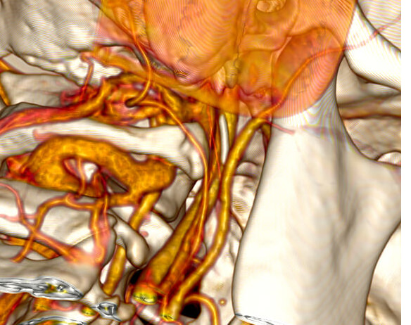


 There should be an open groove like you circled on the other side but is encased and it looks crowded. Of course this is one slice.