So I can finally see whats causing me HELLLLL!!!! Can someone tell me if its on my artery??? I cant find it but it looks like something is getting squished....can u see it? I got tons more pictures if anyone needs more too see it better, I just don't know what too show u...lol
89-ct1.jpg (56.8 KB) 90-ct2.jpg (42.9 KB)another one
96-ct3.jpg (49.8 KB)It's best to speak to the doctors about that, we're not experts on here, but they're certainly pretty long- looking at the first pic the styloid process on the right is touching the other bones (?is this the C2 vertebrae?), which would be very painful. They're certainly very close to the blood vessels...it's hard to tell if it's compressing them or entangled, but either way it's good that you've got them now, and if you can find someone to help you it'll save time when you see them. How's the doctor hunt going?
Well I found one waiting too here back from them… Everything seems too take too much time while I’m laying here suffering. I have no idea what’s it’s touching but in my side view it seems like it’s floating there and not touching anything! How do u see it touching vessels? I can’t see it lol was I born with these lengths? Or do they grow every year?? Lol because why am I in so much pain now?!
Doctors still don't know the exact cause- there's several theories! Research looks like they might grow, but also that there could be an inherited tendency towards having longer SP's, like some people have longer fingers etc. I guess. But they can also grow or become inflamed because of things like tonsillectomies, or injury etc. to the neck. It is weird how it can suddenly flare up- mine were like that, then got a bit better, then got a whole lot worse. I had other neck problems- a slipped disc- which I think perhaps altered the angle slightly and made it worse. There's other research which shows as we get older elasticity of the tissues can alter then angles of the SP's too. A lot of people on here have had endocrine disorders, like you mentioned thyroid problems, although I haven't found any research into that.
Ya I'm just trying to figure out what caused it....no tonsils out or neck injuries.....hmmmm....I think inflammation and thyroid...only closest thing i found possible...And my inflammation markers were sky high when I started seeing the doctors about my pain....they are normal now....
I think the ct would show it compression the artery but i had a ultrasound done on my Carotid artery after styloid removal thinking it might of been pinched but nothing id definitely recommend for the styloid to be removed it helped me alot but still have symptoms
Well did you look at the ct scan pictures I posted above? I’m not sure where the artery is?
Deleone said:
I think the ct would show it compression the artery but i had a ultrasound done on my Carotid artery after styloid removal thinking it might of been pinched but nothing id definitely recommend for the styloid to be removed it helped me alot but still have symptoms
I did not see how long they are? About the artery...didnt radiologyst told you and showed you or the surgeon.....they should! I dont know either...nice CTscann....go back and ask them ..did they not write a report...or what? hugs my dear xxx
They wrote a report… But they said anything about the arteries.
I would presume that the blood vessels are in red... but which are which... you could look at images of blood vessels in the neck and compare them, I guess, but asking your doctor is the best bet, as they're the experts (supposedly!!)
 Found this on the internet: it shows the main ones, but it's not easy to compare the two!
Lol ya u think… Just got another call back by a different doctor and he doesn’t do eagles surgury. I just don’t know wut to do anyone!! I was referred in Toronto because he does the surgury but he is only taking cancer patients now…do he referred me too he associate and he doesn’t want do it cause in too far away…
That's rotten for you... I don't know what you can do next Kelx- but it might be that you have to travel further? It's crazy that there's no-one else in Canada to do this? Otherwise you could research skull base surgeons- there must be some!- it's the area they work in so in theory they should be okay with removing styloids. The surgeon who operated on me was a skull base surgeon and otolaryngologist.
Ya totally rotten! I can look outside canada but if they are concerned I’m too far away in canada why would someone want too do it in the states! And it’s a whole other process to get stuff one there… Lots of paper work and appovals first by the health board… Geez how can this be so hard?! Aren’t doctors trained in certain areas like this know how to do it?! Makes no sense!
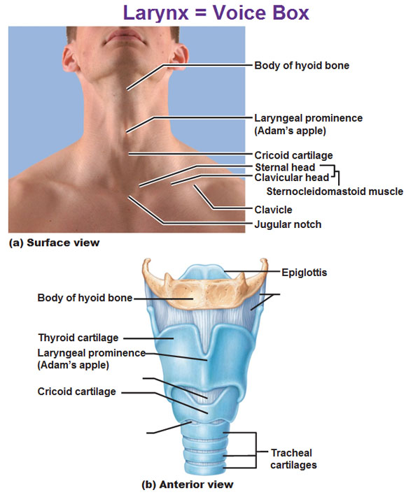
Hi Jules! Can you help me find my thyroid cartilage on my CT scan....Im having severe severe pain behind it....Soooo bad....Man I'm having problems with this neck of mine...I probably have another condition of my thyroid cartilage....When I press behind my cartilage...I have too press it from the side I can feel something sharp.....Maybe more calcification but there now?!
Jules said:
It's best to speak to the doctors about that, we're not experts on here, but they're certainly pretty long- looking at the first pic the styloid process on the right is touching the other bones (?is this the C2 vertebrae?), which would be very painful. They're certainly very close to the blood vessels...it's hard to tell if it's compressing them or entangled, but either way it's good that you've got them now, and if you can find someone to help you it'll save time when you see them. How's the doctor hunt going?
You would have to ask your doctor about it, I've no experience with that. I've added a labelled diagram to show where the thyroid would be, compare that to one of your side view CT pics- you can see the hyoid bone on those, and work out where the thyroid would be. I think it looks like a blue fuzzy area on the pics- but don't know what it would normally look like on a CT, so can't compare, like I said it would be best to get a proper medical opinion.... sorry I can't be more helpful!

Apparently, it can be difficult to tell what is Definitely getting squished as what isn't through a neutral position CT.
Without an obvious bulge, which I think very few of us on here could accurately identify in your scans, it's kind of a symptomatic based diagnoses.
I'm going through a gamut of follow up images this week because of this particular issue.
Your styloids are obviously weird, and long, and in the general area of your vasculature and cranial nerves. However, it would still be hard to give a definite "yes" or "no" as to if these are causing compression or irritation in part because Many people have similar anatomical variants with no symptoms.
Right now my doctor is super grossed out by my hyoid spike and stylohyoid elongation, but without a CT confirmation of a compression he's hesitant to give any kind of positive diagnoses or hope for relief after surgery. All he is sure if is it's not supposed to be like that, and there's a medical record of people having similar problems.
I just did a CT scan with me in a symptomatic position to try and capture the horror for sure, but the time I'm most symptomatic is sitting up so there's no promise it will produce results. I have an ultrasound later this week to try and confirm the same thing. Something's up, it's positional, and they want to know how bad it is.
The doc told me that it's hard to understand the venous in these 3d images, and things that Look tied up can be far apart, and those that look far apart can be crashing together. Because of this, my new images are only serving as a map while he's still planning exploratory surgery to get rid of all the weird BS growing in my neck.
I think you have a pretty good case, imaging wise, to take to Dr's and petition to these suckers being cut out.
I've tried to label some of your anatomical structures, but my time at a keyboard is limited due to.. ya know.. neck spikes.
Also, I hope my anatomy is up to snuff. I make my living taking dead thing Apart, not keeping them together.
Hope this helps! I always feel better when I have an idea of what I'm looking at.
Blue- Approximate location of the thyroid, with cloudy partial visual of the thyroid and thyroid cartilage (3d programs are based on density parameters, sometimes things get lumped together. The blue haze indicates a different type of tissue)
Purple- External Jugular
Green- External Carotid
Yellow- Cervical veins
Black- Vertebral artery
Thanks SnappleofDiscord, that's really helpful labelling- I'm not technical, so can't do anything like that!
My CT pics were at a totally different angle- sections from the top of the head looking down through my neck- the venous compression could be seen really clearly both sides that way. Hope that the new images show something for you (so that the docs are confident of surgery helping, I mean!), or the ultrasound. Is it a normal ultrasound, or Doppler one- that can sometimes be helpful if arterial compression is suspected.
