I have no idea how to read my scans but I know I have Eagles syndrome...help!

Hi, I hope I am posting this correctly as it’s my first post. I have to start by saying thank you because I think this forum is what has given me direction and hope. I have been told for years that I am fine…when I am most definitely not. It’s a nightmare isn’t it… being told you are a picture of health, but you feel the complete opposite like every step is a struggle.

Approximately four years ago I started experiencing the weirdest symptoms. It started with periods of odd fatigue, headaches, brain exhaustion. One night approximately 6 months after this started, I woke up from a dead sleep to lightning like pain in the left side of my face. Besides this sudden facial pain, I also felt exhausted and just ill. I went to the ER, they did an MRI and said I was fine and should follow up with neurology. They diagnosed me with complex migraines (I’d never had migraines before). This started a period of years of neurology visits and many ER visits to be given things like the migraine cocktail (which made me feel absolutely horrible) or to have eye pressure tests, etc. The lightning pains ended shortly after they started but ever since this the left side of my face has been numb. It feels like I had lidocaine at the dentist on the left side every day in varying degrees. I also started having ridiculous brain fog, dizziness, eye pressure, eye watering, fullness in my ear…. everything left sided. Over time, my voice changed, I feel like something is in my throat, I can get short of breath and have the sensation I can’t swallow. After a long while, I realized I could not sleep on my left side because something gets compressed and I would wake up to a day of headaches and brain fog. I saw over 50 doctors during this time from every specialty. My primary wasn’t helping me so I was determined to figure it out. I could go on and on but something tells me my story is similar to so many so I will try not to bore you. The important points are:

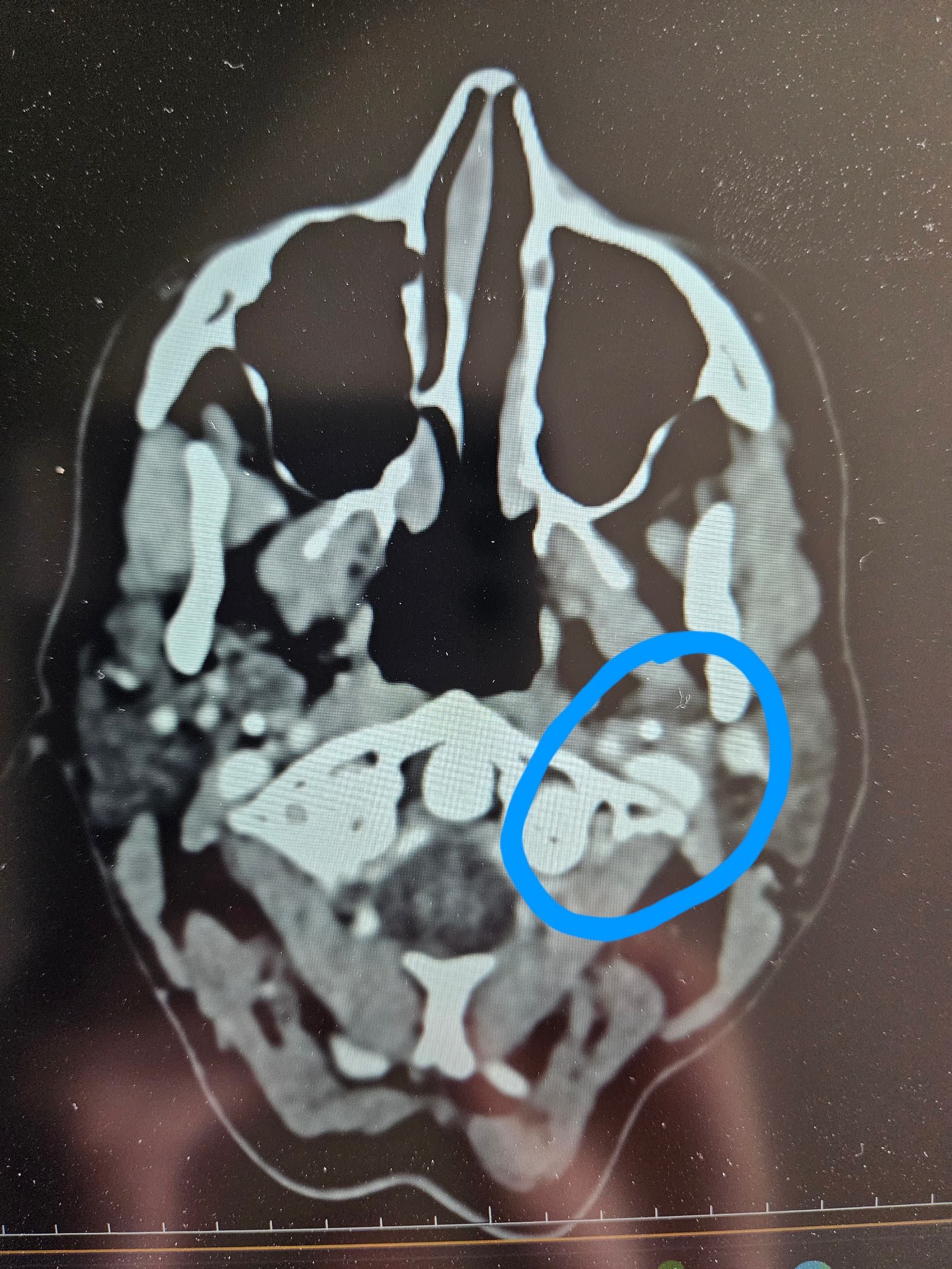
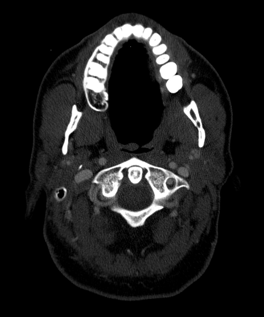
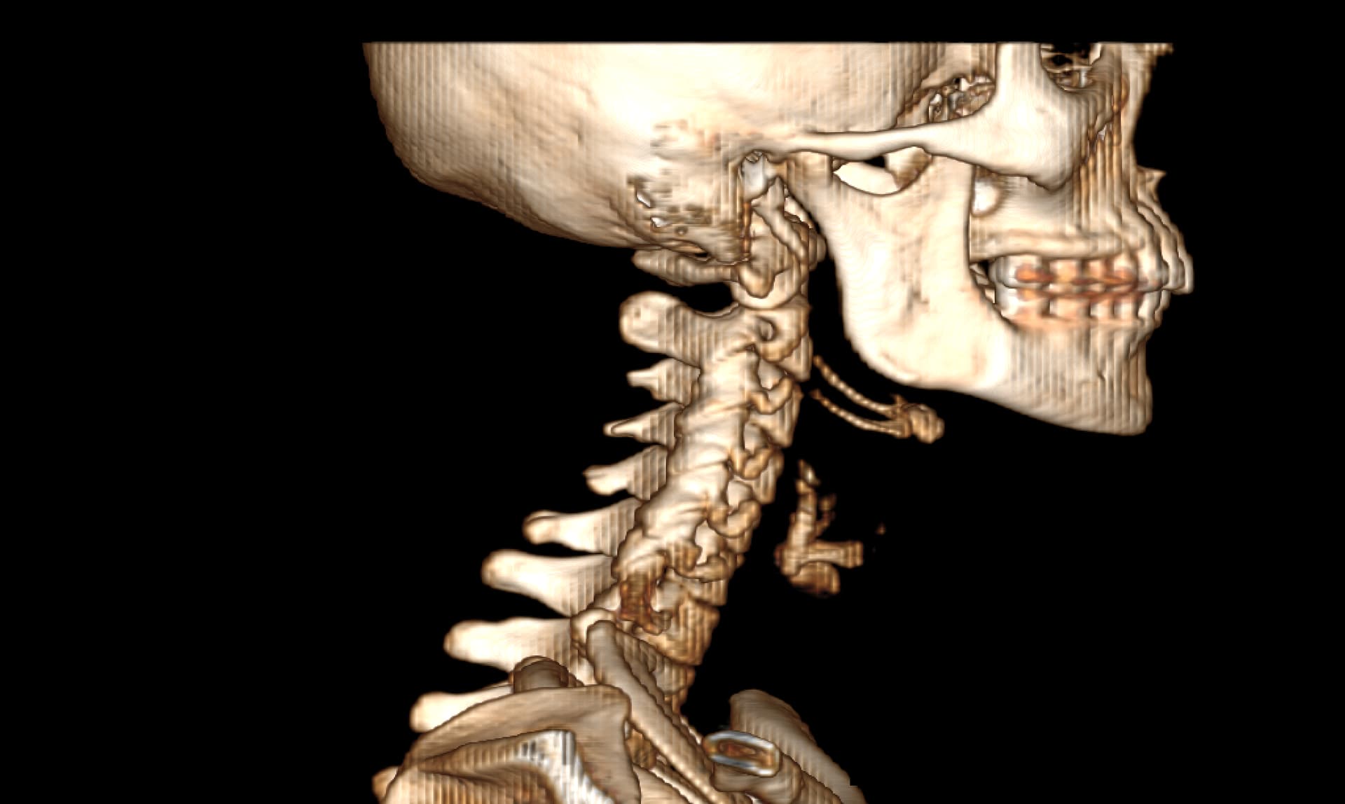
I did have an MRA done at Mayo that showed severe blood flow compression on my left side with my arms up. Because of this test, I went back to UCLA and did tests for Thoracic Outlet Syndrome. The limited tests they can do for this seemed to help. So, I had a left rib resection 2 yrs ago. Before this surgery, I did do a CT w/wo contract to look for Eagles and the report came back saying my stylohyoid was normal. So I went ahead with the thoracic outlet surgery. That surgery got rid of the daily headache I was dealing with. It seemed to have relieved pressure headaches and some of the loss of feeling in my arm. It never helped with the facial pains and dizziness though that I live with. Months after this surgery, I also developed Frozen Shoulder. I found it odd I got this so long after the surgery and when I was in PT. It seemed like the more we tried to use my arm and mess with my neck, the worse it got. I then went back to Mayo and had a head and face MRI that showed compression of my vagus nerve with an artery around my brainstem. It also showed a blood vessel touching my trigeminal nerve which is what they look for with trigeminal neuralgia surgery. So I took this and followed this lead. After visiting three trigeminal neuralgia surgeons, most of them said since I didn’t live with shooting lightning pains, I wasn’t a good candidate for the surgery. I was referred to some neurosurgeons regarding the vagus nerve compression at the brain stem. I almost had surgery booked for this and then the dr said something more is going on here, let’s hold off. This led me to circle back to Eagles Syndrome. Even though my CT hadn’t shown eagles on the report, I took it to Dr. Osborne. He indicated I had a calcified ligament. I almost moved forward with surgery, but I am concerned about the vascular issues I know I’m experiencing. I worry that I need to ensure we target these issues in my surgery. In the past few years, I’ve had near fainting spells, dizziness (I no longer can drive), BP issues that were similar to POTS but not, heart palpitations, horrible brain fog, and a complete inability to concentrate. I also get left sided body numbness and weakness. My left leg can even be impacted. It’s not to where I can’t walk but it feels like I’m living on a cruise ship trying to balance myself and my left side is weak. I feel like I need to confirm I have vascular issues and make sure I care for this in surgery. I tried to get a second opinion with an ENT at UCLA and they said they saw nothing wrong with my stylohyoid but that they see something wrong with my hyoid bone that they might be willing to operate on.

I really need help reading my images myself. I have anonymized them, but I don’t even know how to provide the right image to view here. I want to be able to advocate well for myself but I need to know what I’m looking at. I feel like the group on here know more than any doctors at this point. I would love to get back to normalcy…driving my kids around, actually getting sleep, being able to think clearly, being able to physically exercise, to be thriving and not barely surviving. Things I took for granted before this, I won’t again. Pls help if you are able. I can post my images following this first post though guidance on steps would be appreciated.

3 Likes

Hi @PhoenixRising I can help you find the images that would be helpful for others to give their opinions. @TML is really good at looking for vascular compression around the C1/Styloid and also the hyoid. If you have access to your CTA or CTV with contrast, go to the axial views and see if you can find the following slices. The first two are the hyoid (horseshoe shape bone). The last one is the C1 (vertebrae with wings). If you can upload these. @Isaiah_40_31 & @Jules will be able to give you more details with regards to your symptoms.

2 Likes

@PhoenixRising sorry to hear about you long, exhausting, and scary story. I think you may have come to the right place.

Glad to hear the TOS surgery provided some relief from the pressure. Your IJVs could still be compressed between your styloids and C1 though, causing more pressure.

Your dizziness may be coming from IJV issues. But I am more concerned about your hyoid bone. Particularly the left greater horn. If it’s in contact with your left ICA or the carotid sinus area it can cause dizziness, unilateral weakness, pain, collapsing episodes and more. It essentially causes carotid sinus hypersensitivity symptoms.

As @Rosie has mentioned, we need to get the axial view of your hyoid (specifically the greater horns), and your styloids/IJVs (at C1). The CT with contrast would be best. I see @Rosie has uploaded some images so you know what to look for. Here’s a few more since everyone’s anatomy looks a bit different.

C1:

Greater horns of hyoid:

1 Like

Hopefully you can upload the images okay, if the site wouldn’t let you earlier you should be able to now.

It is a tricky one & I feel for you with the dilemma of surgery- some of your symptoms sound like possible IJV compression, some potentially carotid artery or carotid sinus compression, and also vagus nerve compression, so there’s alot potentially going on! As well as the ES symptoms like swallowing issues. And also some less common ES ones like possibly spinal accessory nerve compression given the issues you have with your shoulder, & perhaps hyoid bone syndrome too!

So if we can help at all with your imaging maybe we can give you some pointers, especially as you’ve had differing medical opinions…

So sorry that you’ve been having these symptoms for so long, & with little ones to look after too :hugs:

2 Likes

Thank you for all of the replies and helpful feedback. The morning after I posted, I felt relief that people were trying to help me. I woke up with a sense of relief and felt more informed. It took me so long to reply partially because I was really trying to get the images shown and also because I’ve had a few pretty rough days. After spending probably 10 hours trying my best to get the images shown, I think I’ve come to the conclusion that either I’m using the wrong software (I’ve tried 3 programs) or that the imaging I got was poor. I can’t seem to get an image of my hyoid bone fully like the examples. I couldn’t catch one full image of the bone entirely. I don’t know if that’s just indicative of the issue I have or if it’s totally user error.

If you can let me know if I’m at all on the right track or some general feedback about the Imaging that would be super helpful. I anonymized the file if anyone is willing to help view it in the program they use and see if they get better results.

I’ve had a few days lately where I feel like I could pass out or am very dizzy so I’ve had to limit activity. So, if I don’t reply asap, it might just be that I’m riding out the rough wave. I am super thankful already for the help and guidance!

3 Likes

Hi @PhoenixRising good to hear from you. It’s not unusual for the hyoid not to show fully on one slice as it can sit at an angle with the greater horns sitting higher. Hopefully @TML and others will be able to give you their views on your images soon.

I’m sorry to hear you have been having a bad few days with dizziness, its a horrible symptom.

3 Likes

I’m sorry, I’m not so good at reading these images, I can’t see any obvious compression of your IJVs which some of your symptoms do indicate could be happening, maybe @TML can see…
I’m sorry that you’ve had some rough days, I hope that these improve :hugs:

2 Likes

@PhoenixRising before I annotate anything, can you download Radiantviewer and open your CT in Radiantviewer and construct a 3D model? Will be helpful in looking at the calcification fully!

2 Likes

Hi, @TML Ty for your reply. Is this what you are looking for? I feel like this is not very clear but this is what I got so far.

I see this view from the back and I found it odd the vessel on the left but maybe that’s normal.

I hope you are having a good day and thank you for any help you can provide.

@PhoenixRising , I did a little annotation in your imaging of what I see. Your carotids on the left are in a different position than I’m used to seeing them so I’m not sure my assessment of what your greater hyoid horns are doing is correct, however, it looks like the left, one at least, is making contact with vascular tissues & from your symptoms I’d guess it is your ICA/ECA or the bifurcation where the two split off the common carotid.

1 Like

@PhoenixRising can we see your right side in 3D? Your right styloid is longer than your left based on the axial view

Right IJV looks great at level of C1. Some compression of left IJV, but uncertain if styloid is involved unless you go up a slice or two

1 Like

@PhoenixRising there could be something going on with your greater horns.

can you get this same slice but have it where there’s contrast in your arteries/veins like some of your other images? Also need a slice or two up to see left greater horn

2 Likes

Hi, here is my right for comparison:

I don’t know, Dr. Osborne had commented something that you have to take into account body frame when looking at the size of styloids and the amount of room in a person’s anatomy. So even though my left is not crazy long, he’d said there was calcification and that it was thick. I think in this image my right is smaller than my left though which would align with my left sided symptoms.

Did I catch the greater horn here? I waited until the hyoid bone was gone and then went a few slices up.

Thanks again with deepest appreciation!

1 Like

Your right styloid is looks to be adding to the compression of your right IJV, & the left greater hyoid horn does look to me like it may be closely associating with your ECA on that side. @TML will be able to give you more accurate info based on the axial slices.

@PhoenixRising yes that’s your left greater horn!

It is in contact with something that is vascular but I’m unsure what. It’s either the ECA or a branch of the ECA. I think more likely a branch because the larger vascular bubble behind it looks to be the carotid bifurcation, and then behind that is the IJV.

Can we see the right greater horn with contrast? It looks to be touching something too.

2 Likes

I had the opportunity to review @PhoenixRising ‘s imaging. See first four annotated images attached, and the remaining four images attached to following post.

Regarding styloids - they don’t seem to actually be doing much. The right styloid does come in contact with the right IJV, but does not compress it against C1. However, the right IJV does become pancaked against the front/right side of C1 transverse process.

Your left IJV is a different story. Left styloid again is not a problem at all. However, your left ICA is compressing the IJV. I have never seen this before. If you follow the left ICA and left IJV, the left ICA actually slips in behind the IJV and switches sides - I have also never seen this before. At the point of where they meet to swap sides, the ICA is compressing the IJV. Not only is this not ideal for the IJV, but also, the ICA becomes closer in proximity to the SCM muscle which could cause mechanical irritation/compression to the left ICA in certain positions. It’s almost like you have a “tortuous” ICA, but instead of it being inwards (which is what we usually see when there is a tortuous ICA), yours is directed outwards.

Further down, both your greater horns of the hyoid are in contact with your ECAs, and both of your thyroid superior horns are in contact with your common carotid arteries. Not sure if these are causing any symptoms though. There is no visual compression in the neutral position, but that isn’t to say there isn’t any compression in other positions. There could also be some scraping while swallowing.

Hope this helps.

1 Like

And actually, if we look at the earlier 3D model posted, you can visually see the left ICA loop around behind the left IJV:

When you say you can’t sleep on your left because something gets compressed, I can almost guarantee it’s your left ICA pushing against your SCM, or your ICA further compressing your IJV, or a combination of both. That or it’s your left greater horn or left thyroid cartilage.

2 Likes

Great observations, @TML! I saw that loop of vascular tissue on the back side of the IJV but wasn’t sure what it was. The axial views reign supreme! Though I still do better w/ the 3D images. :wink:

2 Likes

@PhoenixRising - Based on @TML ‘s findings, it would be very wise for you to see Dr. Nakaji (closest to you) or Dr. Costantino who both do more compliated ES surgeries where vascular compression is involved. Dr. Hepworth & Dr. Cognetti would also be helpful but are booking into next year at this point. We have also recently added Dr. Babu Welch in TX to our Doctors List. We don’t know how much experience he has w/ vES surgeries but he has done surgery w/ a C1 shave for one of our members.

2 Likes

I thought the same!

1 Like