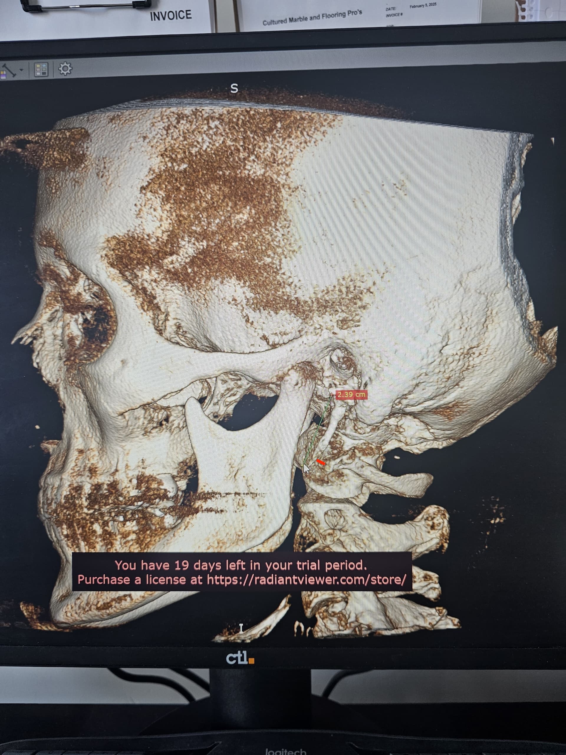
Just wanted to share my “normal” CTA. I don’t understand why it’s so damn hard to get help with this syndrome. I went directly to radiology to get a CD, I went home and converted it to 3D I’ll attach images. This is nuts!
EXAM: CTA neck with IV contrast.
INDICATION: Migraine, evaluate for Eagle syndrome.
TECHNIQUE: Contrast-enhanced CT angiogram of the neck was performed with 1 mm axial images, coronal and sagittal reformats, and 3D maximum intensity projections. Carotid stenosis measurements were made using NASCET criteria.
COMPARISON: Cervical spine radiographs dated 6/20/2024; MRI cervical spine dated 1/28/2025.
FINDINGS:
Vasculature:
There is a conventional three-vessel anatomic configuration of the aortic arch. There is no atherosclerotic plaque at the bilateral carotid bifurcations with without stenosis by NASCET criteria. There is no plaque ulceration or intraluminal thrombus.
The bilateral vertebral arteries appear normal without significant stenosis. Incidental note is made of proximal origin of the right PICA from the right V3-V4 junction.
There is no evidence of dissection.
No evidence of venous thrombosis.
Nonvascular findings:
Lung apices: There is mosaic attenuation of the lung apices, potentially the consequence of expiratory phase of imaging. No focal consolidation or mass is present.
Aerodigestive tract: Normal.
Lymph nodes: Scattered cervical chain lymph nodes are present without concerning imaging features.
Glands: The thyroid gland, submandibular and parotid glands are within normal limits.
Orbits and globes: Within normal limits.
Paranasal sinuses and mastoid air cells: There is sequela of bilateral uncinectomies/maxillary antrostomies. The paranasal sinuses are well-developed and well-aerated. The middle ears and mastoid air cells are well-aerated. Incidental note is made of pneumatization of the petrous apices.
Dentition: No dental caries or periapical lucencies. There is torus mandibularis interna. There are scattered dental amalgams and changes of prior root canals.
Skeleton: No elongation of the styloid processes or ossification of the stylohyoid ligaments. There is mild reversal of the normal cervical lordosis, which may be positional. No lytic or blastic skeletal lesions. No significant spinal canal or neuroforaminal narrowing.
IMPRESSION:
No acute abnormality great arteries of the neck. Specifically, no elongation of the styloid processes or ossification of stylohyoid ligaments to suggest imaging evidence for Eagle syndrome.
(attachments)