@Rosie - ECA compression/irritation does make sense w/ the symptoms you have. It looks to me a bit like your left IJV is a little compressed, too, as @TML noted. Some of your symptoms seem like IJV compression to me as well. It’s rare, but we do have members who have both jugular & carotid compression.
1 Like
Hi all, well I am not quite sure what is going on but I have had an email from Mr Hughes office to say that they cannot see my Styloids on my CT angiogram and want me to go for another scan! I have waited a month to hear that. I have sent screen shots of my Styloids in 3d and circled them, am waiting for a reply.
2 Likes
@Rosie - That also happened w/ our member @McWelly, & hers are significantly long. Gotta wonder if doctors sometimes look at scans with their eyes closed. ![]() I hope you get a reply before another month passes.
I hope you get a reply before another month passes.
2 Likes
@Isaiah_40_31 @Rosie
@Jules @TML
Reading through your posts @Rosie - could be me! How can JH not see them? Tbh, how much time is dedicated before and during a short telecom? Incidentally I also saw PA who was of opinion I needed a neurosurgeon. I’ve spent thousands of hours and £, had extensive dental work - some of which wad probably not needed.
It started in a dentist chair ![]()
![]()
![]()
2 Likes
That’s so frustrating @Rosie , especially having paid for the consultation and waited a month, it’s ridiculous! Maybe see what reply you get from your email , & otherwise try Mr Axon? Although very frustrating having to fork out for these appointments ![]()
2 Likes
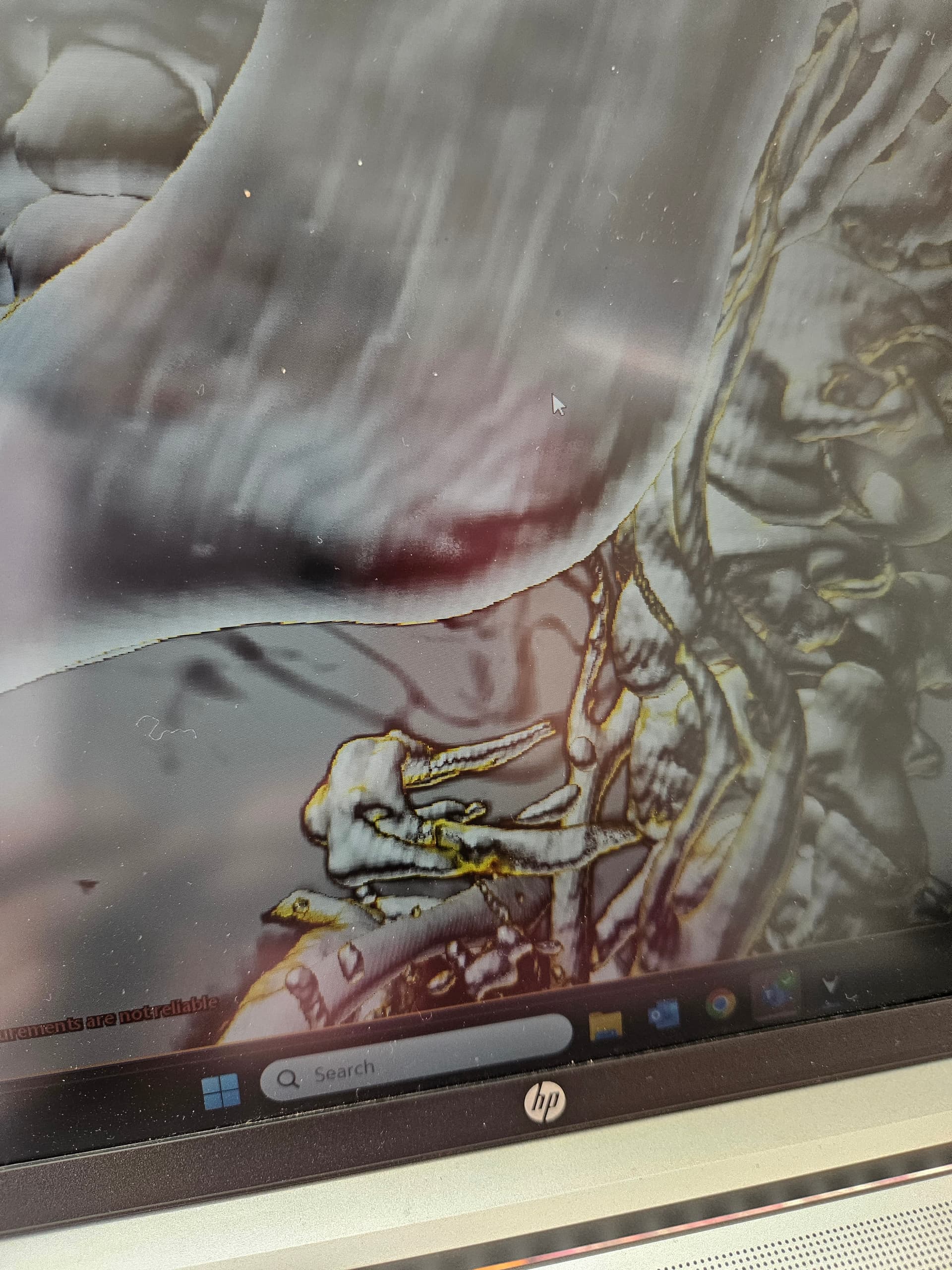
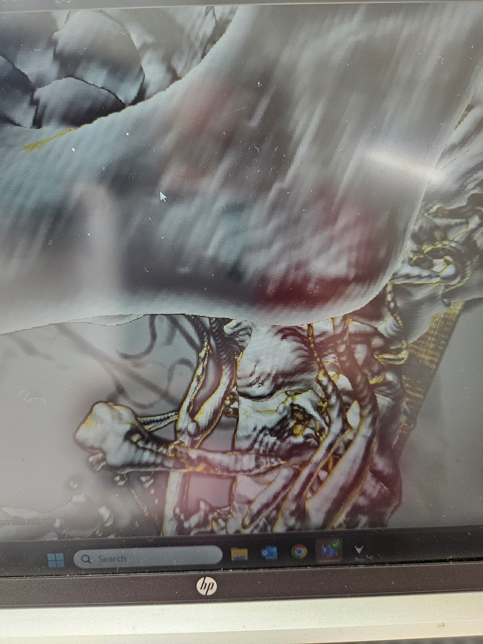
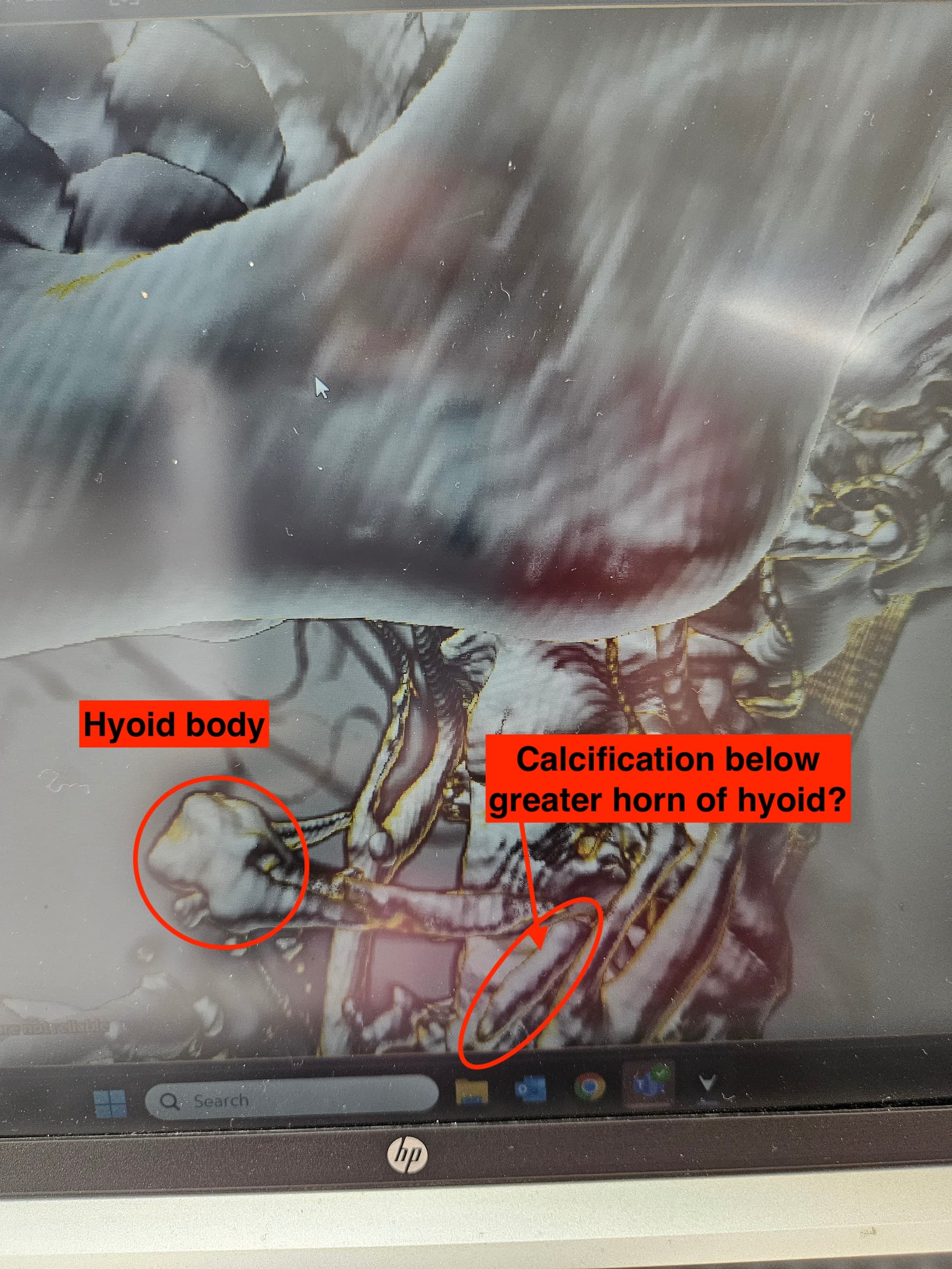
Good Morning all, @TML I have managed to get a couple of extra 3d shots of my hyoid, its difficult to get the right angle, but there appears to be a piece of calcified thyroid cartilage just below the greater horn of my hyoid. I cannot see any obvious compression of my left ECA but maybe the clicking when I swallow is the horn against the cartilage. Its very annoying and uncomfortable. Maybe having the styloid/ligament removed would help this. Any theories on this greatly received.
1 Like
@Rosie which program are you using to convert your CTA imaging to 3D? Are you able to use Radiantviewer? I think it may provide clearer modeling for us. I’ve attached my own CTA imaging that I converted to 3D via Radiantviewer. I find the program is great for showing vascular structures (and styloids!).
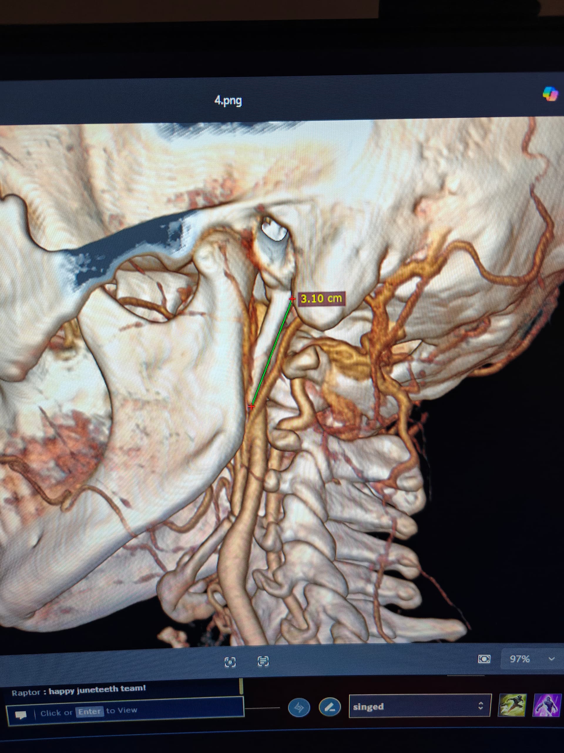


@TML thanks for your reply. The file was sent via NHS and the viewer recommended was Weasis. I am not very good with IT and my employers IT department sorted it out, so unfortunately I don’t have access to use anything else. I cannot see anyway to view my IJV in 3d on the program only the Carotids seem to show.
1 Like
@Rosie you have access to all your CTA imaging on your computer, correct? I can help you with Radiant viewer.
Step 1: download radiant viewer onto your computer - https://www.radiantviewer.com/ (click download now for windows)
Step 2: once it’s downloaded, open the program to install it. I think it’ll ask if you want to start a free trial, indicate yes.
Step 3: once you get the free trial started, open the program and click on the file folder in the top left. Clicking on that will allow you to choose a file from your computer that you want radiant viewer to open. So find your CTA imaging file (wherever that is stored on your computer) and select it.
Step 4: once radiant viewer opens your CTA imaging, it’ll show all the views on the left side (i.e., axial, sagittal, etc). I would click sagittal. Once you have that selected, click “3D” at the top (looks like a 3D cube). It’ll turn your imaging into a 3D model instantly. Then you’ll be able to spin the model around however you like and be able to zoom in and out and whatnot.
If you don’t have your CTA files saved on your computer than you can ignore all this. But I really think radiant viewer will help here. It’ll show your jugulars and carotids clearly (and styloids). I’m guessing you do have your CTA files saved somewhere, since you were able to provide me with the slices from the axial view?
2 Likes
Thank you for help @TML I would have to check with our IT as I am using work laptop unfortunately and they don’t allow unauthorised downloads ![]() . I don’t presently have access to another device. I can rotate the view, so I will see if I can get a better view. Thanks again
. I don’t presently have access to another device. I can rotate the view, so I will see if I can get a better view. Thanks again
3 Likes
@Rosie - The body of your hyoid appears to be very thick, almost like it is excessively calcified though I can’t say if that’s contributing to your symptoms. I also see the bit of calcification below your left hyoid greater horn & agree w/ you that it may be causing or contributing to the clicking.
I’ve annotated your second image to make sure I’m looking at what you think might be calcified thyroid cartilage below your left hyoid greater horn.
1 Like
Thank you @Isaiah_40_31 yes this was my thinking. Looking at the hyoid body straight on it appears more calcified on the left, so I will bring this up once I manage to get a further consultation. Hoping I may hear something from Mr Hughes privately next week and have spoken with Mr Axons secretary who is looking out for my NHS referral, everything takes so long, its painful… literally ![]() .
.
2 Likes
A couple a years ago I had blood tests which came back very low for Vitamin D3 and have been taking a supplement since, levels back to normal now. Does make me wonder if I was low then because I was growing this spike etc.
1 Like
Interesting thought, Rosie. Maybe someday we’ll know a lot more about the mechanisms that cause styloids to elongate/s-h ligaments to calcify & how those affect our bodies in other ways.
1 Like
Hi well I have eventually heard back from Mr Hughes office by email today and “my styloids are normal” that was it. I’m gutted, to say the least. So no explanation for my symptoms again…
I’m so sorry that he wouldn’t help you! They don’t look normal to us! Unfortunately sometimes doctors decide not to offer surgery, we don’t know why…I don’t know if it’s possible financially to see Mr Axon, given it looks as though you might have some IJV compression? Although he doesn’t treat hyoid bone syndrome if your hyoid is involved…
Sending you a hug & thinking of you ![]()
2 Likes
Hi @Jules thank you for your reply. I booked an appointment yesterday with Mr Axon privately for next Friday, as I had given up hope hearing from Mr Hughes, but I feel so deflated as I was sure that my left styloid was causing my problems. The pain is wearing me out, constant ear pain, temple pain, headaches, tinnitus and throat pain. I will see what Mr Axon says but if my styloid is not the issue, then what is.
1 Like
It certainly sounds like it’s ES, but so frustrating he disagrees…hope that it goes better with Mr Axon, let us know how you get on ![]()
![]()
1 Like
@Rosie - It seems Mr. Hughes diagnoses ES based strictly on the length of the styloids which is incorrect. As we’ve mentioned on the forum the shape, angle of growth, thickness, curve, if it’s twisted or has bony outcroppings, etc., can all cause symptoms w/o the styloids being long. If Mr. Axon says you don’t have ES due to styloid length, bring up the other physical features of styloids that can cause symptoms to see if he will think “outside the box”.
1 Like