Question About C1 Shave

If I could put into a picture how my VES symptoms make me feel, it would look like a combination of these: a ton of pressure, asymmetrical head/neck pain, and one wonky eye.

The pillow compression picture sums it up well, but the others add emphasis, especially Bill the Cat’s crazy left eye. For clarification, I am not a cat. :wink:


ImPxeX1_d
1341295-btc

1 Like

Hahahaha you are not a cat :joy::joy::joy:. Humor gets me through life :smiling_face_with_three_hearts:

2 Likes

So sorry that you’re feeling so rough, but good you can laugh :hugs:
I hope that the doctors you’re seeing soon will help- if not maybe see a veterinary surgeon- my hubby & I often say we think they’d probably do a better job than some of the doctors we’ve seen!

2 Likes

Love the pictorial explanation of how you’re feeling! Made me laugh, too!! The pictures are so apropos for your situation! I hope your surgery date sticks tight so this will be behind you SOON!!

1 Like

One of my girls goes to the vet on Wednesday. Maybe I’ll hop up on the exam table with her. At least at the vet’s office there’s no hassle with health insurance companies. I can just hand over my credit card, have surgery, and return home with a cone around my neck.

2 Likes

Again, it’s been a rough couple of days. Last week ended with updated CT scans, and the results received earlier today read as follows,

“Styloid processes: Both styloid processes are elongated on the order of 4.3 cm (image 22 of series 304, image 17 of series 305). There is slitlike narrowing of the internal jugular vein between the styloid process and C1 on the left and right sides (image 177 and 181 of series 5, respectively). The distal tip of the left styloid process contacts both left internal carotid artery at the base of C2 level (images 210-216 of series 5). Just slightly proximal to this, the left styloid process abuts the left external carotid artery (image 211 of series 5). The right styloid process tip is 0.4 cm away from the right internal carotid artery and 0.2 cm from right external carotid artery at base of C2 level (images 216-219 of series 5).”

The good thing about the doctor ordering new scans is that I went to a different imaging company this time around. The report is more detailed than the one I received last summer, and 3D images were immediately uploaded and accessible. Though they were requested, I never received 3D images from the August 2023 scans. I’m sharing 2 of the images where finally the size, shape, and positioning of the styloids can clearly be seen.

When I shared the images with my sister, she said they look like Predator. She removed both feet from her mouth when I told her that’s MY skull. The expression on my mother’s face made her look somewhat like Bill the Cat. She finally gets it now that she’s seen the 3D images.

Next week I see Dr. Taka. Onward!


1 Like

They are real daggers! And good to get the clarification on your CT report this time, very detailed, how unlucky are you to have jugular compression and artery compression/ irritation! Certainly explains why you feel so rough :hugs: I’m glad that your family finally get it!

1 Like

Made me laugh again! Love your sense of humor, @CoHDa!

You do have a couple of really nasty looking spikes. I’m quite impressed by the thoroughness of the radiology report you got. If you don’t mind sharing the name of the radiology lab & radiologist who read your scans, I will make a note of that on our Doctors List as those will be a helpful resource for others who live in your part of the US. I expect at least part of the reason for such a thorough report is the specific info your surgeon requested to be reviewed in the scan so kudos to him, too!

You will fell SOOO MUCH BETTER once those spikes are outta there! I’m also glad the imaging gave credence for your complaints to your family.

2 Likes

@Jules Typically when I get sick, it’s to the nth degree. Thankfully, the scans and report bear that out. The diagnosis part of the journey and wondering why I feel like garbage is over. Now, having seen actual images of the styloids, I feel a growing sense of disbelief and shock about the fact that I’ve been walking around all this time with the equivalent of sewing shears or outside calipers in my neck that at any moment - with a hard fall or bump - have the potential to do even greater harm than they already have. It makes me think I really do need to have a cone around my neck until these things are removed.

2 Likes

@Isaiah_40_31 I’ll let you know what the vet says after my appointment tomorrow. :wink:

So, the scans were done by Borg & Ide Imaging. I rarely use them, because I’d always thought they were inferior to the University of Rochester Medical Center (URMC), where I’ve had the majority of imaging done for just about everything over the years. But I’ve become very displeased with URMC. I learned that one of Borg & Ide’s locations is in the next building over from Dr. Coniglio’s office, and being that he is not affiliated with URMC there was no reason not to give Borg & Ide another try.

I’ve had tons of scans with contrast done, but never before had I been taken to recovery following a scan and given the once-over by a nurse, who had me sit for 10 minutes to make sure I was okay before being “discharged.” I was given a choice of beverages to enjoy while I sat, and then was checked once more before being escorted to the exit by another nurse. I was lucky if I got a simple “bye” from URMC staff when I had scans done there in the past.

By Monday, Borg & Ide had uploaded all of the images, 2D and 3D, as well as the report, which was written by Dr. Bradley Cole. In addition to what I’ve already shared about my styloids, Dr. Cole also provided a summary of his findings about my tongue and sublingual space, pharyngeal space, carotid space, lymph nodes, epiglottic region, laryngeal structures, major salivary glands, thyroid gland, esophagus, upper chest, and spine. He noted in his report that his findings are in comparison to the URMC report from August 2023.

For reference, the images were presented in 15 “Series” clearly labeled with the “view” and including the number of images in each series. In the first picture I provided, you see Series 12-15 listed. Then in the second picture you see Series 11 and some of the 40 images that appear when clicking on the hamburger button in the right margin. I’m not sure if this is similar to what other imaging labs do, but it is quite the different presentation compared to what I got from URMC last August.

I hope this information helps.


2 Likes

Thank you for the information. Dr. Cole was really thorough & it’s great to know he took the time to compare the current images with those from your 2023 scan.

1 Like

Again, my apologies for not replying/posting sooner. Like most folks here on the forum, I am worn out for a number of different reasons.

To get to it, I saw Dr. Taka last week and was very pleased with the encounter. I won’t go into all the details, but I will say that I was pleasantly surprised by the manner in which he conducted himself and his thorough evaluation of my situation. One of the first things Dr. Taka did was clarify that he won’t be doing the C1 shave. His role in this process is to perform an invasive angiogram to survey and draw up a roadmap of my vascular system in order to help better formulate a surgical plan of attack. He reviewed the CT scans with me pointing out indications of fibromuscular dysplasia (FMD) as well as venous collateral pathways.

FMD causes arteries (mainly the carotid) to alternately narrow and balloon, so the artery resembles a strand of beads. This rare disease (yet another one) adds to the degree of complication of performing the angiogram and any other procedure. Dr. Taka was glad to know my cardiologist already has me on a daily regimen of a low dose aspirin to mitigate the possibility of an aneurysm or stroke.

Venous collateral pathways essentially confirm compression of the jugular vein. When there is an outflow obstruction, blood is going to find its way back to the heart through extrajugular pathways generated by the body to reroute blood around the primary pathway that is impeded by the compression. The invasive angiogram that Dr. Taka is going to perform will provide more information about the extent of the collateral pathways that in turn provide more information about the outflow obstruction.

I will add that one thing that really surprised me - actually two things - is Dr. Taka genuinely wanting to know if I’m okay with the plan to proceed with the angiogram. I had to laugh and asked him if he’s okay with it. He is. The second thing that almost knocked me off my chair was Dr. Taka taking out his phone, looking at his calendar, and asking me if I’m available April 4. Again, I had to laugh and told him I’m available whenever he is. I can only think of one other doctor who actually took the initiative to look at his calendar and schedule a procedure himself, instead of passing the responsibility off to a secretary to follow-up on in the next day or 2 or 3 or 4.

Anyway, that’s the long and short of it. If it turns out that I do need the C1 shaved, I don’t know at present who’s going to do it, but if they’re anything like Drs. Coniglio and Taka, I’m not worried.

4 Likes

@CoHDa, look, now that’s getting really cool and interesting. So did Dr Taka mention why the FMD might have developed in your case? Could it be something related to the connective tissue in general, and affect not only the blood vessels but muscles too, stability of the spine, ligaments etc? As then it would make sense why the body may have “decided” to start “implementing” various mechanisms to address it.

E.g. I would imagine it going something like that: certain posture makes the body feel dizzy as the arteries can’t hold the shape like “healthy” arteries can, so the body subconsciously changes the position of the head to make less compression on the arteries, sacrificing veins. Eventually muscles just transform to make it permanent, and causing the C1 compressing the veins, but keeping the arteries free of any significant impact.

What do you think?

But overall, I’m impressed and very happy for how your appointment went.

3 Likes

@vdm, wow! You’ve posed a very interesting and quite plausible theory.

Dr. Taka didn’t actually run through details at great length beyond indicating that FMD is a connective tissue disorder. For whatever reason, healthy cells within the artery or arteries are overtaken by fibrous cells. The result of the fibrous cells is arteries that become either too stiff and rigid or conversely too malleable and elastic. FMD can occur in various parts of the body, so it is possible that it’s affected not only my carotid arteries, but my brain and my kidneys as well. Woo-hoo!

I don’t know if FMD is a byproduct of VES or if it is a primary disorder that is wholly separate and unrelated. What I do believe is that a sick and chronically ill body is likely to continue to break down as the body’s various systems are no longer functioning at optimal levels and incapable of maintaining homeostasis. The body just becomes an environment that cultivates cellular mutations and a multitude of subsequent illnesses, related and unrelated.

Anyway, that’s my two cents for now. April 4 is when the fun actually begins. Thanks for weighing in in the meantime. Stay tuned for Dr. Taka’s report!

3 Likes

Take care, and thanks for sharing all the info with us!!

3 Likes

I’m glad that Dr Taka was so helpful & will pray that it goes well on 4th April… Thanks for updating us :pray:

1 Like

What a breath of much needed fresh air your appointment w/ Dr. Taka was. Thank you for sharing the details with us. It’s a huge blessing that you were able to get your angiogram appt. w/ him so quickly! That’s next week!! I hope if C-1 needs to be shaved Dr. Taka or Dr. Coniglio can refer you to someone who will do it for you.

I really appreciate your updates. Your self advocacy has paid off big time!

2 Likes

I made it home from the hospital yesterday afternoon relatively unscathed. Needless to say, it was a long day, but the procedure went well. Dr. Taka performed a cerebral angiogram to help Dr. Coniglio determine how to proceed.

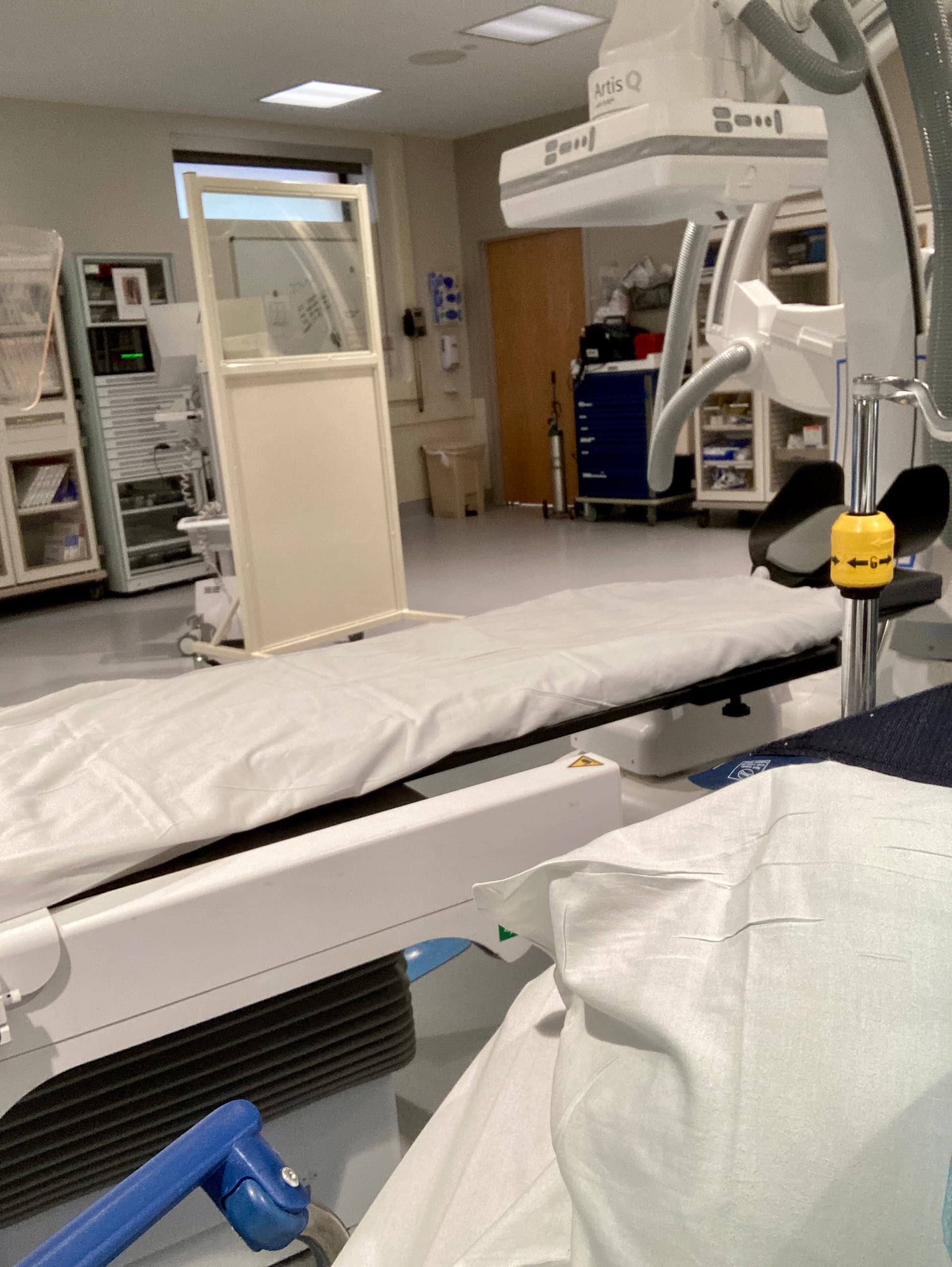
For those who don’t know (I haven’t seen anyone on the forum discuss this in detail), an invasive angiogram involves making an incision in the femoral artery in the leg and then threading a catheter with a camera to the site in question. Typically, it’s the coronary arteries, but in the case of VES, it’s the arteries in the head and neck. I was wheeled into the procedure at 10:00 AM and into recovery at 11:27 AM.

Invasive angiograms almost always require anesthesia to knock patients out. I, however, remained awake, because Dr. Taka needed me to be able to turn my head left and right as well as nod my head forward periodically during the procedure. To accomplish the feat, I was given an intravenous cocktail of Versed and Fentanyl. I felt nothing, and the procedure seemed to float by in what seemed like 10 minutes.

While Dr. Taka was examining the arteries in question, he was also capturing radiographic images with the use of two large cameras in the room, one positioned overhead and one beside me. This procedure further helped capture things that all the previous MRIs and CTs missed.

I had a 4-hour stay in recovery following the procedure, because the incidence of complications with the femoral artery significantly decreases after that point. Dr. Taka came in during this time (the majority of which I had to remain lying flat) and told me he found exactly what we expected. My left side is completely compromised due to severe narrowing and drainage of the internal jugular vein as well as the vertebral artery. He found that venous collateral pathways on my right and posterior sides were functioning to compensate for the blockages.

FMD was also confirmed in both my carotid and vertebral arteries, so I will take a daily low-dose aspirin for the rest of my life to mitigate the increased possibility of aneurysms and strokes.

To be on the safe side, I was instructed at discharge to have someone stay with me overnight in the event that the vascular closure device, Angio-Seal, used to seal the femoral artery doesn’t hold. I did and thankfully, it did. (Apparently, before the use of the device, someone in the operating room had to apply pressure to the femoral artery for at least 15 minutes to stop the bleeding. Now a nurse or technician simply inserts the device into the incision, gives it a good poke to close it, and sends you on your way with a bandage to recovery. The device eventually dissolves and is absorbed into the body in about 90 days.)

I am still pretty sore today. I’m not going to lie, it feels like I got cleated by Abby Wambach, so it was a rough night, but I’m walking with a bit less pain this afternoon, albeit slowly. Now as I recover from that, I wait to hear from Dr. Coniglio following Dr. Taka’s report.

2 Likes

Thank you for the update & thorough explanation of your angiogram procedure, @CoHDa. What great news that all went well. As expected, Dr. Taka was very thorough. I guess your next step is surgery w/ Dr. Coniglio. I hope that gets scheduled very soon. :hugs:

2 Likes

Hope you feel less sore soon, great to have the info about what’s involved! The new Angio-seal sounds excellent! ( My hubby had a cardiac ablation done through the femoral artery & it was quite a worry going home with him after that!)
I hope that you don’t have to wait too long for the results, and can get a clear idea of what’s going on & a plan for surgery :hugs:

2 Likes