Symptoms persist after bilateral surgery

Hello everyone,

I’m glad I found this group because my story has been long, confusing, and honestly overwhelming.

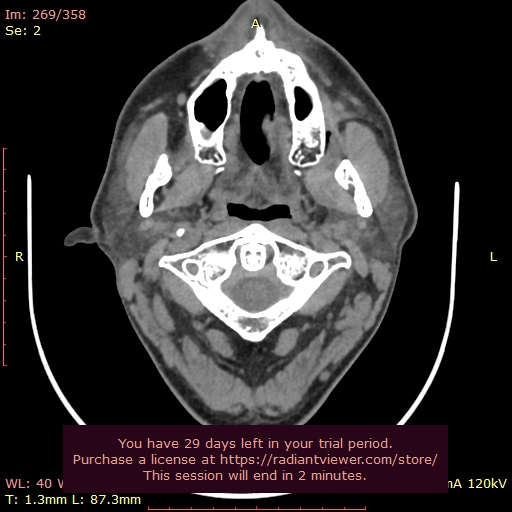
A bit of background: a few years ago I started experiencing strange symptoms — constant tinnitus (always left-sided), ear pressure and fullness, difficulty equalizing, throat pain at one specific point, dizziness, hot flashes, general malaise, and even sweating episodes. At first, it felt like something was “out of place” in my throat and face, and over time the symptoms just got worse.

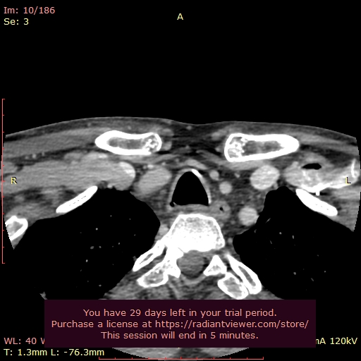
Eventually, I was diagnosed with Eagle Syndrome and had intraoral surgery here in Spain to shorten both styloids. The surgeon reduced them a lot (between 1,5 and 2cm left), but unfortunately my symptoms did not resolve. In fact, they shifted in character. Since then, I’ve been caught in between two possibilities:

  • Residual styloid stump compression (especially on the left, where all my symptoms are).

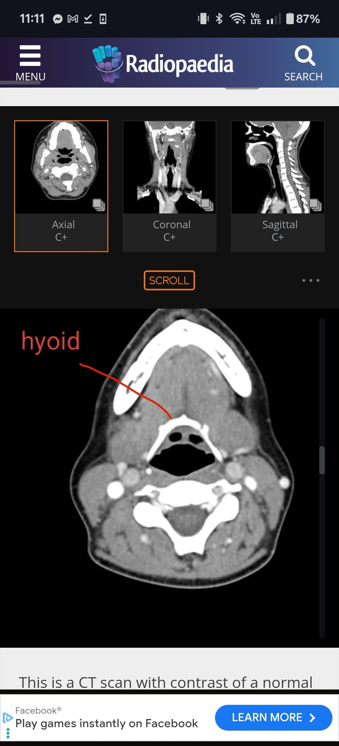
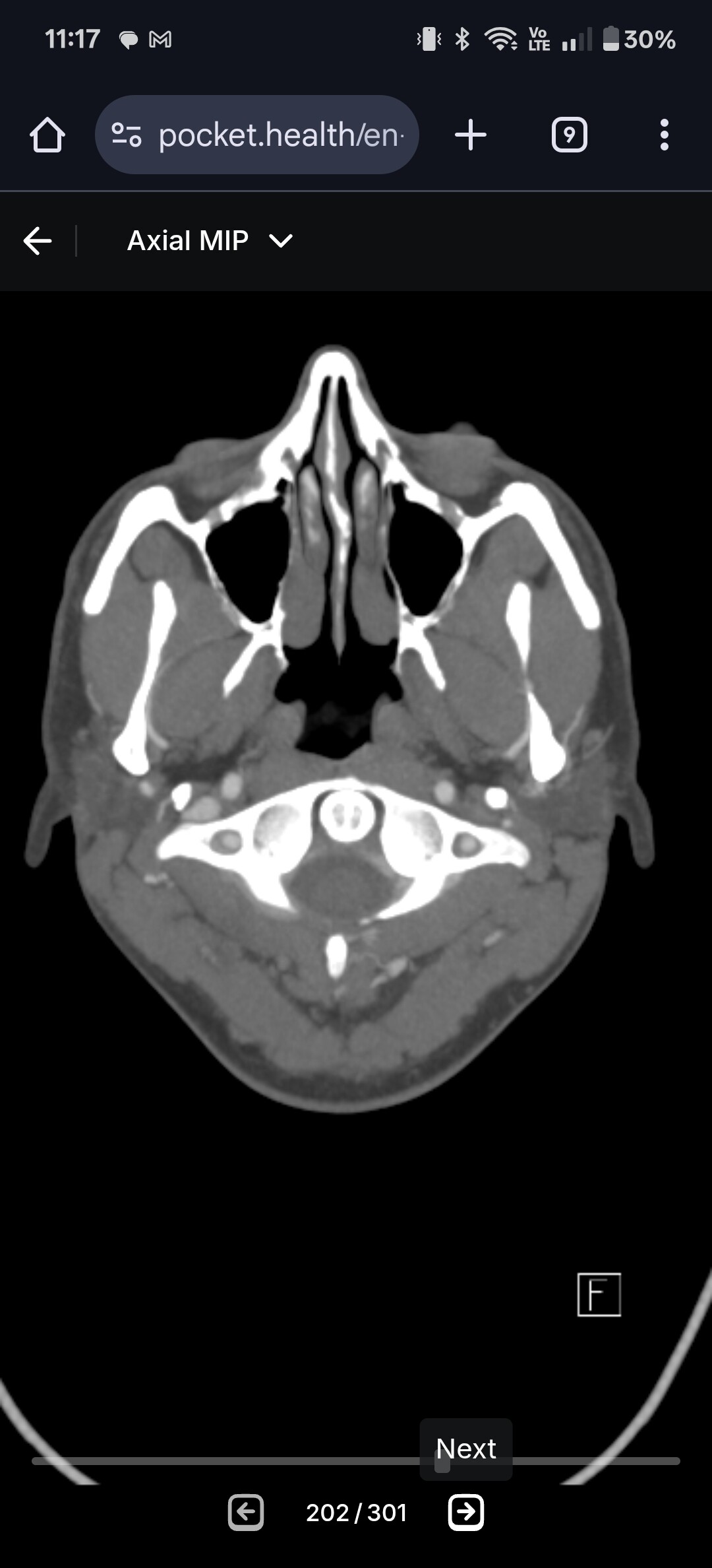
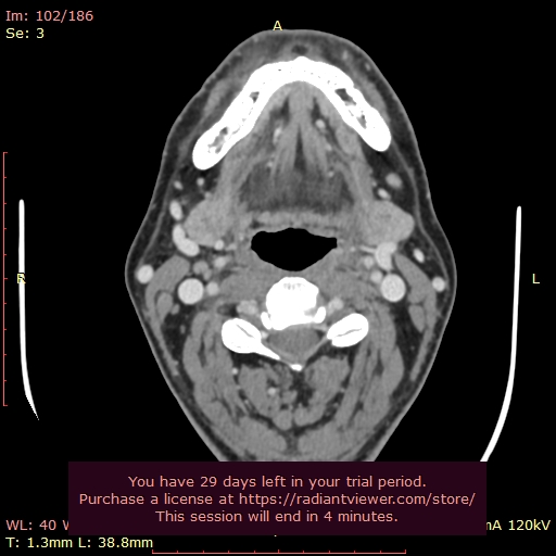
  • Misaligned hyoid bone (the greater horn sitting too high or angled, possibly pressing on nerves or vessels).

This has made things very confusing, since the CT scans show both structures could be implicated. Sometimes I feel like my jaw and face don’t “fit” together, as if something needs to click into place, and other times I feel choking sensations at the hyoid level, especially when there’s mucus or throat irritation.

Of course I got in contact with my surgeon, but unfortunately there isn’t a lot he can say. I actually mentioned the hyoid bone possibility before my surgeries, but he just ignored that and didn’t even look at my CT scan.

So now I’m stuck between wondering if my symptoms are due to what remains of the styloid, or if it’s actually the hyoid bone causing the problems.

My posture is a real problem, I have a sitting job and when I’m not working, I feel a lot better. From the moment I sit down, after 30 minutes, all hell breaks loose - increasingly so. I’ve been training myself to sit correctly, bought a desk that I can move in height, I’ve got a good ergonomical chair… still working on it…

I work with people over webcam, and the hotflashes, sudden sweating, suddenly feeling miserable is making doing my job harder and harder. Thank God I work from home, so at least there’s that!

I’d love to hear if anyone else has had symptoms shift after styloid surgery, or if anyone has experience with hyoid misalignment being mistaken for Eagle Syndrome.

Thanks for letting me join — I’m hoping to both learn and share my journey here.

I’ll include some 3D pictures of what the situation is now, I know you’re not doctors but I would love to hear your opinion! (I can’t seem to upload any pictures?)

I’m so sorry that your surgery didn’t help with your symptoms… It could be that there’s still compression from the styloid above where it was removed- it sounds as if possibly the vagus nerve is being compressed , it emerges from the skull base through the jugular foramen along with the internal jugular vein and internal carotid artery, so between the styloid & the C1 process, so where you’ve ringed the styloid. It’s not so easy to tell from that angle, but does look as if there is compression of the IJV.

Again, not easy to tell from the angle of your imaging about your hyoid, but the greater horn does look as if it has a ‘joint’ or elongated section on the left, & is quite thick, the right side looks as if it could have an extra bit of calcification too. There are quite a few posts about hyoid bone syndrome you could search for, the symptoms are often clicking, choking or strangulation feelings. Although the hyoid is a ‘floating bone’ it is connected to the styloids through the stylo-hyoid ligaments, which are detached from the styloid when it’s removed, so it may be if yours was already a bit out of alignment before surgery, it’s perhaps shifted a bit more afterwards? Most people don’t notice any difference with the hyoid after surgery, but I guess if there’s already an issue it could aggravate it a bit more?

The site doesn’t let new members post images immediately but I tweaked yours so you could, have you got any more images?

As for what you do next, that’s a tricky one given that we don’t know of any doctors experienced with this in Spain, some members have found out about doctors in their countries by looking at FaceBook groups, others give up & resign themselves to having to get imaging looked at & treatment abroad, I don’t know if either of those are an option for you?

2 Likes

Thank you Jules!

Someone on a Facebook group looked at my scans, before my surgery, and already commented on the hyoid. Since it’s all connected together I understand that there’s a domino-like effect.. I do recognize the symptoms you mention of Hyoid bone syndrome.

I know of some doctors outside of Spain that could maybe help me, but unfortunately I can’t pay for the surgery (I already lost a lot of money on the first 2…) at the moment, and since I’m not really sure what the problem is, I’m very hesitant to have a new surgery done anyway.

I asked my surgeon about the ligaments, and he basically didn’t come to the point or gave me a real answer! I mean, did you detach it or not? He just cycled around it and I never got an answer…. I’ll upload some more pics, I have an entire folder full :smiley:

It does look like it is more the styloid compressing the IJV & not the C1 process, but obviously not doctors here!

So rotten that you’ve paid out already for surgeries that didn’t work , & certainly you’d want to try & clarify whether it’s the styloid or the hyoid causing issues, although it sounds as if it could be both? :cry:

I’ve reached out to several professionals and their answers are very mixed… some told me they don’t think the stump that is left can actually compress much, and others told me that the stump is still the issue and I need a full cutting… They’ve also mentioned the hyoid being wonky and possibly compressing… So I guess it might as well be both :frowning:

I have been doing manual therapy with a gentle osteopath that works on the hyoid bone slowly, and I’ve actually found SOME relief, and it’s affordable. But of course, if the stump is the issue, it will have to go sooner or later…

1 Like

@quediusara the best view for the hyoid is the axial view. I can help you locate the greater horns. Sometimes they are in the same frame as the main body of the hyoid, other times it’s up a slice or too due to the horns being angled. See attached examples below. I’ll take a look once you upload.

1 Like

Thank you!

I’m not very good at this, but these are the images that I found that look most similar to what you showed. I never saw it in this view and I’m not an expert or anything but that doesn’t look right to me, wow!

@quediusara can you go up one or two more slices so we can see the very ends of these horns? Just follow thr bone until it disappears and go back to the slice where you last see them. I’d say we are still half a cm or so from seeing the very ends of them!

Like this?

@quediusara right side looks good. See annotated imaging below. For left side I think you need to go a bit higher, maybe slice 93 or 94.

Here you go! That white little circle on the other side doesn’t disappear until slide 101!

I’m in really bad shape now, like someone hit me with a fist on the left side of my face, and the throat and neck feeling is so weird. It feels like everything is ‘twisted’ and upside down inside…

Thank you again for taking a look and guiding me!

@quediusara the axial view revealed something that we couldn’t really see in the 3D model unless you looked with a magnifying glass.

See attached annotated imaging.

The lower portion of your left stylohyoid ligament is calcified. Your right side appears normal length. I missed the calcification in the 3D model because it was hiding behind veins. So that’s the power of the axial view. And not only that, I can see in the axial view that the calcification is not pressing against any important vascular structures so that’s good. However, id say it’s definitely possible that the calcification is irritating nerves. This could explain some pain on the left side.

1 Like

@quediusara there’s one more thing I’d like to check. I want to see in the axial view how compressed your IJVs are at the level of C1 and your styloids. I’ve attached my own imaging showing you what C1 looks like in the axial view. It’s the very top vertebrae that has wings.

@quediusara & @TML - I’m very sorry but I had to delete most of the images because they contained quediusara’s full name & birthdate.

@quediusara - Please make sure to remove your personal information from your images before posting them. This is to protect your privacy.

I will say @quediusara, your styloid stump is pretty long & thick so it may have grown some since you had your surgery. Your surgeon didn’t cut your styloids back very short. We’ve learned over time that having the styloids cut back as close to the skull base as possible helps prevent regrowth & can also stop vascular compression in some cases. I’m glad that @TML found the greater horns of your hyoid aren’t causing vascular compression but I’m sorry the s-h ligament is calcified at the lesser horn of the hyoid & may be also causing your current symptoms.

3 Likes

Thank you! I honestly have no idea what I’m looking at, so here are my best guesses, and the other slides higher up you asked for…

@quediusara see attached your left greater horn area. Everything looks good. So we’ve confirmed that neither of your greater horns are likely causing any issues. But we did discover your left lesser horn/ bottom of your stylohyoid ligament is greatly calcified and could be causing your left sided symptoms.

As for C1, you don’t quite have the right slice uploaded. I’ve attached a photo circling what to look for. It’s your very top vertebrae that has wings and a white circle in the middle at the top.

1 Like

No worries! I didn’t even realize my personal info was on it, I’ve made the images anonymous now.

I know, apparently it’s currently not possible to cut it down more using the intraoral technique. It’s strange though, because another person I know that had the same original length as me, and was operated by the same surgeon, had hers cut back to under one centimetre. When I look at mine, it’s close to 2cm, and it does look rather thick.

I was a bit better after surgery, but then slowly symptoms came back. In December, I spend the entire month in the hospital with a bilateral pneumonia, I was in a coma for 2 weeks in the UCI. Full of antibiotics, prednisone and of course, minimal movement, and I want symptom free for the entire month after that. Symptoms came back with a vengeance, especially the ‘vascular’ ones… hot flashes, feeling super weak suddenly, sweating, general malaise, left side of my face and lips going numb… incredibly uncomfortable :frowning: It feels like I’ve been hit by a bus on the left side.

Is there anything I can do about the s-h ligament? I found it so weird that my surgeon basically didn’t give me a straight answer, and I see the question of it being cut or not in surgery come up regularly.

Thank you for your time!

2 Likes

@Isaiah_40_31 thanks for catching the identifying information! I didn’t even notice. Too locked into the imaging :joy:

3 Likes

What about this?

BTW, if the ligament is calcified as you mentioned, what can be done about it? Does this still fall under ‘Eagle syndrome’? Please tell me I can break it myself :smiley: