Thank you so much for that comment, I was seriously concerning prolotherapy, now will put this idea aside.
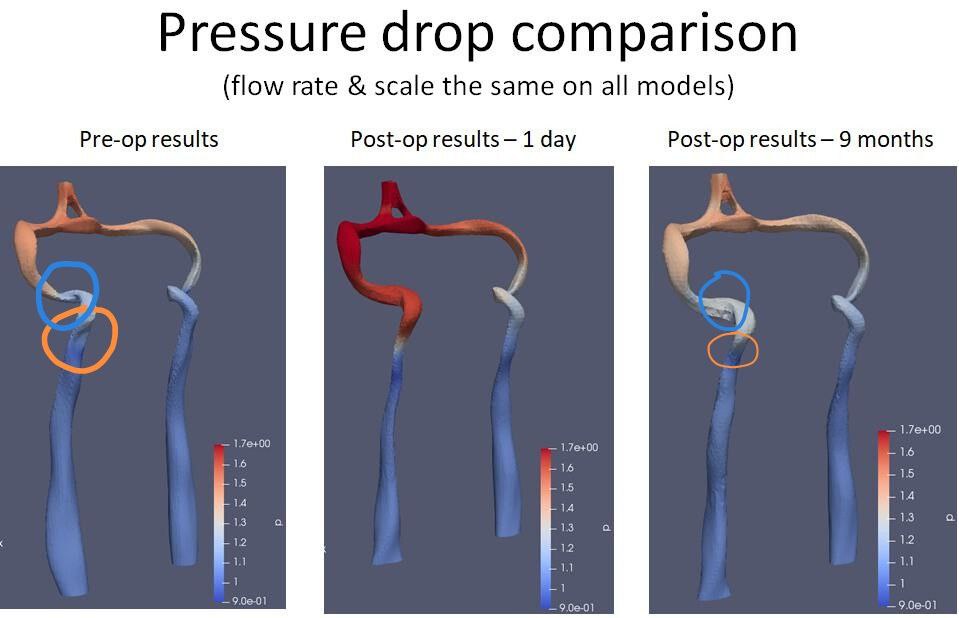
But what I can’t understand, the place of the compression between C1 and styloid is at the place of the orange circle, whereas the vein got wider at the other place, at the blue circle, and got thinner at the compression site… How come? ![]()
The vein gets thinner at the compression site because it’s being squashed between the styloid process & C-1 or by some other means at that location. It’s thicker above the compressed area because the blood is backed up there since it can’t flow normally through the squashed area because of the smaller size of the opening in the vein.
3 Likes
Well the model isn’t accurate, because sucking through the straw gives negative pressure and bulging occurs due to the reasons related to the positive pressure*, so there wouldn’t be any bulging no matter if the straw is pinched or not.
More accurately it would be to describe it as trying to inflate a balloon. The bottleneck (narrow balloon’s part) gives resistance and the cheeks will puff out. But try to simply blow the air out from lungs with the mouth wide open, and the cheeks won’t bulge because there is no resistance (causing positive pressure).
*There are certain aerodynamic nuances that under specific conditions lead to the negative pressure causing positive pressure in case of various air vertexes, pulsed form of negative pressure at certain air flow velocities, quantities, and forms. But that’s not the best place to discuss about it ![]()
2 Likes
Thank you for the correction @vdm. I knew my illustration was faulty, but I couldn’t figure out a more accurate example. I’ll delete it so it doesn’t confuse anyone. What you wrote is a much better explanation (& accurate). ![]()
3 Likes
This is a great explanation @vdm. As someone who has played a lot in the lab with microfluidic devices, certainly sounds about right
3 Likes
The compression site is at the orange circle, right? Why after the surgery the site at the blue circle got bigger, not at the orange circle, as expected?
What a lucid example with the baloon, thank you!) But still, according to the pressure drop comparison model of M_UK, if we apply this, that would mean, there was no compression at the site of C1, but it appeared after the surgery… Given the compression site is at the orange circle…
@Irina777 - I understand your question but am not sure of the answer except that perhaps the size of the veins is somewhat dynamic/flexible to accommodate different neck positions, rates of blood flow (i.e. at rest vs when moving), etc. Maybe @vdm or someone else will have an opinion that answers your question better.
2 Likes
So there can be many reasons why compression at the specific spot happens. Best is to have a look at the CT scan/MRI. Could be scar tissue, lymph node, swollen muscle, resected muscle hanging free, permanent inflammation or anything else…
2 Likes
Hi, did you have your operation private or on the nhs?
@markp - I’m in the US, & the surgery was for bilateral hip replacement not ES or IJV decompression. I have medical insurance that covered the cost, however, 2 years after my surgery, the insurance company decided the surgery wasn’t necessary after all & demanded the doctor return the insurance payment. This didn’t affect me, but I was devastated that the doctor had to return what he was rightfully owed. I was told by someone in the medical field that US doctors pay for insurance that covers them in such cases as mine so they get their money one way or the other.
1 Like
That’s terrible! What a hassle for your surgeon, I hope he was insured & got his money back okay ![]()
1 Like
Was this Medicare? Clawing back an approved surgery would seem to me be very difficult unless it was the govt .
Hearing that makes me shiver. I’ve tried to deal with insurers on my own before and it can be a nightmare. I ended up getting lawyers involved. I’m in Australia and thankfully we have universal healthcare. My situation is neurological and although the first surgery was to be the ‘Fix’, that fix didn’t happen and I’ve required a further 5 neurosurgeries to manage it all. If they withdrew funding for any of them ![]() I’d hate to think of the costs involved for one, let alone all 6
I’d hate to think of the costs involved for one, let alone all 6 ![]()
![]()
![]()
Scary stuff.
Merl from the Modsupport Team
2 Likes
Absolutely Medicare! The Medicare doctor who reviewed my case 2 years after my surgery said I wasn’t disabled enough to need bilateral hip replacement, yet the surgery had been approved by Medicare after 3 different orthopedic surgeons said I needed both hips done. I firmly believe if I’d done one at a time, the surgeries would have been covered, but because I had both done at once, it was later denied. I figured it would cost Medicare less to have one big bill than two medium sized bills, but maybe I was wrong.
After being through two separate ES surgeries & recoveries, I decided if I ever needed another surgery that had a bilateral diagnosis, I wanted both done at once thus my decision to go that route w/ hip replacement.
2 Likes
I’m sorry for all you’ve had to go through, Merl, but am thankful you’ve had medical coverage to allow for those surgeries, & especially that the funds paid out weren’t demanded back at any point. I think that should be illegal whether gov’t funded or private insurance.
2 Likes
Hey @Isaiah_40_31
Absolutely. Coverage should all be sorted out prior to surgery and have no such thing as a clawback. Here, for some ‘elective surgeries’ there can be a patient component or co-payment and some specialists charge an additional co-payment, but costs need to be disclosed prior to the appointment.
Merl from the Modsupport Team
2 Likes
That’s the way it’s supposed to be here, too, but obviously, there are some exceptions to that rule that are out of a doctor’s or patient’s control. Sigh…
1 Like