Just wondering if anyone is still dizzy 2 months after IJV decompression?
2 Likes
Most likely, your dizziness was not related to the veins. Perhaps your dizziness is related to the vertebral arteries,or something else.
Oh, how would I check that?
Do a head-turning DSA angiography. And an MRI scan to exclude formations in the brain.
1 Like
@Anita It took about four months after my styloidectomies for my lightheadedness to improve, although it did not completely resolve. @anon30378544 is correct that there can be other vascular possibilities as well.
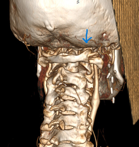
After Dr. Liu unbridged my arcuate foramen last month, the lightheadedness improved significantly, and the constant pressure at the base of my skull has almost completely resolved, although I’m still early enough post-op that swelling could still be affecting things..
For surgical planning, Dr. Chancellor performed a dynamic cerebral angiogram with venous manometry the day before my surgery to assess the vessels dynamically and measure venous pressures. If you move forward with this type of testing, you might consider asking whether venous manometry could be included as well.
4 Likes
Please share your CT scans, before and after the surgery (Dicom).
I did mean arteriography
@Chrickychricky also had unresolved dizziness after surgery and found that virtual reality vestibular rehab at UCSF has really helped, I’m sure she’ll chip in with her experience, here’s one of her posts:
Finally have an answer but it has no solution - General - Living with Eagle
And a more recent detailed post she did:
Surgery scheduled with Dr. Kamran Aghayev - General - Living with Eagle
Two months is still fairly early on in the healing process though 
3 Likes
I was 95% compressed on my dominant side. Could this be why my dizziness may take longer to subside….
2 Likes
@Anita That actually seems pretty rational to me. If you were 95% compressed on your dominant side, I can see why recovery might take longer. From what I’ve read, the longer the compression has been present, the longer it can sometimes take for things to normalize. I know two months feels like it should be enough time to see a big change, but it literally took me about four months before I noticed improvement in the lightheadedness.
3 Likes
I certainly hope it gets better. I’m so anxious over this. It’s been a long 3 years of the not knowing until I discovered the IJV compression. Thanks.
3 Likes
@Anita I completely understand. Three years is a long time to go without knowing what was causing it. Hopefully now that the compression has been addressed, your body just needs more time to adjust and recover. Two months is still pretty early in the bigger picture. 
4 Likes
@Anita You could check this out and see if it holds anything useful for you
3 Likes
Thanks for the info.
1 Like
Though my lightheadedness has been resolved, I still have some positional dizziness after decompression of both internal jugular veins. It was proved to me a couple months ago through a simple physical test by a neurologist that it is due to BPPV, Benign Paroxysmal Positional Vertigo. I was given exercises to treat it, but I’ve since had a lumbar surgery and have not yet been able to perform them.
2 Likes
@Octoberkurt - That’s EXCELLENT news!! I’m so glad your vascular dizziness has gone away!
I’m sorry you had to have lumbar surgery but hope once you’re sufficiently recovered the exercises to treat BPPV help alleviate the symptoms completely.
1 Like
Thats great the lightheadedness has practically gone. Can I ask you what was involved with unbridging the acurate foreman? My daughter had 2nd ijv decompression on dominantbijv this time 3 weeks ago and the lightheadedness is her main symptom which is 24/7, hasn’t budged. She has had vertebral artiogram with head rotation too done and they are fine. Thanks
2 Likes
@Shauna Dr. Liu approached it from the back/right side of my upper neck/skull base using a far-lateral posterior approach (craniectomy approach). He removed the bony bridge over the vertebral artery and also removed some additional C1 bone during the exposure. At the same surgery he removed my right C2 dorsal root ganglion because of the severe occipital/facial pain I was having.
I’m not sure how much of the additional bone removal was specifically for access to the C2 ganglion versus the arcuate foramen itself, so I don’t want to guess on that part.
The surgery was primarily being done to relieve the extreme pressure I had at the skull base. The dynamic cerebral angiogram with venous manometry was also used for surgical planning and did show pressure gradients, but Dr. Liu was already planning to remove the arcuate foramen. I had always felt better lying down, with less pressure in my head, which I was told was atypical, so I honestly did not expect the pressure testing to show much at all.
It was a much rougher recovery for me than the bilateral styloidectomies a day apart, although having the C2 ganglion removed at the same time may have contributed to that.
After my styloidectomies, it took about four months before I saw improvement in the lightheadedness. The only symptom that improved immediately was my facial swelling, so please give it some time before worrying that the surgery did not address your daughters lightheadedness.
I’m very thankful Dr. Liu agreed to remove my arcuate foramen bridge because the extreme pressure at my skull base, which was very painful, is now gone. My lightheadedness has substantially improved but is not completely gone, which could be related to the remaining IJV compression after styloidectomy.
If your daughter does have a bridged or partially bridged arcuate foramen, I would definitely recommend seeing someone who can map the anatomy out carefully beforehand and determine whether it is actually likely to be contributing to her symptoms. It is not a surgery you want to go through unless there is a good reason to believe it will help.
Before and after for visual context below:

3 Likes
Thanks so much for your indepth reply with all that information. And continued healing to you. The scans look incredible. Did Dr Liu do the cat venogram with manometry? Was your pressure measuremts high? My daughter’s looked normal except with head turning there was 2 to 3mmhg difference at c1 level and at c5 c6 on dominant side. There is no neck pressure like you described. Was your lightheadedness there all the time? ThanksDid your lightheadedness stay the same for some time after the styloidectomies or did you notice a difference straight away? It’s such a worrying time. Thank you so much.
1 Like
@Shauna Dr. Liu works with Dr. Breehan Chancellor, who did my testing the day before surgery. It was actually a dynamic cerebral angiogram/venogram with intracranial venous manometry rather than a CT venogram with manometry. Your daughters study sounds like it focused more specifically on jugular pressure measurements at different cervical levels, including lower down at C5–C6, whereas mine was a broader dynamic cerebral angiogram/venogram with intracranial venous manometry that started in the brain and followed the venous system down through about C3, while also evaluating the carotid and vertebral arteries.
If your daughter’s study only measured pressures in the jugular veins at cervical levels and did not measure the intracranial venous sinuses, then it may not have assessed whether pressures were elevated higher up in the brain. My study started in the superior sagittal sinus and followed the pressures down through the intracranial venous system into the jugulars.
My pressures were definitely abnormal even though the angiogram itself did not show a significant jugular blockage. My superior sagittal sinus pressure was 23 mmHg. With head turn it was 22 to the right and 21 to the left, 24 with extension, and interestingly dropped to 9 with flexion. The pressures then stepped down through the venous system: torcula 21, right transverse sinus 19, transverse-sigmoid junction 18, sigmoid sinus 16, and jugular bulb/above C1 13.
On the left, the transverse sinus was 18–19, transverse-sigmoid junction 17, sigmoid sinus 16, jugular bulb/above C1 13, C2 12 and C3 12. They documented a 3 mmHg pressure gradient at C1 on both sides, despite the venography showing no significant narrowing there.
So that was one of the interesting things in my case, the vessels looked patent without an obvious angiographic blockage, but the pressure measurements still showed elevated intracranial pressure and gradients. And this was laying down when I feel my best, would be interested to know what they were while upright. The testing only followed the jugulars down through about C3, though, so it would not have evaluated something happening lower down around C5–C6.
The angiogram also showed tortuosity/redundancy of my right internal carotid artery, described as consistent with a fibromuscular dysplasia phenotype versus hypertensive arteriopathy. FMD and arterial tortuosity are also seen more often in people with connective-tissue disorders, which is interesting given how many of us in this group report hypermobility/connective-tissue issues.
As far as the lightheadedness, mine did not improve immediately after the styloidectomies. For quite a while it seemed essentially unchanged, and it was probably around four months before I really noticed that it had substantially improved. Mine was much more noticeable upright and I generally felt better lying down.
One other difference is that my angiogram/manometry was done awake with local anesthetic only, not under general anesthesia. Anesthesia and sedation can affect venous and intracranial pressures, so that may be worth keeping in mind when comparing pressure measurements.
5 Likes