I’ve been struggling with weird neck, throat, and neuro symptoms for many years after whiplash from a car accident plus a surgery where I woke up with the worst neck pain of my life.
-
I started getting a really flushed face from bending/looking down that would take a few hours to resolve, and nearly passing out from looking up.
-
I have constant neck and throat pain, it hurts to swallow and feels uneven, my voice is usually hoarse and it hurts to talk. My ears are always ringing and feel full.
-
Tongue ulcers
-
I have had visual static for as long as I can remember but it’s been getting worse.
-
The most debilitating symptom is that whenever I talk for a moderate amount of time or laugh heartily, I get pins and needles in my face and arms, a dull pain in the back of my neck, and tunnel vision with lots of visual snow. It seems vagal-related because it doesn’t happen when laying down, and elevating legs helps.
-
The second worst issue is weird episodes of feeling a massive drop in energy, like my blood pressure has halved and I have little blood in my brain along with visual disturbances - greying out, tunnel vision, smearing, blurring, confusion, followed by a rebound with skyrocketing blood pressure and heart rate (including SVT). My neck often hurts, and sometimes my ear, during these episodes.
I’ve given up on hanging out with friends and family, I don’t participate in meetings at work. I can’t live like this.
I also may have some form of connective tissue disorder, meeting the 2017 criteria for hEDS.
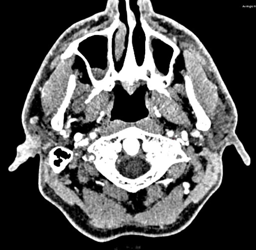
One of the few issues I’ve heard of that could possibly explain the symptoms from talking is ES. I got a CT angiogram for another issue and I’m curious if anything here seems of interest to the intelligent lay people here ![]() .
.
My health provider’s chart system lets me log in and look at imaging but it’s pretty limited. I thought I measured the styloids at around 3.5cm, but a radiologist thought they were normal.
Is there any reason to be concerned about these things on my imaging?:
- Styloids - hard to tell with what views I have available, but seem to be around 3.5cm - are the highlighted white parts in the grey images calcified ligament?
- C1 - C2 - it appears the angle of C2 is rather out of line compared to neighboring vertebrae
- C1 and jugular vein - it looks like C1 may be compressing the right jugular vein
- V3-V4 vertebral arteries - these look quite anomalous. I have pain in that area in certain postures
Fairly clear view of left styloid process. According to viewing software it’s about 3.5cm
Less clear views of right styloid, possible calcified ligament
Umm… that vertebral artery looks gnarly (FYI I have a hair tie on in the image, the rubber is visible here)
Front view - is the highlighted white calcified ligament? I’m not sure how CT MIPS works or if that’s manually highlighted?
Is top right corner of C1 jugular compression?
Rotated C2
Darkish grey material is bone and the highlighted white is calcified ligament?
View of vertebral arteries
Left jugular compression by C1?


