Hi,
I don’t have any diagnosis yet. I have lower jaw under ear and neck pain for more than 10 months now.
I got ct neck scan with contrast. I tried to convert to 3d. I would like to know if I missed any angle. Are the styloids and classification visible here?
Any inputs please.


I’m sorry, but I can’t see the styloids in the images, so I’m guessing that they’re not elongated…maybe something on image 003…we’re not doctors though! Is this a new CT you’ve had done? Did you get a report with it?
1 Like
Hi @Jules ,
How are you? It’s been long since I chat with you here.
Yes this is my ct scan with contrast done in December. I have a report with it. But the radiologist missed to evaluate eagles. I had to talk to him few times to add it. He missed left side, reported only right. Then added left and missed stylohyoid. So many addendums in my report.
I am not sure if this angle is not shown the styloids clearly. I will try another angle in that radiant viewer.
This is my report.
Addendum:
Right styloid process: Not elongated, measuring about 0.3 cm.
Right stylohyoid ligament: Calcified distal portion, which measures
about 0.8 cm. This is located about 2.1 cm from the tip of the right
styloid process.
Left styloid process/stylohyoid ligament: Borderline top normal size of
the left stylohyoid ligament, which is in continuation with the
elongated left styloid process, measuring about 2.4 cm combined. One
cannot differentiate between what is considered the left styloid process
and a calcified stylohyoid ligament in this case. In regards to Eagles
Syndrome, an elongated styloid process or a calcified stylohyoid
ligament can both result in pain. Eagles Syndrome is ultimately a
clinical diagnosis.
Two BB markers were place in the region of focal pain in the right face
and right neck. There is no focal abnormality underlying these BB
markers.
12/28/2020 3:43 PM PXRRR02R
Addendum: The left stylohyoid ligament is calcified and measures 2.4 cm,
which is borderline top normal. This is in continuation of the left
styloid process, which essentially can be considered elongated.
12/24/2020 4:26 PM RAADOCS006
Addendum: The right styloid process is not elongated. The calcified
distal portion of the right stylohyoid ligament measures 0.8 cm and is
located about 2.1 cm from the tip of the right styloid process.
12/24/2020 3:47 PM PXRRR14R
Addendum:
Additional clinical indication: Evaluation for Eagle’s syndrome.
The left stylohyoid ligament is calcified and measures 2.4 cm, which is
borderline top normal. The right stylohyoid ligament is mildly calcified
along the distal aspect on series 4, images 34-35.
Narrative
CT SOFT TISSUE NECK W CONTRAST
TECHNIQUE: CT images of the neck were obtained with intravenous
contrast. Isovue-370 was injected without complications. Dose reduction
techniques were utilized for this examination. DICOM format image data
is available to non-affiliated external healthcare facilities or
entities on a secure, media-free, reciprocally searchable basis with
patient authorization for at least a 12-month period after the study.
CLINICAL INDICATION: Lymphadenopathy, neck;Localized enlarged lymph
nodes;Cervicalgia.
COMPARISON: Neck ultrasound August 28, 2020.
FINDINGS:
Aerodigestive Structures: The nasal cavity, nasopharynx, oral cavity,
oropharynx, hypopharynx, larynx, and included trachea and esophagus
demonstrate no masses or abnormal enhancement.
Lymph Nodes: No pathologically enlarged, necrotic, or otherwise abnormal
lymph nodes.
Vasculature: Normal.
Salivary Glands: Normal parotid and submandibular glands.
Thyroid: Normal.
Visualized Intracranial Structures: Normal.
Included Orbits: Normal.
Paranasal Sinuses/Mastoids: Predominantly clear.
Included Lung Apices: Normal.
Bones/Soft Tissues: No aggressive osseous lesion. No acute soft tissue
abnormality.
Unfortunately I am unable to see the styloid processes as well with the images that you’ve provided. The styloid process looks to be within normal length within the write up, 2-3 cm in length is within normal range. However as we all know here that the styloid and calcifications with the the ligaments can cause irregularities within the vascular system or still cause focal pain.
I had left styloidectomy in September that caused TOS (Thoracic outlet syndrome), panic attacks, and focal pains. I have been very fortunate and extremely grateful that I do not have anymore symptoms after surgery. I have my right side scheduled in June this year. I have completely different symptoms on the right. Focal ear and eye pains, feeling of heart burn or stomach cramps, and of course the pressure in the hypoglossial nerves.
Eagles Syndrome has notably caused an array of symptoms which are extremely hard to diagnose. It’s rather frustrating. I also had many addendums added to my CT scans as well. It seems the doctors don’t really know what they’re looking for.
1 Like
It sounds as if there is a small amount of calcification on both the ligaments, but not that much, which I guess is why the radiologist was being cautious with his report? It’s a shame after all that struggle to get your CT done that the CT wasn’t more conclusive…It could be enough to cause your symptoms but not as clear cut as some cases are  It might still be worth sending the CT to Dr Samji for example to get his opinion?
1 Like
@Jules ,
I did send my ct scan to Samji office in Jan and waited for 3 weeks. They replied me back that they rejected my case as I am not meeting Samji’s criteria. It should be more than 4cm it seems and he is not considering calcification. They said I am not eligible.
I think the radiologist is not aware of eagles much and don’t know what to look for. I had to email him after every addendum to check the other side, hyoid bone etc to add in the report. He did every addendum after my emails
What I noticed on the ct scan, your neck seems very straight to me. That might cause some of the symptoms too Military Neck (Cervical Kyphosis): Causes, Treatment, and More
3 Likes
I’m afraid I’m not competent to elaborate on this… Probably the best would be to see some orthopedic doctor? Physiotherapy specialist?
1 Like
That’s an interesting suggestion & a good spot!
1 Like
So sorry that Dr Samji couldn’t help you…although it does sound from the report that the radiologist has evaluated the CT as required when you asked him. I think it’s been mentioned before that there are places online you can send your CT to for evaluation; if you’re really not confident of his opinion that’s something to consider? I wouldn’t have a clue how much it would cost or anything, but a quick search online shows places. That is if you still think it could be ES & want to explore all avenues?
1 Like
@Jules are you living in the US??
The Radiologist added those when I asked him and have more addendums and nothing clearly mentioned. He told me that eagles is the clinical diagnosis, so take this report and get the disc and see the ent.
What places are available online for evaluation of CT?
Do you mean other eagles providers??
I have contacted Dr.Hackman, Hepworth, Annino office. They don’t accept out of state patients now. I thought of sending my reports to any one of them and have a phone visit. But that didn’t happen.
Who is your doctor?
Good observation vdm!
1 Like
Normal cervical spines have a lordotic (inward) curve to them. Your neck is lacking that curve. It can be restored by a PT who knows proper neck exercises.
Normal cervical posture presents with a lordotic curve of approximately 43 degrees measured from C2 to C7. Without this normal lordosis , most often the balance of the weight of the head is tilted forward and thus creates increased wear and tear on the intervertebral discs and the vertebral bodies.
I will add to that statement that shoulder pain & mid to upper back pain can also be present. Though I don’t think that’s what’s causing the nerve pain in your face, it could be contributing to it.
1 Like
I’m UK, I’m afraid. I did mean online radiology services; if you google them it comes up with quite a few, but if you’re happy with your radiologist’s report then no need for that. I know that Dr Cognetti does phone consults, but not sure about the others.
1 Like
Thanks @Jules . Why are you afraid ? I don’t get it.
I am not completely satisfied with my Radiologist report. I have to see a reputed Radiologist who is very well versed in reading ct for eagles diagnosis. Have to browse through.
I will check with Dr.Cognetti office. I read from the group that he won’t consider calcification and dismiss that. So my report did show some Calcification. So I want someone to check those too.
Thanks for the information about lordotic curve @Isaiah_40_31.
To be honest, I am very bad in anatomy and not sure what you are talking about. I will read more about it.
One thing I understood is, my neck is somehow contributing to the pain. I have forwarded head posture and slouching upper back.
You mentioned 43 degrees. Is there a way to measure those by ourselves at home?
I printed a quote from Google. I think the 43º measurement is an average not an exact number. You’re correct in your assumption that your lack of a neck curve is probably contributing to your pain.
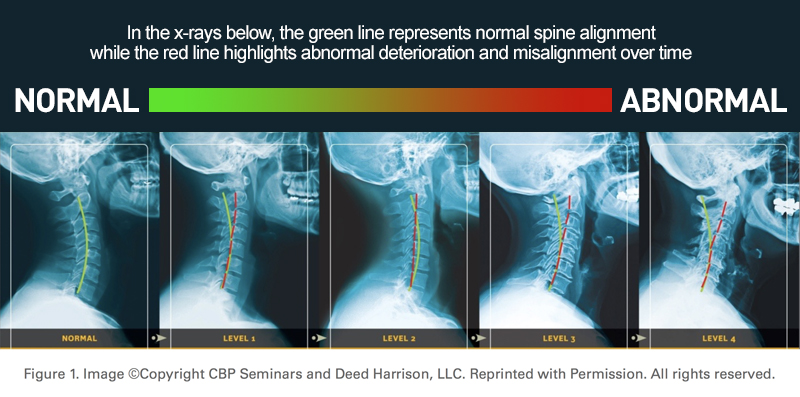
Here is a chart that shows a range of normal to abnormal curves of the cervical spine (neck vertebrae).
2 Likes
@Newuserhere
After reviewing the comments I have to agree with the observation that you have a very straight neck. In the image that shows a posterior view you can clearly see the jaw impinging the nerves along the side of your neck. C6 and C7 vertebrae have rotation as well, indicative with your shoulder pain.
I am medical massage therapist with over 20+ years of experience and when I was diagnosed with Eagles I decided to seek out alternative therapy for nerve pain relief. I stumbled across upper cervical chiropractic.
I posted about it here in this forum, the thoughts I got were not necessarily in the positive direction. However I still pursued the therapy with hopes it could provide relief. I keep saying I had a very fortunate journey with my diagnosis of Eagles Syndrome, the chiropractor I went to also has Eagles. He deals with a lot of the same symptoms we have and we have learned a lot from each other in this process. I will post a link for the type of therapy he does in a private message for you.
I did get relief from the anxiety and nerve pain after the adjustment, but there was an added bonus to being adjusted. The pains I had in my neck, Rt. shoulder, Rt. Hip, and right knee were GONE! Pains I’ve been dealing with from a very young age.
To be clear for other readers here. Upper cervical chiropractic is not your conventional chiropractic treatment. It’s a very small movement with very little pressure applied to the C1. No risk to breaking or damaging an elongated styloid.
3 Likes
Thank you so much. Appreciate it.
I haven’t been to any chiro or upper cervical chiro before. Are they same as Nucca chiropractor?
@Isaiah_40_31 , @Jules , @ESinLV , @vdm
I have got 3D images from my imaging center. I couldn’t get the proper view of my right styloids in radiant viewer. So I asked them to give a 3D view. I got few pictures. Please let me know if anyone could see something on the right.


