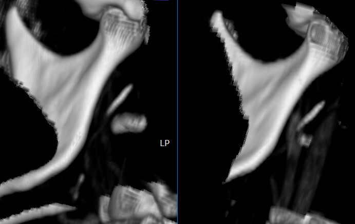
Second image shows new CT snap at 1.25 mm slice size.: 1. styloid process (short), 2. tip of mastoid bone (for ref), 3. tip of c1 transverse process (for ref), 4. upper calcification, 5. lower calcification.
Changes are apparent in the lower calcification. It is now 4 piece chain there, whereas it was a 2 piece chain 1.5 years ago.
I am in a LOT of pain from that lower area. By a lot, I mean cold-sweating from pain and it feels like my tongue is being cut up from underneath. I feel like I am being operated on live, by my own body .
I don’t know how much longer I can carry this pain, TBH. I am going to seek virtual appointments with doctors mentioned here (and maybe a 2nd one w/ Dr. Osborne) and also some diagnostic nerve blocks, but in the meantime it is HELL.
I’m sorry that you’re in so much pain, it sounds awful…good you can see the calcifications lower down now, so that needs addressing too by the sound of it, you know now what you need removing ! I hope that you can get somewhere with the doctors and can get surgery soon…
I also hope you can get appointments soon, @sbincamp. Be sure to ask to be put on a cancellation list at each office where you make an appt.
Have you tried icing your neck to see if that helps reduce your pain? OTC Lidocaine patche (5%) can also be helpful if you’re trying to avoid taking nerve pain medication.
Thanks, it’s difficult for topicals to reach past the muscle (platisma?) and get to the affected area. Benzocaine lozenges cannot reach from inside, either. I cannot press on the area with hot or cold, plus head movement and swallowing just re-inflames the whole thing.
Since the symptoms are relatively new (month?) I’m hoping and praying they just return to normal, so I can continue to search for surgeon.
My main issue (re: surgeon) is that, even within Eagle Syndrome group, I’m not classic. I actually have Stylohyoid Chain Syndrome. Self-explanatory, but existing surgeons are used to snipping off elongated styloids, not searching for calcified sections of stylohyoid along its length.
2nd issue is that everyone is out of state. I can only consult virtually, for now.
My stylohyoid muscle is also calcified, and I don’t have a long styloid process. I’ve tried surgery and asked a neurosurgeon to remove my stylohyoid muscle, but he didn’t remove it. You can view the rest of my CT and MRI scans in my forum thread.
@Filya - You’re correct that your styloids aren’t long, but you have a long, broken chain of calcified stylohyoid ligament calcification on each side plus the stylohyoid ligament is quite calcified coming off the lesser horns of your hyoid bone. I can see in your axial images that the greater horns of your hyoid (which are very thick) are impinging on your carotids at the bifurcation. It also appears your hyoid is tipped lower on the right so isn’t sitting level in your neck.
You have a lot going on - both ES & Hyoid Bone Syndrome. Have you found a doctor in your country who will remove the calcified sections of stylohyoid ligament plus the lesser horns of your hyoid & shorten the greater horns of your hyoid? I believe you will need all those things done to get relief from your symptoms.
I don’t see that your stylohyoid muscle is calcified, but you do have a lot of calcified spots along both of your stylohyoid ligaments which effectively cause the same types of symptoms as elongated styloids because they reduce the ability of the hyoid bone to move as it needs to & can also irritated the same nerves elongated styloids do.
In each picture the right side is now. The bottom segment now has 4 bones, also thicker. Upper segment just got thicker. Continuing calcification may indicate body depositing bone because of trauma to nearby nerves or tissue. I am calcifying nowhere else, so my best bet is that once the trauma starts it just keeps going and getting worse.
Tomorrow I talk to Osborne. I will show him the full pictures. I will be in disbelief if he still says he may not find the lower segment (the cluster).
Based on your images, there is no convincing evidence that it has become larger. The difference in the images is most likely due to the contrast adjustment in the viewer program.
Frustrating answer for you from Dr. Osborne, @sbincamp. It’s good he provided an honest answer as it would be terrible for him to go for it w/o warning & have you end up w/ a lot of nerve damage & no positive end result.
I’m actually in a similar situation. I have a form of dehiscence in my left ear that’s the result of a surgery I had in 2019 which has left me w/ chronic loud tinnitus in that ear. Dr. Hepworth thinks it can be fixed, but the otologists I’ve seen both said no. I revisited one of them, & he ultimately said he could do the surgery to patch the hole in my skull base that’s causing the problem, however, he gave the caveat that surgery could help, or it could make the tinnitus worse. I’ve lived with the tinnitus for 7.5 years, & the thought of it possibly getting worse has made me rethink pushing to get this done.

 .
.


