Ct scan results are back. They did not order it with contrast and refused. I understand this isn’t ideal- please don’t mention it, I’m already upset as is. But these are my results.
I’ll be sending to Samji asap.
Are these worth surgery???
Styloid process on the left measures 1.5 cm (602/75). Styloid process on the right measures 1.5 cm (602/37). Calcified stylohyoid ligaments bilaterally extending from the styloid processes, ossification on the right measuring 4.1 cm extending to the level of C2-C3 (601/54). Ossification on the left measures 4.6 cm, extends inferiorly to the level of C3 (601/48).
@SeekingInfo glad to hear the radiologist noted the calcified ligaments.
Any chance you have access to the CT? I can help see how close your calcified ligaments are to C1 which will help determine how much IJV compression could be occurring
I have the disc but honestly have no idea what I’m even doing with it or what I’m looking for. I really hope that they ordered the correct scan, they said they don’t do a head/neck, it’s either brain and/or neck/soft tissue. The images I’m looking at don’t show anything, but I’m hopeful that because the radiologist was able to measure them, those images are somewhere, I’m just not seeing them on the disc. Fingers crossed. Really terrified of radiation (it’s a legit phobia I have, so even doing these scans was a huge deal for me).
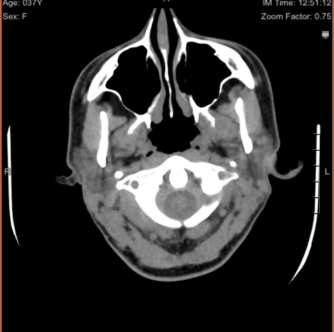
@SeekingInfo there should be a series of images called “axial”, but sometimes it’s called something else. What we want to see is the axial view of C1. I’ve uploaded my own to help you know what to look for. It’s the top vertebrae with a white circle in the top middle:
Your styloids are average length, but your calcified ligaments are very long so not surprising that you’re getting symptoms of ES! It’s certainly worth looking into getting them removed, frustrating that it wasn’t done with contrast, but I presume they did the CT of the neck as they’ve commented on the styloids & ligaments. Hopefully you can find out some images to upload here so we can have a look- make sure you remove any personal info if you do though
Thank you. Hopefully he will agree to see me with the scans and records I have. I’ve also reached out to Dr. Hepworth with hopes that he may be in my insurance network. Naturally, traveling with a toddler is going to be extremely difficult (not to mention, expensive).
If, let’s say Samji were to do the surgery and there was in fact vascular components to my condition, what are the risks? it also sounds like Dr. Osbourne does a lot of styloid surgeries but he also doesn’t do vascular cases…. If 80% of eagle syndrome cases are vascular (I read that stat on the Facebook page), wouldn’t Osbourne not be nearly as busy? Hopefully I’m making sense.
That’s an interesting statistic! I wonder where they got that from? I think there is more awareness now of VES, but from what we see on here I’m not sure it’s that high…
So any of the surgeons members use regularly are experienced , so the risks of surgery should be lessened; the area around the styloids is very crowded with blood vessels & nerves, so it’s always tricky & needs skill to avoid blood vessels whether the styloids are compressing them or not. The issue with VES & where a doctor isn’t experienced / aware of this is more from the compression of IJVs by the styloids, as often there is compression quite high up near the skull base, and sometimes not enough of the styloid is removed to remove the compression. Not all doctors remove the styloids right at the base of the skull, and sometimes it not always safe to do this depending on where the nerves are, so if a small stub id left it might not stop some compression. Also the experienced VES doctors often check to see if the IJVs re-open , & sometimes balloon then to open them if they don’t, & look for other compressions like muscles, and some will do a C1 shave if that’s needed too, which Dr Samji & Dr Osborne wouldn’t do. But for you, if it looks like from your scans that it’s the calcified ligaments causing your issues rather than the styloids, then I would have thought that either Dr Samji or Dr Osborne would be good options for the surgery. Lots of members have had successful surgeries with both doctors…
I’ll be sure to post any updates here regarding Samji and Hepworth.
I am preparing a list of questions to ask both doctors in addition to my local vascular specialist and neurologist.
Since my styloids are average length and the calcifications are the issue, I hope I’m not being overly optimistic or naive thinking that I will not need my actual styloids removed. I’ve read a lot about folks needing their styloids removed completely, and I recognize how complex that is in terms of procedure and selecting the right surgeon.
Jules, am I understanding that IJV occurs more often with elongated styloids and not calcifications with normal styloids? Thank you again for your time and guidance in all this, seriously, you all are angels helping guide folks like me who are completely lost.
@SeekingInfo - You are correct that IJV compression occurs most often when styloids are elongated. I don’t recall seeing it w/ normal length styloids. The vascular tissues more likely to suffer compression from calcified stylohyoid ligaments are the carotid arteries. Stroke-like symptoms are often the result of that. Because the carotids (interal/external) get compressed lower down in the neck, the surgery to decompress them is less risky than for the IJVs.
Yes, it does seem to be that IJV compression occurs higher up and close to the skull base/ C1 process. It can occur with short styloids if they’re particularly thick though, but you would be symptomatic if there was much compression - like head and ear pressure, off balance/ rocking boat feeling, brain fog, fatigue, pulsatile tinnitus…
We have a list of questions to ask if members are seeing a doctor & they’re not necessarily that experienced with ES, obviously Dr Samji & Dr Hepworth are, so it might not be helpful but they’re in this info section: ES Information- Treatment: Surgery - Welcome / Newbies Guide to Eagle Syndrome - Living with Eagle
I was able to convert some images from my scan. Hopefully these are helpful- @Isaiah_40_31@TML
I am consulting with Samji and Osbourne this month and trying to navigate a gap exception to be seen by Damrose in Stanford, as well.
I see a vascular doctor next week to hopefully rule out any vascular issue with an mri or ultrasound. Thank you all again… being a parent of a toddler and navigating this while working full time and recovering from a cardiac ablation 3 weeks ago has been pretty awful. Really appreciate everyone here and the advice and support. I hope to pay it forward once I have this all under control
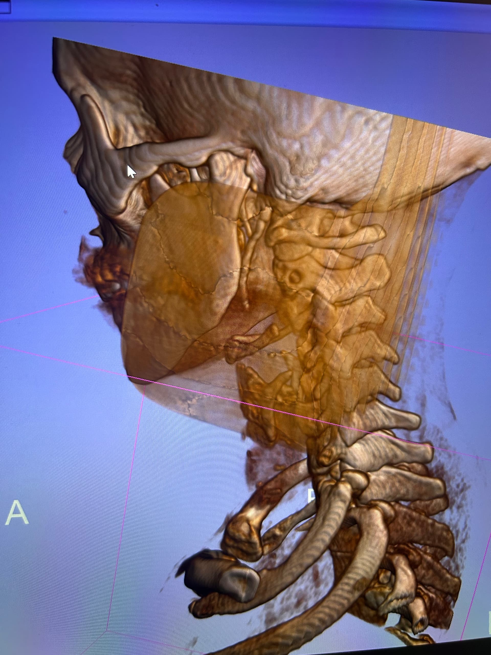
Hi @SeekingInfo knowing the length of the calcified ligaments is something, but seeing it in 3d just gives you a better picture of just how long they are and also the shape. If you can find the axial slice that @TML has mentioned previously then he can give you an idea with regards possible vascular compression between you styloids/ligaments and your C1 vertebrae.
Hi! Would the axial slice be from the 3D image or just the regular images? I found the axial photos, but there’s several, so I’m unsure what to include. Thanks again for your help!
Hi @SeekingInfo yes they are the ones. Both your Styloids are extremely close to your C1 transverse process. I would assume therefore that you will have vascular compression probably IJV. Hopefully other members will give you their opinions shortly.
I think it would be really beneficial to get a CT with contrast, but I am fairly sure you will need the calcified ligaments removed anyway.
Looking at your 3D images, your styloid on the left is completely calcified with the ligament very close to the C1 process, and looks pretty thick at the top, so I think would need to be removed at the skull base to stop IJV compression. On the right your styloid looks very long, plus extra calcified ligament at the end, again, it’s very close to the C1 and wide at the top. Your hyoid processes look quite thick, and also your thyroid cartilage looks pretty chunky…
I’m sorry that I can’t do labels, but on your left side, to the left of your styloid at the side of your jaw it looks like there’s something calcified, curved back towards the styloid process- I don’t know what that could be, I’ve never seen that before!
So I think looking at these images, it does change what you do next & which doctor you see, I do think that you need to get a consultation with one of the VES doctors, like Dr Nakaji, Dr Hepwort or Dr Costantino. So not as simple as you were hoping for
@SeekingInfo this is perfect, see attached annotated image.
Your styloids are extremely close to your C1, practically touching. This is important because the IJVs and vagus nerves (and also spinal accessory nerve) run between the styloids and IJV.
Research shows that the average space between a styloid and C1 in a healthy individual is about 9mm. I’d be surprised if you have more than 1.5mm between your styloids and C1.
Here is a research paper by one of the main ES surgeons in the US, Dr. Cognetti, explaining that the styloid-C1 space is the cause of ES symptoms and that styloidectomies with the patients resolved the symptoms:
I would recommend getting both styloids removed and to make 100% sure that whichever surgeon you decide to go with cuts the styloids above the level of C1. If they do not cut them above C1 you are going to continue to experience IJV compression symptoms.
I agree with @Rosie that a CT with contrast would be beneficial. However, most ES surgeons (the reputable ones) are going to look at the CT you already have and just know that those daggers need removed. And if they remove them above C1 then it’s all good. Some surgeons require the contrast CT though, so maybe they won’t even look at your CT without contrast, but it depends on the surgeon.