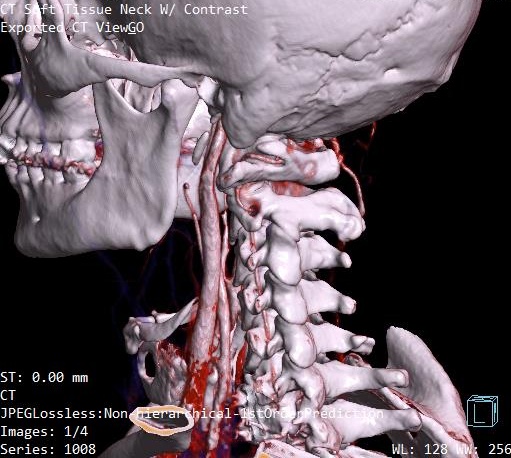
I have some fresh images from a CT with contrast I had done a few weeks ago. Those of you who are good at this stuff, feel free to give me some feedback on what you see. I’m not good at studying images like these, but it looks to me like between the styloid and C1 the blood vessels on the right side appear to be compromised. Let me know what you think.


3 Likes
Hi, I’m sorry but I deleted your images because your personal info was on them! would you be able to take your personal info off them & re-post?
2 Likes
I can work on it and see. For now, you can go ahead and delete the post. Thanks.
1 Like
The personal info has been removed and the images reposted. Thanks!
2 Likes
@jrhode2873 axial view is the best to see the IJV compression. If you go to the axial view, look for C1. I’ve attached my own imaging to help you find it. It’s the vertebrae that has wings!
It’s often hard to tell if the styloids are at play with IJV compression in the 3D models. Sometimes it just ends up being C1 causing the compression, but can never know for sure unless I see the axial view!
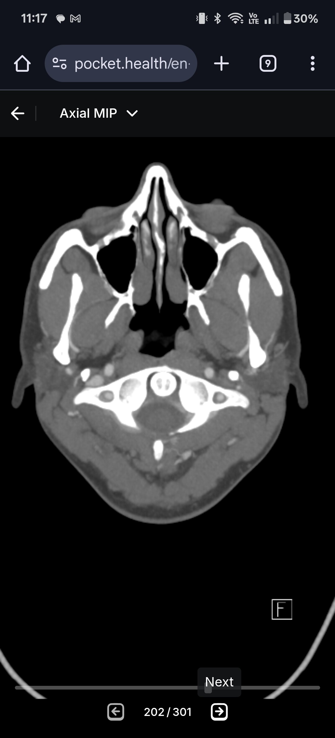
@jrhode2873 - Your right IJV is definitely compressed & it looks like it’s predominantly being caused by C1. The left also has some contact w/ C1 but doesn’t look like it’s got the serious compression that the right side has.
The greater hornse of your hyoid look somewhat long which means they could be making contact w/ your carotids in some head positions. Do your carotid arteries show up in your imaging or was this just a CTV? If you have any carotid pics & could give us some views of your hyoid bone w/ respect to the vascular tissues around it, that would be great.

You, like so many of our members have lost the lordotic curve in your cervical spine. That brings the styloids & potentially the greater horns of the hyoid closer to the nerves & blood vessels in the neck.

2 Likes
Ate there visible styloids?
There may or may not be depending on length. That’s why the axial view is beneficial. If you find your C1 in the axial view like I showed above, I can help you look at compression
1 Like
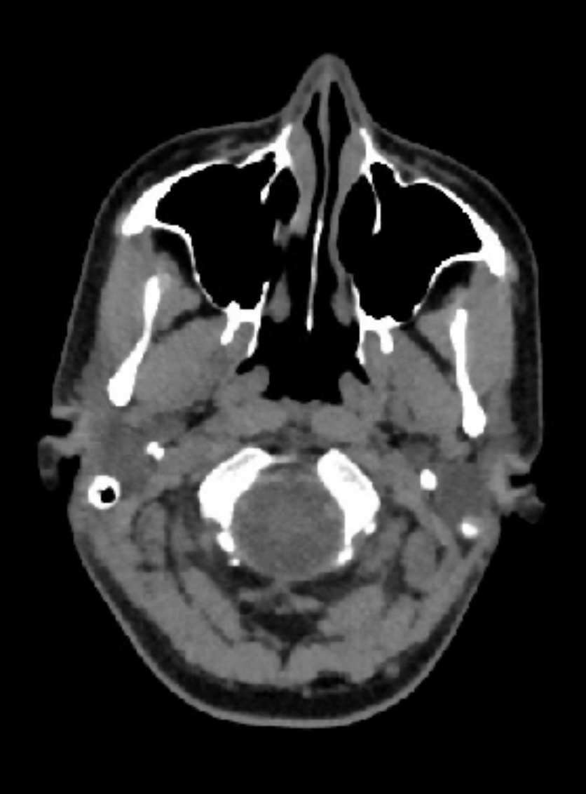

@Jete see attached annotated image.
Appears both your styloids are in direct contact with your IJVs, and so your IJVs are wedged between your styloids and C1. It’s hard to say for certain the degree of pinching since there’s no contrast but it’s definitely happening. Do you happen to have an older CT with contrast?

1 Like
Thank you.
No I don’t, but I have symptoms, so I try to figure it out. ![]()
1 Like
I would definitely see if you can get a doctor to order you a head and neck CT with contrast.
I will also mention that the compression on your left side appears slightly more than the compression on your right.
2 Likes
I have posted a new post with more images.
I am allergic to contrast. ![]()
1 Like
I saw the post, but the images that we managed to get to in this forum are better.
That’s unfortunate about the contrast allergy! I’m not sure if there is any other imaging techniques that would be better than what you have then.
What are your symptoms? It might prompt us to look elsewhere in your CT scan
2 Likes
Pain in my right ear. Pain radiates down to neck, I have constant feeling that something is stuck in my neck. Headaches, constant pain in my right aye, jaw, cheekbone even sometimes in my teeth. Tingling in face, nose, tongue. I am choking on food. Dizziness (swaying, rocking, floating), flashes and spots in my vision.
At night I wake up from pain. Brain fog and difficulty to think. Sometimes I feel like my head is full with pressure.
Symptoms may worsen or decrease but never goes away.
The most scary symptom is when I feel like I will pass out.
2 Likes
Thanks for the helpful annotations. Please refresh my memory: who are the best surgeons for a potential C1 AND styloid problem? I know Dr. Osborne and Dr. Hackman won’t touch C1.
1 Like
Definitely sounds like IJV compression and vagus nerve compression symptoms. Are you able to open your imaging with the Radientviewer software? It might give us a better idea on styloid length and will be easy for us to navigate to hyoid bone too just to ensure your greater horns aren’t digging into anything.
2 Likes
I can, but I don’t know where to measure…
@Jete if you’re comfortable with it, you can put your CT DICOM files all into a google drive and private message me a link to the drive. I can open the imaging on my computer and measure the styloids, send you images, then delete the files off my computer. Likely easier than trying to explain how to measure them properly and whatnot
1 Like
Closest to you would be Dr. Costantino in White Plains, NY & Dr. Cognetti in Philadelphia, PA. Dr. Cognetti is booking into next year already but you should be able to get in to see Dr. Cognetti this year. Dr. Nakaji in AZ & Dr. Hepworth in CO would be the other two.
•Dr Peter Costantino, 4 Westchester Park Dr, 4th floor, White Plains, (914) 517-8056
http://www.nyhni.org/find-a-physician/Peter-D-Costantino-MD,FACS .
Does do online or phone consults we believe.
•Dr David Cognetti, Thomas Jefferson University Hospital, Philadelphia 215- 955- 6760 (Has done many successful surgeries on members). Only removes ligaments if calcified. Works with Dr Heller now to do C1 shaves
David M Cognetti MD | Jefferson Health Does do online or phone consults.
2 Likes