Hello Everyone, I am new here but experincing manyt overlapping symtopm,s with ES.
From my very amateur 3D skills I made this.
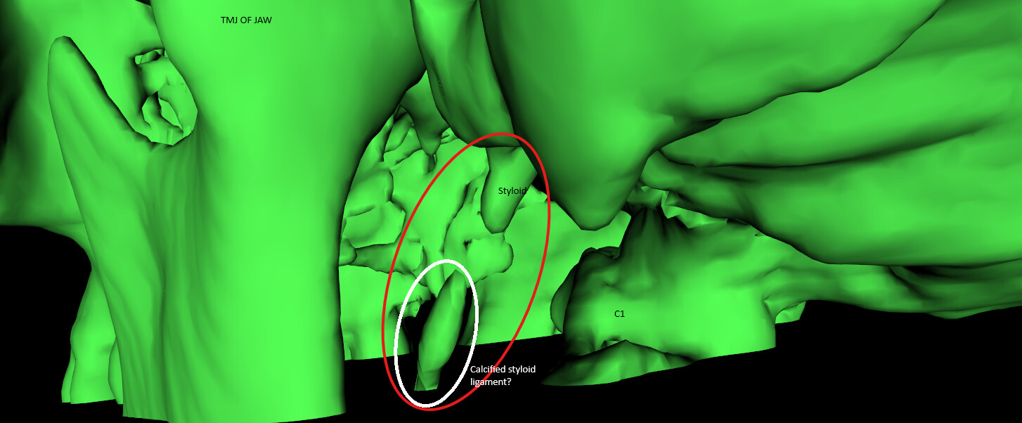
MRI of Brain and Neck, and unfortunately only a sinus CT, which only just reaches the lower level of the skull base. I have 3D reconstructed what I have available, and from what I can see, the styloid itself is not elongated, however there seems to be lower styloid ligament calcification? Unfortunately this is disappearing off the edge of the CT thus I’m not able to fully see the extent further down.
@LouGatchar - Please try uploading your images again since @Jules has confirmed that your site trust level has been elevated so you can. New members are prevented from uploading images until they post as a spam prevention device in our software. Once a post is made & replied to, trust level is automatically updated so going forward uploading images & links is possible.
It’s well known on our forum that styloids don’t need to be elongated to cause problems. The angle they’re growing, how curved, thick, twisted or pointed they are can cause symptoms even if they aren’t especially long. One member even had little bony “barbs” growing off her normal length styloids which caused symptoms. Calcification of the stylohyoid ligaments below but not connected to the styloids can also cause the same types of symptoms as the styloids can.
It’s likely that this is a section of calcified stylo-hyoid ligament; the styloid can occasionally get fractured but there’s quite a gap so I’d plump for calcified ligament, a shame you can’t see how long this is… It looks from this image as if there’s a reasonable gap between the styloid & C1 process, but without a CT with contrast and looking from other angles you can’t see for sure… If it is the styloid process, then as this is very short (the width & angle can be enough to cause IJV compression) , if you wanted to try surgery you would need a really experienced & skilled surgeon as it would need removing at the skull base- so probably Mr Axon in England would be the best person to do this…
Are you able to push for a CT with contrast do you think? It’s probably the best way to either rule jugular compression in or out?
You might find this video from Dr Costantino interesting, he explains symptoms and shows the anatomy quite well: Dr Costantino IJV compression and surgery - Symptoms and Treatments / Doctor Information - Living with Eagle
I can see the calcification, however as this is produced from a sinus CT, we loose the calcified ligaments off the end of the scan.
However I do note that the styloid process itself is not elongated, in fact it seems rather short, especially on my right side, ~10mm. I do wonder if this has been fractured in the past?
The section we can see in this imaging, from the base of the styloid process to where we loose it, is around 25mm.
I can also identify a boney ‘spur’ to the internal side of the right styloid process. Not sure if of any significance? however as I’m aware even the shortest, but thick styloids can cause issues, maybe this ‘spur’ also has a role to play.
I have emailed Dr Higgins today. I’m not a religious man, but God knows I’m praying this leads to resolution of my symptoms.
I’ve included some main parts of the letter below:
About 10 years ago I began experiencing the following cluster of symptoms:
Tinnitus ringing
Tinnitus pulsatil e
Hyperacus is
Headaches/head press ure
Light sensiti vity
Pain in trapezoid area when moving my neck to the side
Pain/tightness in my neck under m y ears
Bruxism
Vision disturbances – seeing heartbeat in periphera l vision
Feeling heartbeat in throat
As it currently stands the working diagnosis of the audio vestibular team in Cardiff is PPPD with tinnitus and migrainous features. I have been prescribed pizotifen. I have also been advised to have CBT, which I have already heavily engaged with in the past 10 years However, the PPPD diagnosis does not appear to match my symptoms and furthermore I have attended a neurological rehabilitation centre (January 2025) where it was confirmed that my condition does not appear to be a functional disorder and therefore could not be trea ted there.
@LouGatchar - It’s hard to tell from the pics & video you posted exactly what the styloids could be affecting, but I know you know it’s not an ideal scan for detecting ES or vascular compression that the styloids/stylohyoid ligaments might be causing. Your symptoms do sound like you potentially have IJV compression & as @Jules noted your styloids/s-h ligament calcification is quite angled so could be affecting your IJVs or internal carotids.
That looks to me to be the leading edge of the right s-h ligament calcification which could continue toward your hyoid bone if the calcified section of ligament isn’t removed.
I hope Dr. Higgins is helpful. If not, try Dr. Axon as he more specifically deals w/ IJV compressions.