Well, I finally figured out how to create 3D images. In the lateral (side) views, I’m wondering if the right styloid is elongated and partially calcified. I actually couldn’t see the styloids in the posterior (back) view.
Thoughts?
Well, I finally figured out how to create 3D images. In the lateral (side) views, I’m wondering if the right styloid is elongated and partially calcified. I actually couldn’t see the styloids in the posterior (back) view.
Thoughts?
They aren’t the longest styloid processes we’ve seen, but still a little longer than they should be…They are definitely very angled though which can cause symptoms, & the left side is pretty thick which again can cause problems. It may be if you see a doctor who doesn’t know that much about ES that they don’t consider the styloids long enough to cause ES & to need surgery, but there are research papers which state the angle can cause issues, so if you do see anyone it might be an idea to print off one of the papers & take it with you. (There should be links in the Newbies Guide Section) Here’s one for you though:
Evaluation of the length and angulation of the styloid process in the patient with pre-diagnosis of Eagle syndrome | Kosar | Folia Morphologica (viamedica.pl)
Are you able to tell how long the styloids are? In one of the lateral pictures, the styloid has an odd jaggedness to it. What are your thoughts?
I think what you’re seeing as a jagged area is an artifact on the image. I base that on how that styloid looks from the back view. I do agree w/ @Jules that your right styloid looks somewhat elongated & very angled & your left one is angled & thicker than normal & both of those features can create ES symptoms & thus an ES diagnosis. It also looks to me like you might have some calcification lower down on your stylohyoid ligaments, but what I’m seeing may also be insignificant artifacts.
Thanks a million to you and @Jules for your help and information. I’ll certainly be more prepared for tomorrow’s doctors appointment. I realized in my previous post, my pictures were from December 2020 from a CT angio. I will attach pictures to this message from a CT scan (that was done 2 months ago). To me, the updated pictures look a bit more pronounced in terms of thickness, angulation, and length. Of note, I have a congenital fusion fo c2-c3 spinous process, which probably makes me high risk for ES.
I have a few questions…things that are important for me to understand for tomorrow’s doctors appointment.
Again, thanks a million for your input!!! Included below are more recent CT (without contrast) images from a couple months ago.
First, here’s an annotated version of one of your first images that shows where you might have stylohyoid ligament calcification:
Interesting about your congenital spinous process fusion. It’s hard to say if it could predispose you to ES but it is possible since it limits the mobility of those two vertebrae which could put stress into that area of the neck & cause styloid elongation/s-h ligament calcification.
Question answers:
The angle of styloids that you see in other images are sometimes based on head position during the imaging & may not accurately portray the actual styloid angles. In your case, it does look like your left styloid is angled inward more than the right, & it is thicker than the right but shorter. The right styloid looks pretty long, also thick & could be angled a bit upward & inward as opposed to other images you’ve seen on this forum.
I’m not able to measure your styloids, but the doctor you see should be able to (they often measure using the slices not the 3D image), or you can ask for the radiologist who wrote your report to please revisit your scan & measure the styloids & amend the report adding styloid measurements & comments about ligament calcification if seen.
Styloid thickness can indicate excess calcification though styloid thickness does tend to vary between individuals.
See image above.
Yes, your right styloid looks long & I’m not sure about the left. It just looks thicker than “normal” to me. Again, I’m not sure what’s normal for you, but it looks thick enough it could be causing some nerve irritation.
I hope this helps.
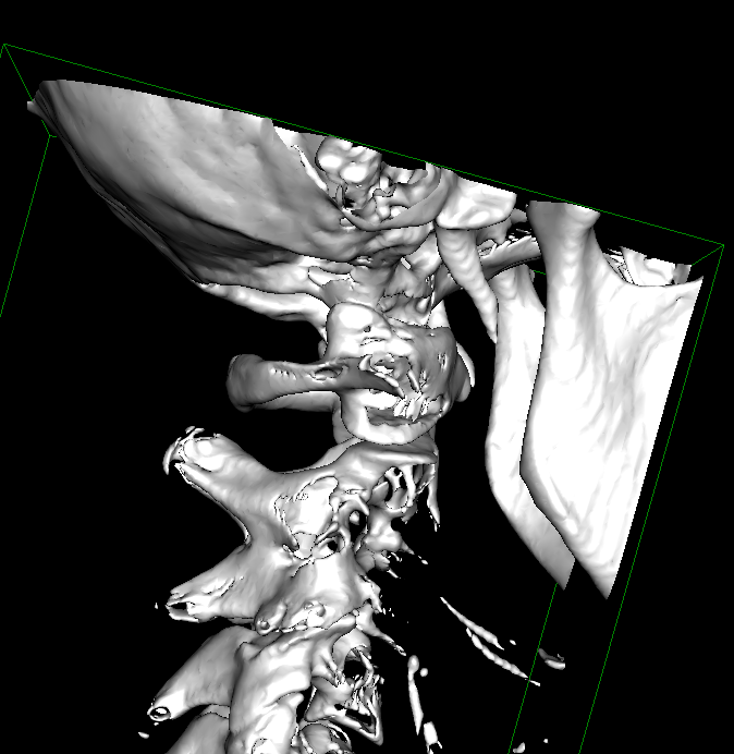
The area you’ve highlighted looks like hyoid bone to me, but hard to say without being able to rotate and adjust the thresholding.
@coldbear @Isaiah_40_31 @Jules
I manipulated the 3D images. I think the red circle is the hyoid bone and the blue circle is calcified stylohyoid ligament in both images. In the second image I caught an angle where you can see the greater horn and lesser horn of the hyoid bone. Please tell me if you think the blue area could be calcified stylohyoid ligament…if not, then what could it be? Many Many thanks for your opinions.
@1speechpick These are different parts of the Hyoid bone. The red circle on first pic is the Hyoid body and the blue circle are 2 greater horns of the hyoid bone. If there is a calcified ligament, it will be diagonal and will join to the Styloid process. I drew a “cyan” line to show you hypothetically where the ligament would be.
This image is from the internet to show you different parts of the hyoid and where the ligament is. You can see Stylohyoid ligament as “dots” toward the styloid.

Hope this clarifies it.
@KoolDude has it right. The hyoid bone is a horseshoe shape made up of the different things you’ve circled. The stylohyoid ligament would be a straight line connecting the middle of each horseshoe side to the styloid process. I’ve drawn it in red in the photo here.
Thank you all so much! This helps tremendously!!
Thank you @coldbear & @KoolDude for correcting me. I wasn’t sure that was calcified ligament since I couldn’t tell exactly where the hyoid bone is. Guess I shouldn’t try to get a job as a radiologist, eh?! ![]() You guys are so incredibly helpful with this stuff!!
You guys are so incredibly helpful with this stuff!!
@Isaiah_40_31 It is difficulty to tell which is which, particularly with this kind of images where there are fragments of bones pointing different directions. I make these mistakes all the time so it has nothing to do with reading it properly. Even trained radiologist miss many things that we detect here so it has nothing to do with being radiologists. I believe the points (1 - 4 ) you raised here are still valid. I am pretty sure, for example, thickness can be an attributed to excess calcification. To Err is human and To forgive is divine as the old adage goes.
Thank you for excusing my ignorance. I will learn not to comment when I’m not certain about what I’m seeing in the future. ![]()