Hello I am a french woman and have both styloides about 40 mm with great pain. All begin in April 2025 (even if I think I had a such episod in 2021 but less strong) after dental care.
My symptoms are:
pain throat +++° as I something stuck in the throat ( the more often on left but something right too)
dysphonia( some days I can’t speak)since September
increasingly blurry vision
viscous and translucent secretions
great vertigo in 2024 (only one in 2025)(in 2024 ORL said it was cristals in the ear but gave me treatment for potentiel AIT during 1 year
pain tongue (but now not too much)
I would like to know if I have compression on my jugulars or(and) carotids? None doctor in france look at my scanner and said eagle syndrom can give compression!!! They all tell me I need depressive treatement . I am so painful and tired ..
On January the 6th I finally will have an”angioscanner des TSA” scanner des troncs supérieurs aortiques.Will it be more helpfull because it will be not dynamic…
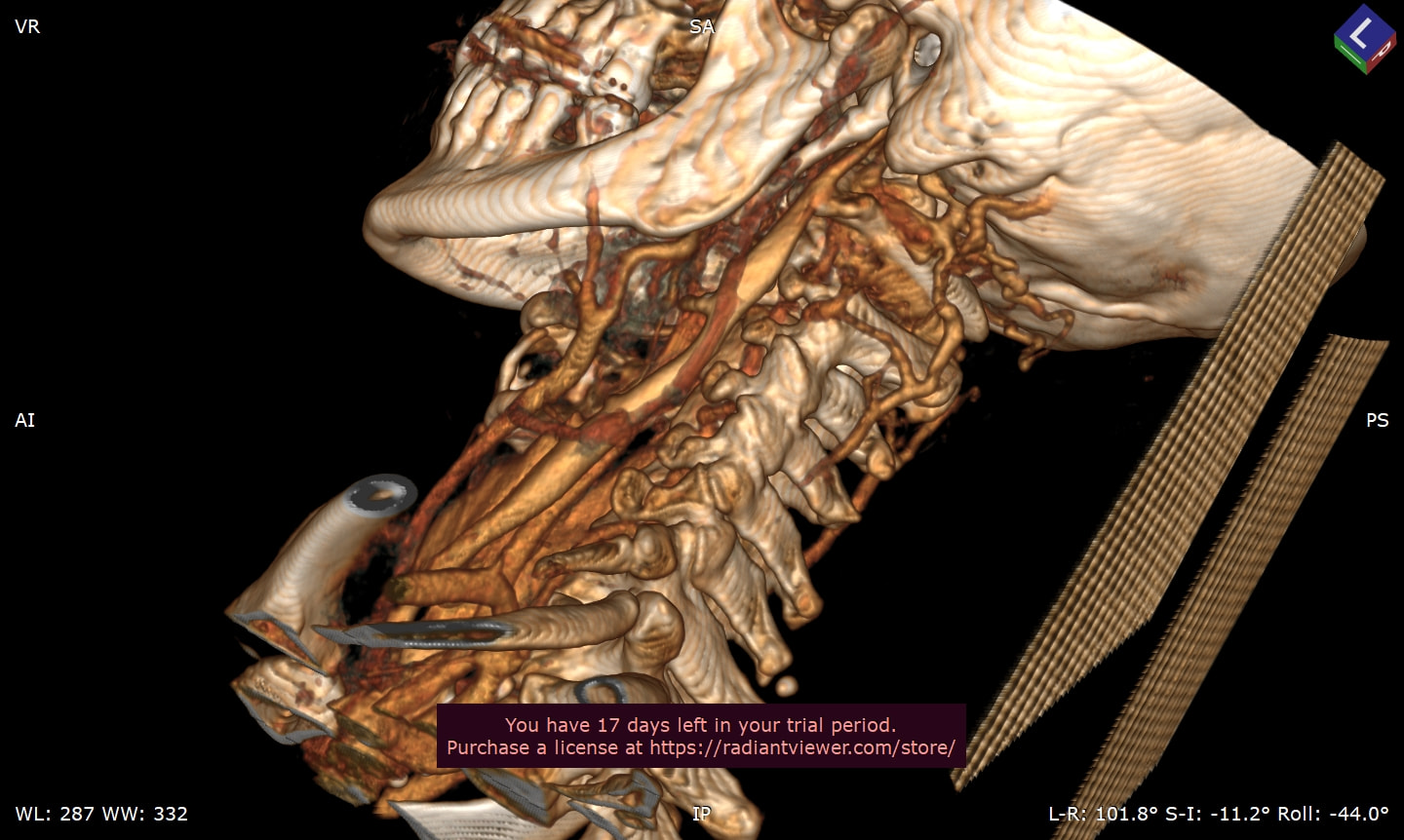
I looked at your imaging & what I see is that your styloids are both elongated & very thick up at the skull base. Your left IJV looks like it’s being compressed above C1 by the styloid, but perhaps also by C1 in certain head positions. You also have collateral veins on the left side. Your right one doesn’t look compressed to me in the head position in which the scan was done, but you have collateral veins
on the right side as well so it’s likely there is also IJV compression on that side.
Collateral veins are veins that enlarge on the back of the brain & skull to help drain deoxygenated blood out of your brain when the IJVs aren’t working properly. They can cause occipital pain, upper neck pain & headaches.
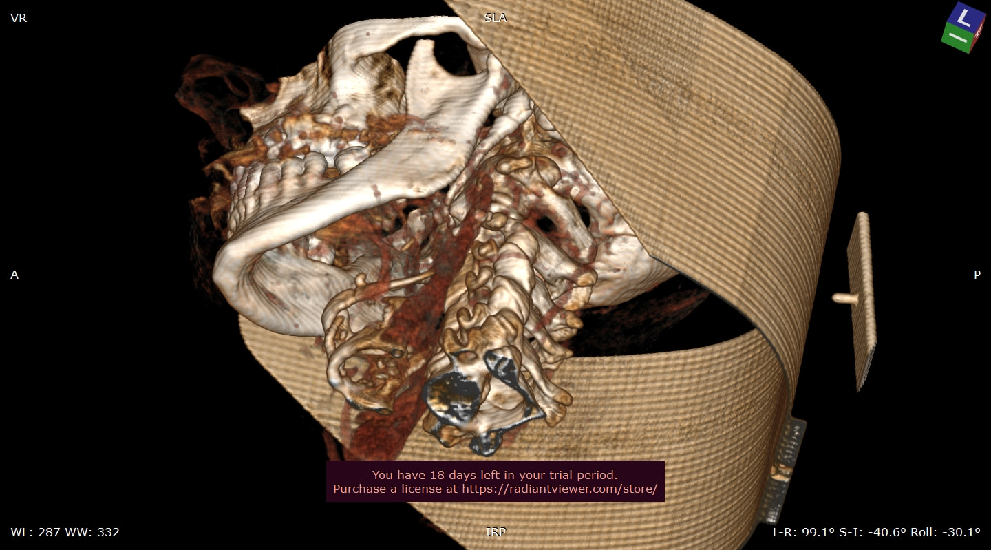
From what I could see of your hyoid, it looks like you may also have elongated greater horns on your hyoid, but I couldn’t tell for sure. If you do, these can poke & irritate your carotid arteries. It’s good you’re getting another scan to look at your arteries. If you can request for your internal/external carotids to be checked in the area of the hyoid bone, that would be helpful to see if they are suffering from any compression or contact with the greater horn(s) of the hyoid bone. In the last image, I’ve circled your hyoid bone in red. It looks very oddly shaped which could also cause it to make contact w/ nerves or blood vessels in that area.
I agree with @Isaiah_40_31 's comments and labelling of your images… I’m sorry that I can’t do labelling too, but this image is interesting as it shows the hyoid better, and the horns look really long & needle-like:
Also your thyroid cartilage looks quite calcified too, which we’re now realising can also cause some symptoms. It might not be for you, but worth considering…
@Jules thanks so much Jules for your help.About thyroïd my scanner said’ two nodules” and the doktor said “it is bebause you are old(66 years) ,come back in 2 years to check)
@Veroguilnec - I am also “old” (isn’t age more a state of mind? ) and have nodules in my thyroid which get checked each year. There are a number of our members who have thyroid nodules alongside ES. We’ve wondered if they are somehow related.
I was 58, @Veroguilnec. I had a 3rd surgery in 2024 when I was 68 to do a revision styloidectomy & decompress my IJV, & I actually healed from that surgery more quickly than the first two.
I had my angioscanner last Tuesday…I can’t find the courage,the energy to read it alone. Tomorrow perhaps..because if tonight I read so worse news I know sleeping again will so hard… I come back to tell you soon. Thanks so much for your great generosity with all members.
@Isaiah_40_31 Hello I have well slept that night .I think it is because I have taken (my doctor doesn’t agree with it) solupred in the lsaturday morning (anti inflammatoire) but I had so much pain…I just get up today at twelve and go back in my bed to 16 PM
When I have good rest it is better with pain.
Of course I come back to show you my report. For now I don’t know if radiant 3D will be accessible. I have used it more than 30 days ago.
I have my angioscanner des TSA(tronc supérieur aortique) but no report to say the result. I suppose I have to put images in radiant viewer for you can perhaps help me? So difficult for us who are not radiologist doctor Thanks again
I can look at the images, @Veroguilnec, but I have never used radiant viewer so I don’t know how to make 3D images that way. I hope you’re able to make the 3D images & post some on here.
It’s the easiest way to be able to upload them I think if you still have a free trial, but I’m not very tech savvy so not the best one to advise you! You could maybe send a link in a private message to @Isaiah_40_31 & myself otherwise, but don’t put that in the open forum to protect your privacy.
hello thanks so much. I am very disappointed because my doctor asked “scanner des tsa” to see compression jugulars and carotids. The radiologist has performed a brain scan and only carotid are notified. I will try to see if radiant 3D is always accessible in free version for me. As you agree I will send you the report and the link of my angioscanner with MP.Thanks thanks so much!