I am new to this group. I’m deaf. I will try my best to summarize. Diagnosed with bilateral ES in July 2024. The partial left styloid was removed with the CC1 transverse on April 10. My husband said the ENT surgeon or one of the ENT team told him he had only removed a small partial styloid. We were baffled and thought the styloid would be removed completely. Now, I am at the hospital ER and staying for observation due to ES symptoms, plus syncope episodes, choking sensation, dizziness, head to ears to neck to hands/arms pain, pressure, numbness, and tingling. My chest is in pain while breathing. My heart feels palpitations at times. Now, I am feeling worse. I also feel the nerve irritation radiating from the neck to the head and down the neck. If I move my neck to the left slightly, I will gag, and something compresses the outside of my left/right throat, and the left side is weak. If I move to the right slightly, something pulls and compresses toward the outside of my left/right of my throat, and I gag. If I move my neck up, something pulls and compresses/gags toward the outside of my left/right throat and my neck. I can feel the swelling behind my ears and neck. It is painful and hurts to breathe. I have a terrible headache. I can feel the pressure and pain behind my ears to my neck sides while swallowing, which is painful. My ES symptoms are no fun. I had a left partial styloidectomy (5 months post-op). I thought it was supposed to remove the styloid completely. I still have an elongated styloid on the right side. I also get syncope episodes. My head, ears, jaw, and neck/throat are sore and painful. The base of my head and neck is giving me a headache: numbness and tingling.
Tests done so far:
U/A with Micro - abnormal
Urine Grey Top
Urine Clear Top
Troponin I, High Sensitivy, Plasma (two times)
Type & Screen
CT Angiogram Chest Dissection with 3D Reconstruction
CT Angiogram Neck W WO Contrast
CT Angiogram Head W WO Contrast
CT Angiogram Abdomen Pelvis W WO Contrast
CT Brain/Head WO Contrast
XR Chest AP Portable
CBC and Auto Diff W Reflex - abnormal
CMP - abnormal
Lipase, Serum
B Naturietic Peptid (BNP)
Prothrombin Time W INR PTT
CBC With Auto Differential Sysmex
MRI W WO Contrast
Now, I am havintlg sudden, sharp headache, snyncope episodes, and dizziness with nausea when moving neck and lying back. i will see the doctor (I think my neurosurgeon) from Neurology. My ears and neck are pressure and sharp pain. I am in tears and want both remaining styloid and elongated styloid to remove. Now I feel imbalance with styloids and whatever inside my neck.
Any input or suggestion before I see the doctor(s) today?
Oh no, poor you, that is rough! It sounds like you have vascular ES- the syncope episodes could well be pressure on the carotid artery, although it could also be if the vagus nerve is irritated. The vagus nerve can also cause breathing issues, sweating & stomach issues; it exits the base of the skull along with the carotid artery & the internal jugular vein right next to the styloids, so it could be that if your styloids are thick or angled slightly then even having been shortened it could still be causing issues on the left side, as well as the right which is elongated.…Headaches and dizziness could be carotid artery compression, or otherwise jugular vein compression which can cause increased pressure inside your head (intracranial hypertension)… given the choking and swallowing issues, this could be from your elongated right styloid remaining, or otherwise it could be that your hyoid bone has shifted… Pain in the skull base or behind your ears I wonder if could be collateral veins, if your internal jugular vein is compressed?
You obviously have lots going on, were the CT angiograms you had before or after your surgery? You could post those on here & we could have a look & see if we can give you some ideas of what to ask your doctor? And if you haven’t had a CT done since your surgery in April I would request that this be done again- a CT with contrast to show both the arterial and venous phases of your head & neck, from the skull base down to the hyoid bone. It would also be helpful to see how much of your C1 process has been shaved- usually a small section is shaved which shouldn’t make much difference to the stability in your neck, but if more has been removed then this might have made the whole C1 shift a little.
So to clarify if this isn’t too late, I would try & find out from your Neuro exactly what was done during your surgery, & ask for more imaging to be done if you haven’t had that already. I would then consider (depending on what you find out) looking to have surgery for the right side with a doctor who has more ES experience & who can remove more of the styloid process, and possibly a revision surgery on the left side.
@bdys - I’m so sorry for what you’re going through & hope you can get some symptoms resolution! If you didn’t see one of the doctors on our Doctors List for your initial surgery & IJV decompression, you will do well to contact one of them for an opinion at this point. The closest to you who isn’t booked out to next year is Dr. Costantino in NY. Dr. Nakaji in AZ may have openings even sooner than Dr. Costantino. Both will want a current CT scan w/ contrast in order to offer you a consult. Dr. Hackman in NC removes the styloids to very close to the skull base & will do both in one surgery. He doesn’t specifically do the vascular decompression procedure though, but sometimes the vein(s)/arteries that are impacted by the styloids open just fine on their own once the styloid has been cut back beyond where it’s contacting them. All 3 doctors do phone consults for patients who are out of state.
@bdys If your symptoms are not being caused by remnants of the styloid I think it’s possible it’s your hyoid greater horns. I am getting my greater horn removed since it’s pressing against my ICA and causing stroke-like symptoms.
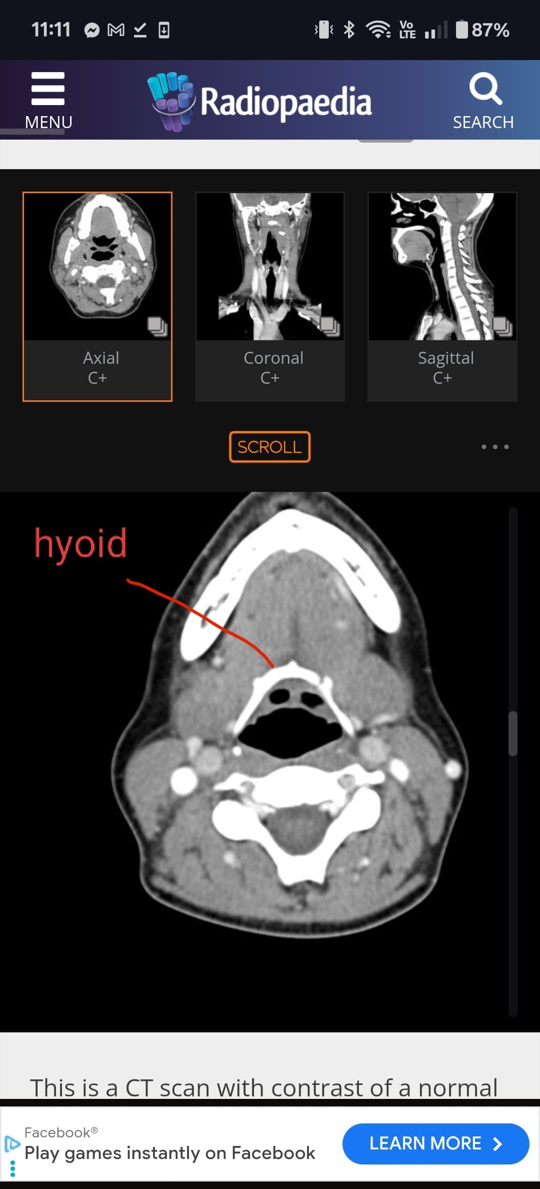
Do you have access to your CTA of your neck with contrast? I can help you locate the hyoid.
Thank you for your response. I have a lot of going on. I will talk to neurology team, maybe my neurosurgeon. The general neurologist came and listened all of my symptoms. She said that I have an appt with movement disorder doctor in Nov and the possibility of the Botox. I told her that the Botox is a temporary, masking treatment. It is only last for two to three months and then come back for more. I told her that I want the fixable solution, not repeating with medications and conservative treatments.
@bdys can you go into axial view and go to your hyoid bone (horseshoe shaped). I want to see if the greater horns are near any of your carotids. Ive attached a few images to help you locate it. Sometimes the whole hyoid can be seen in the same slice, but often the greater horn tips are a slice or two higher because of the hyoid being angled. We are interested in the tips of the greater horns.
Your syncope episodes make me think it’s your carotid sinus. Carotid sinus hypersensitivity is most prevalent in those around age 60 and older. A greater horn can easily irritate the carotid sinus. I’m scheduled to have my greater horn removed because of this.
Obviously not a doctor, but it doesn’t look as if your IJV is too compressed in those images, this can look very different though in the axial imaging that @TML has been helping us with… The hyoid does look quite close to the bifurcation of the ICA & ECA in this image:
@bdys one thing that I could do if you are comfortable with it is go through your imaging. I’ve helped many people on the forum this way. Typically involves putting your files into a google drive folder and private messaging me a link to the folder. I can go to the hyoid and make some annotations. If you don’t feel comfortable with that, I do have an axial CT tutorial on the forum that could be helpful.
@bdys I posted a couple images earlier of how to find the hyoid bone in the axial view. Sometimes the view is not labeled “axial” but there should be a view that slices your body head to toe. That’s the view you want. Then it’s a matter of locating the hyoid by trying to match what I posted earlier. Every hyoid looks slightly different, but almost always a horse shoes shape midway down the neck
Sorry, I mean the axial view of your head and neck CTA, the one you have been uploading 3D images of. If you go to the original CT imaging (the black and white imaging that you turned into 3D) there should be a series of images labeled as “axial”. It’s a series of slices that go from your clavicle area all the way to the top of your head.
Can you help me with images? I tried to figure. I’m sorry. The case manager came to me early this evening that he is working on getting me a room. I’m here in the ER hallway. With ES symptoms, it is hard for me to find comfortable. I was here before the other patients came in, but they got their rooms. I m baffled.
I am very sorry you got put off when you were in the ER first. That just isn’t right. I hope you didn’t have to wait long for a room.
I agree w/ @Jules that your IJVs look compressed & especially the left side where it just disappears. I can’t tell about what your hyoid is doing though. Your styloids both look quite long, but I understand your left side has been partially removed so is shorter now. I expect it is still putting pressure on your IJV which is causing some of your symptoms. I’m sorry the doctor who did your surgery didn’t do a great job.
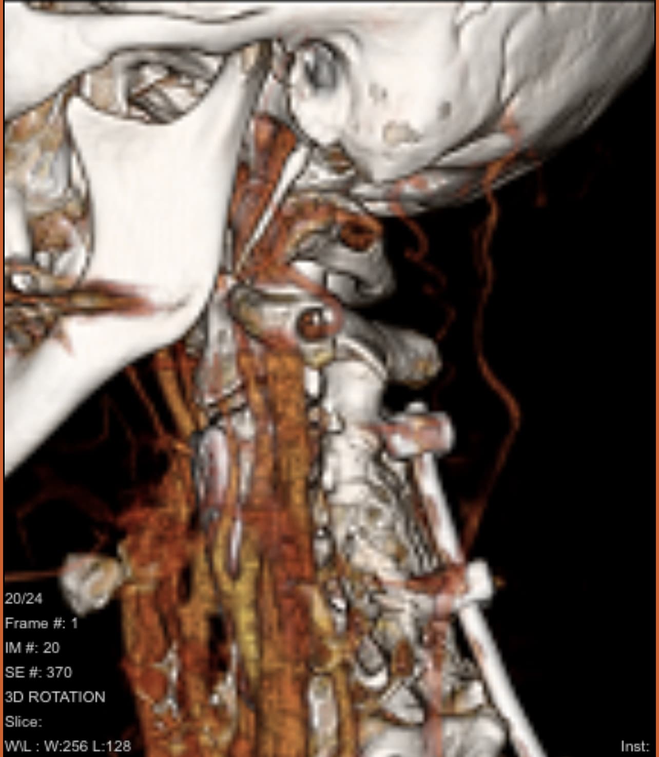
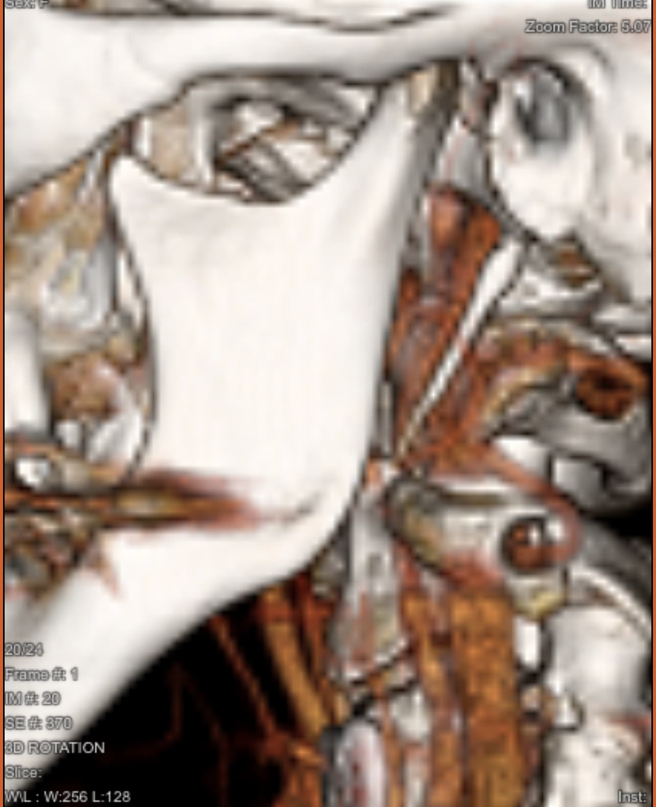
Here are two of your images that show your hyoid bone. I marked them so you can see where it is. One is from the front & the other is from the right side. Unfortunately, there isn’t a picture of the hyoid from the left side. In the picture of the right side, your IJV blocks the greater horn of the hyoid so we can’t see if it’s poking your carotid arteries there. Can you also upload a picture that shows your hyoid from the left side?