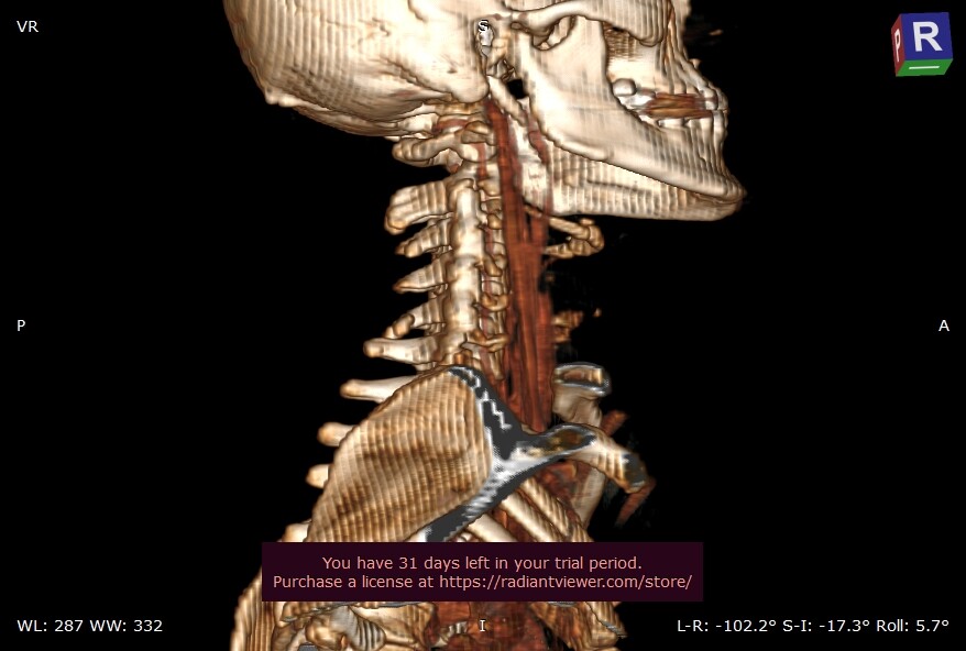
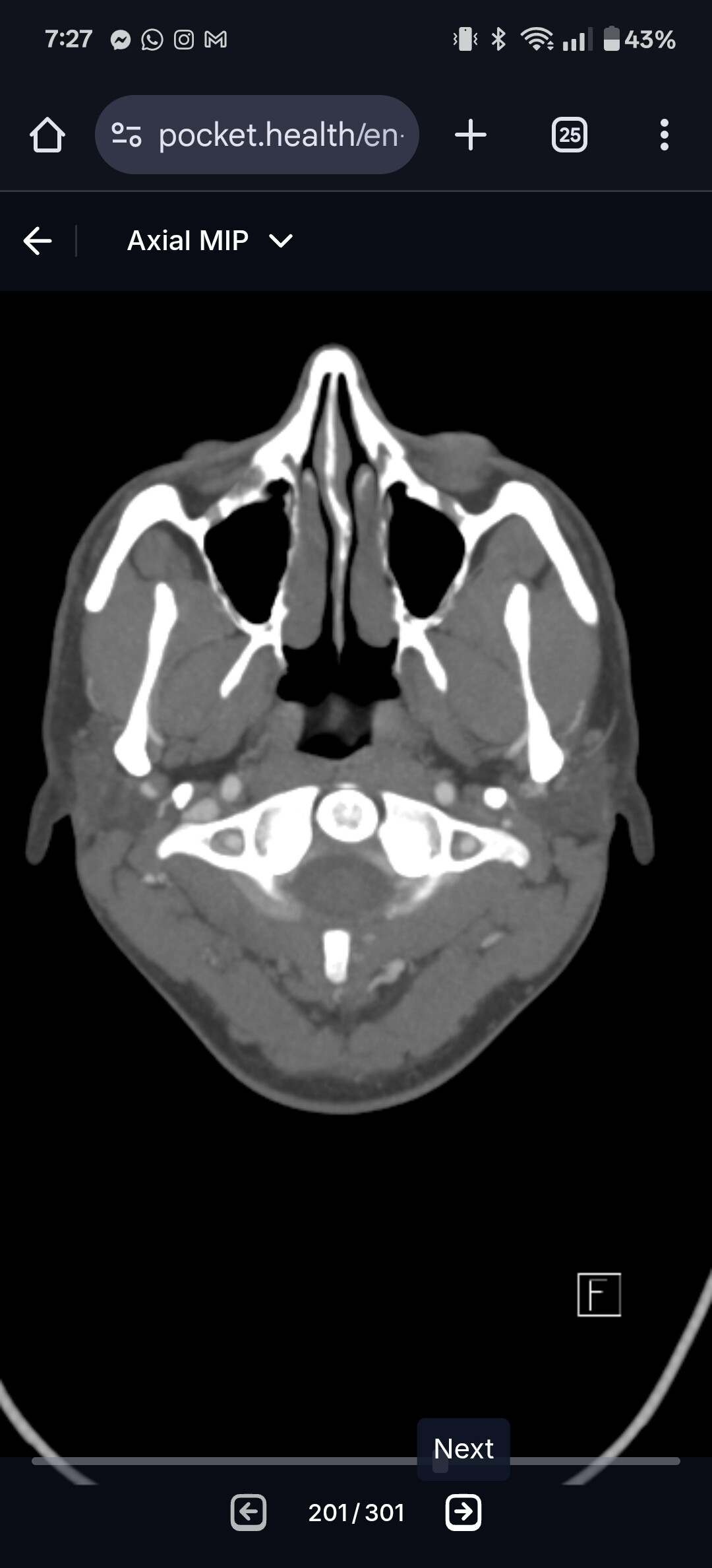
Hi all, new here. Ive been sick for over 4 years. My most debilitating symptoms are sleeping all the time and high heart rate. A sleep study revealed I dont enter rem sleep due to having arousals. They haven’t been able to find what wakes me up during my sleep. I few other symptoms of mine include vertigo when looking up, left side neck pain, low csf neurotransmitters (bh4), and extreme brainfog/confusion. Im only 25 and have been trying to figure this out since I was 17. Due to my persistent left side neck pain I came across eagles. I created a 3d reconstruction from my a neck ct with contrast that was reported as all normal when they were looking for any problems in my airway. Would love to know if anyone has any insight or thinks the styloids look rough. In particular on the left side? Thank you all so much.
@6tdog6 are you able to make the 3D model in Radiantviewer so we can see your veins and arteries? Will help us determine whether we think there’s anything being compressed.
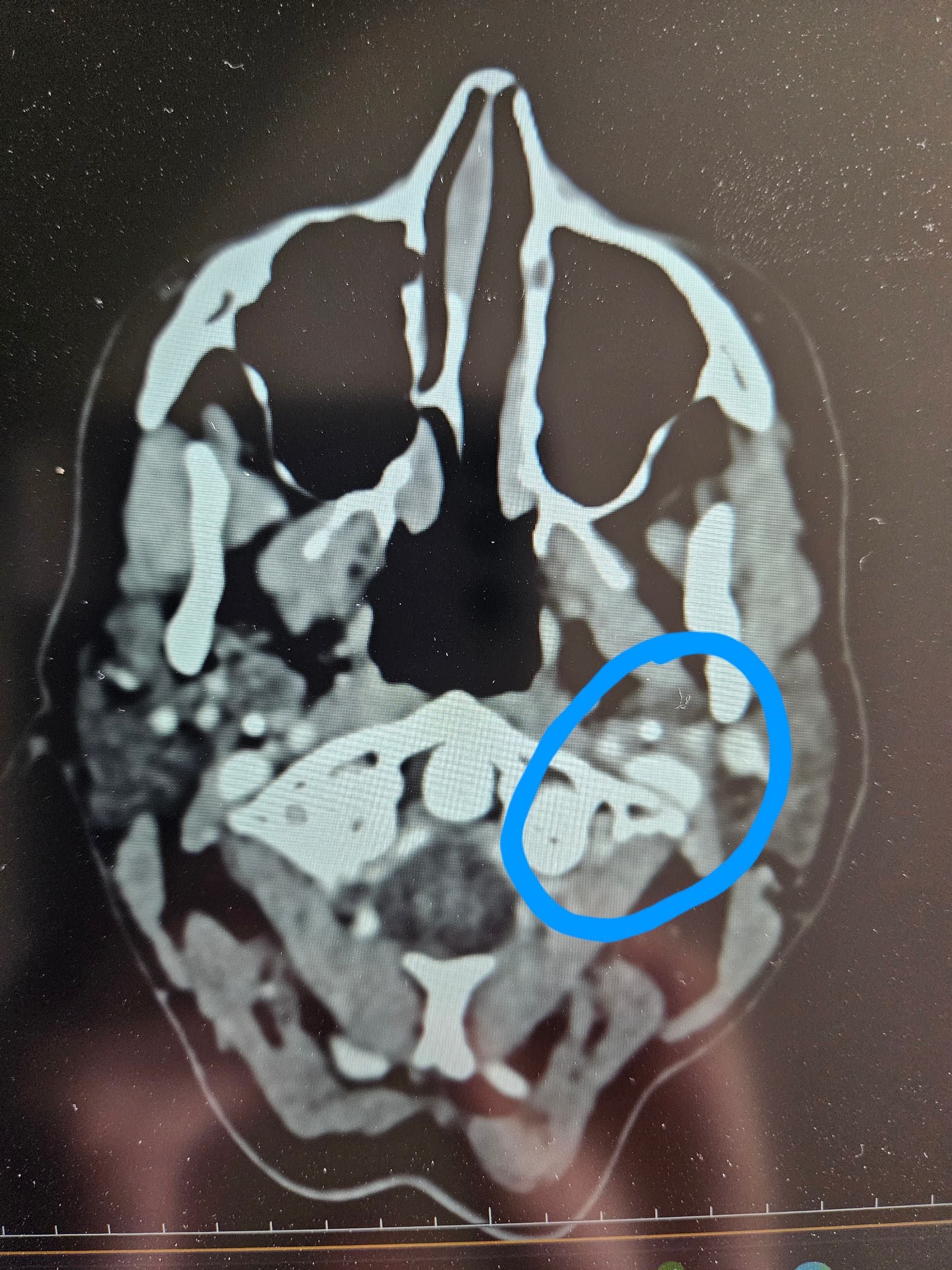
Additionally, can you go to the axial view and go to C1? I can see if your IJVs are compressed. I’ve attached my own imaging to help you know what to look for.
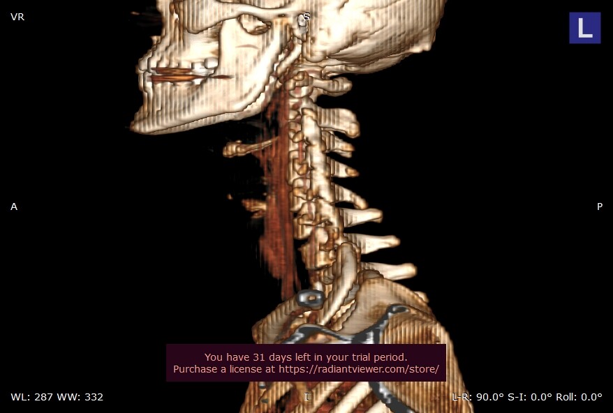
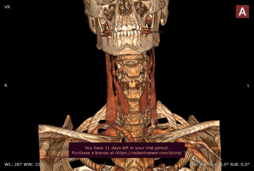
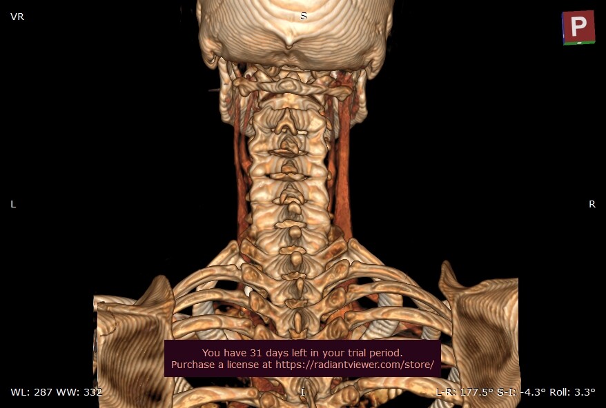
@6tdog6 - Can you also put up a 3D image from the front & from the back if you have them.
Your styloids are elongated & look very “solid” & somewhat thick. In the images you posted they aren’t terribly close to your C1 vertebra, however, the left styloid looks like it may be significantly curved inward. Since you had your scan done w/ contrast, including images w/ your veins & arteries would also be helpful.
For the moment, at least, you know that your styloids are longer than normal & there’s a very great likelihood are causing or contributing to your symptoms.
Another observation is that you’ve lost or mostly lost the lordotic curve in your cervical spine (it looks in the image w/ your right styloid like you still have some curve). Having a straight cervical spine brings the styloids & hyoid bone into closer proximity to vascular & nerve tissues in the neck so sometimes, restoring the curve through PT & gentle neck exercises can help reduce symptoms though surgery is usually ultimately warranted when vascular symptoms are present. Your sleep issues may be due to vagus nerve irritation or compression by your styloids as the vagus runs through the same space between the styloids & TPs of C1 as the internal jugular vein so if your IJV is being squashed (as your symptoms may indicate), then your vagus nerve is likely suffering, too. Another option w/ your symptoms is the internal carotid artery could be compressed or irritated as vascular symptoms such as dizziness & brain fog can be ambiguous as they’re common for both IJV & ICA compression.
Hi @6tdog6 you are a little high on you axial slice, if you scroll down a bit more you are looking for the C1 (bone with wings) then @TML will be able to read this for you. I have attached mine and circled what you need to find.
Hi yes that’s better. I believe your left IJV (right side of image) is being compressed by your C1. I have exactly the same problem.@TML is best at reading axial views so I am sure he will label everything for you soon.
I agree with the others that your styloids are a bit longer than the ‘average’, and quite thick… It looks on your images as @Rosie says that the C1 process might be causing some compression on your left IJV also.
Vertigo can be a symptom of IJV compression, as can brain fog. I used to get a weird sensation of sort of sucking/ like my brain was being rolled up (hard to explain!), which I would get mainly when I was dropping off to sleep, it was horrible & scary, & caused by IJV compression. The vagus nerve can be affected by ES so could possibly be affecting your sleep. Others have had sleep issues too, so might be worth having a search using the magnifying glass for this in the past discussions.
Given that your C1 could be an issue as well as your styloids, it would be an idea to get a consultation with Dr Costantino in NY, as he is experienced with vascular issues and does also treat compression by C1 processes:
@6tdog6 I agree that left IJV looks compressed. Prior to have my surgeries I used to get ‘startled awake’ throughout the night and had terrible insomnia. Since IJVs are the main source of drainage when laying down I just figured my sympathetic nervous system was just doing its job and alerting me that my brain wasn’t getting enough oxygen or nutrients due to the compressed circulation. I’m happy to report that since the surgeries my sleep is great. Good luck to you.
I agree with @Rosie . Your left IJV is pancaked against C1. Not only does this reduce the outflow of blood causing intracranial hypertension symptoms, but it also likely means your vagus nerve is compressed on this side. Note that your left styloid is well clear of the IJV, meaning that your left styloid isn’t causing IJV problems, just strictly C1. On your right side, your styloid is in contact with your right ijv (your dominant ijv based on it’s larger size) in the higher up slice. From what I’ve seen this can be fairly normal because the IJV comes out of the skull base right beside the styloid. Not sure if this is enough to cause symptoms. Your right IJV does have some compression against C1. And your right styloid is fairly close to your right ECA in the lower down slice.
Your right styloid is your longer and thicker one (which is why we can see it but not your left styloid in the further down image). It is possible that a bit further down the tip of the styloid is displacing or compressing an artery (i.e., ECA and/or ICA)
I have also noticed that your C1 looks slightly rotated and its appears your skull is slightly tilted looking at your 3D image from the back. I have absolutely no idea if any of this is relevant, but others maybe able to comment. I also suffer from awful Insommia, head noises & pressure especially when laying down, pulsatile tinnitus & brain fog with IJV compression.
Pretty common for C1 to be rotated! I’d say most people have it slightly off center. You’ll typically see C1-c3ish rotated slightly together and then the lower vertebrae bring things back to center and sometimes the opposite direction. You can see it as a very nonsignificant scoliosis pattern. Vertebrae with often shift and compensate for things like ES as well to give you the best fighting chance until things can’t shift anymore.
My C2 looks to be out to lunch, but apparently it’s normal. But then again, radiology seems to drop the ball a lot when it comes to normal vs abnormal. Especially in our ES world.
@6tdog6 - I agree with what everyone else has said. You now have a good foot to stand on to argue that your scans are NOT normal & you have significant IJV compression on the left side. As @Jules said, Dr. Costantino would be the best & closest doctor to you to consult with for this.
Also, when you post images, please make sure any personal information on them is covered up or removed. I edited your CT 3D images to take off your name & age, but sometimes images get removed from a post so that can be done, then the person they belong to has to repost them.
Thanks @TML I didn’t know that, that’s very interesting, as they say in the UK “everyday is a school day”! @6tdog6 I hope you manage to get to the bottom of your symptoms.
You can tell my c1 is slightly rotated clockwise, my second vertebrae posted above is very clockwise, but then my third vertebrae is pretty centered and then my fourth is actually slightly counter clockwise lol. Pretty funky.