Hi do the images show the styloid at all or are they showing compression against the transverse process?
I don’t have my computer right now so can’t do a tidy annotation but I’ve marked what I think is your right styloid w/ a red arrow. The left one doesn’t appear to be visible.

Have you tried converting your CT images to 3D @markp? That would help us better see what you have going on.
Radiantdicomviewer.com for PC or Bee Dicom Viewer App for Mac
I think red is your carotid, green is your jugular, and black is the styloid.
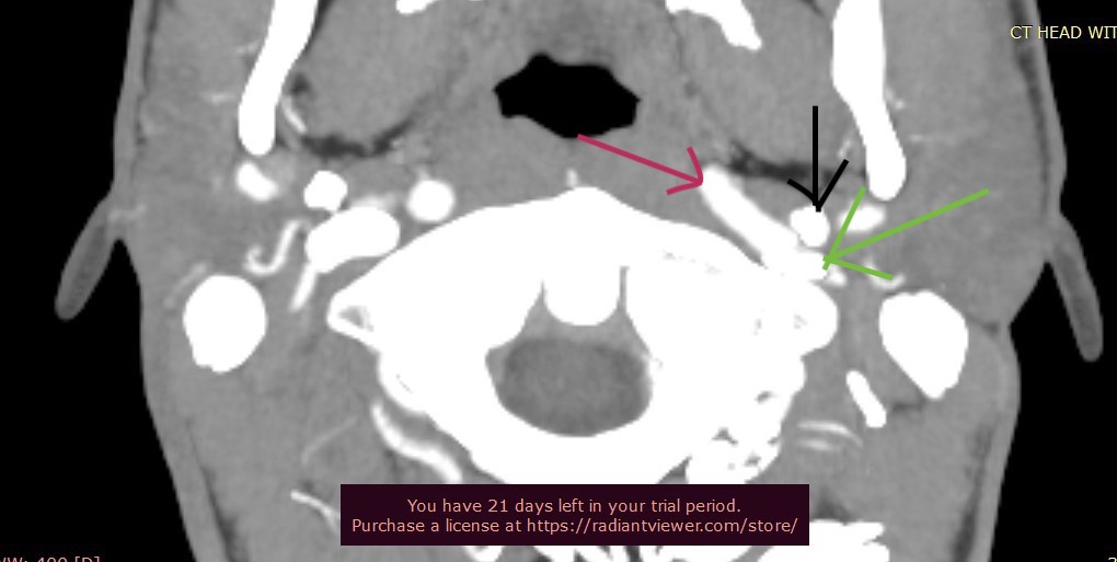
When using the dicom viewer you can select different preset contrast levels, normal the number 1 through 5 and it will become apparent to you which are the vessels and which are the bones.
2 Likes
Thanks for your help @boogs99. I need to learn how to read CT slices better. I can usually tell which are the IJVs & styloids, but not in @markp’s case.
2 Likes
Hi Boogs thank you for your input. Would you sat the jugular is compressed severely?
In my unqualified opinion, I would say it doesn’t look too compressed in this particular slice. If you upload your imaging to www.dicomlibrary.com we can view the entire CT for a better idea.
2 Likes
It looks like there’s a bit of C1 involvement in the compression?
I agree with @boogs99. It looks like the IJV is starting to pancake out but it would be useful to have additional sequences to scroll through to see if it gets flatter (more stenosed), It looks likely that it would continue to be compressed against the c1 transverse process.
2 Likes
hi again, would the right side of the scan be my left?
Most likely that is the case i.e. left is right & right is left.