New, confused about initial imaging

Hi everyone on this wonderful forum, I’m trying to get evaluated for ES!

First, I’m a 53(F) and have a slew of diagnosed health issues, to include Ehlers Danlos, POTS, MCAS, SFN, and CVI (chronic venous insufficiency), PVCs and PACs. These all began as covid related issues, even the hyper mobility had a strange sudden onset. Since covid, I have also developed a mitral valve prolapse, a torn hip labrum and torn knee cartilage. My one condition prior to covid was 25+ year old lumbar spine injury, from a sports collision that left me with a grade 2/3 lumbar spondylolithesis. It’s a lot to manage.

Second, I’ve been having the following symptoms for the past 3-4 years, which are specific to possible ES:

— intermittent L side neck pain (right over the carotid artery, where the pulse is taken). It comes and goes every few months, and can last several weeks to over two months. When it flares, it is unrelenting in it’s presence, but it can vary from a very sharp pain to dull, to a burning pain. When at it’s worse, the pain is about an 8 out of 10, then can fall back to a dull 2-3. It is constant in any position, yet turning to the left or down seems to make it worse.

— it is often accompanied by a strange “catching” feeling, like something in my neck gets caught and then releases, like a pluck on a guitar string

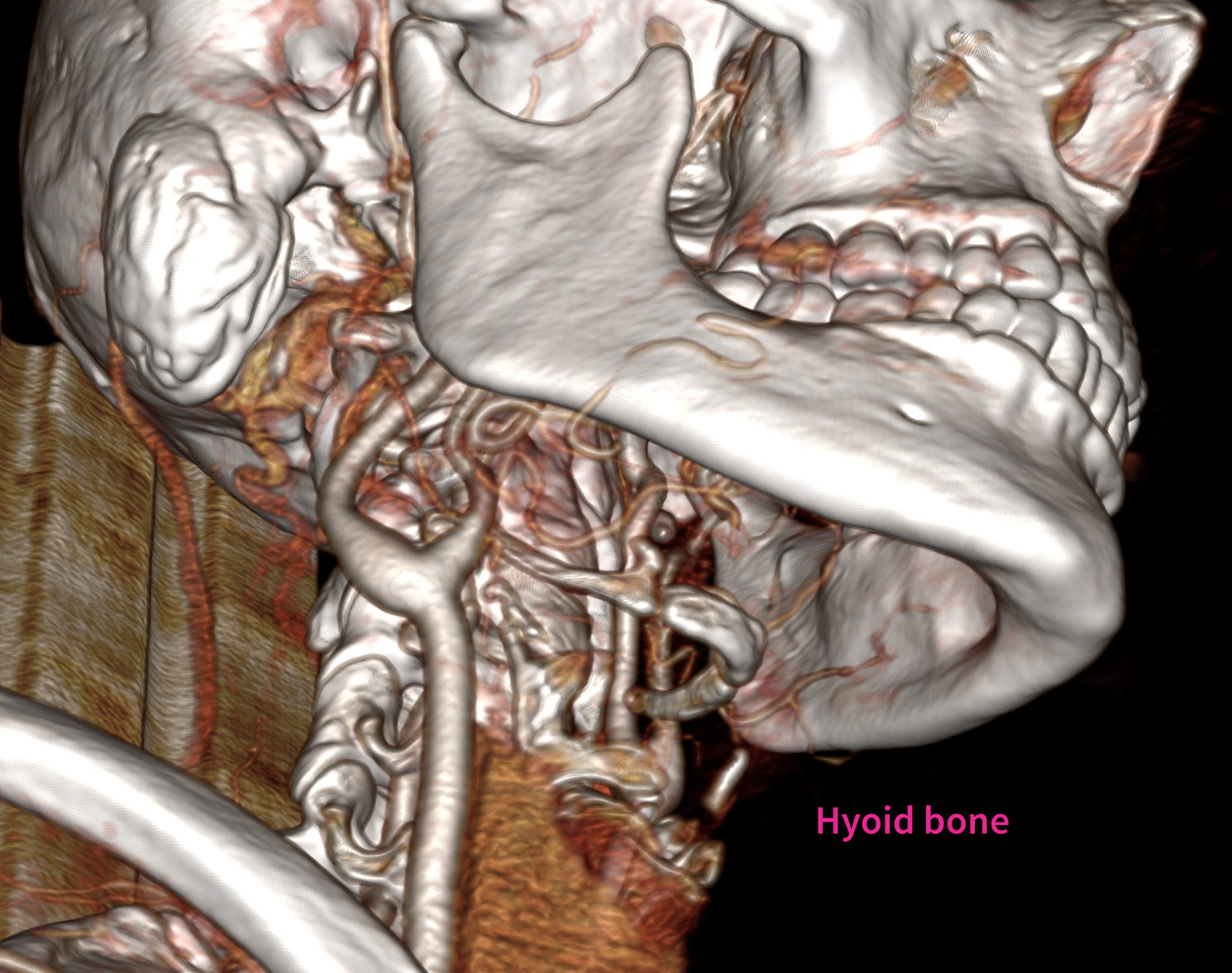
— My voice is hoarse if I talk too long on a phone, etc. I have trouble swallowing pills.

— I had a three year period of constant L sided pulsatile tinnitus. This actually began the day of a vaccine and later reduced a lot when I began treatment for POTS (which also began with a vaccine and got worse with covid). My POTS doctor said I had a 30% blood flow deficiency to my brain and put me on Mestinon to help with blood pooling in my extremities.

— I also experienced strange head issues, where the top rear of my head felt tremendous pressure, and would internally vibrate. Like an internal tremor, but not seen from the outside. Happens most often in a supine position. These were daily for almost three years, but have reduced more recently.

— also had the feeling of “bubbles” or water traveling up the back of my head and neck (CSF leak?) No doctor would take me seriously on this symptom, nor the tremors. (This began after a vaccine and worsened with Covid)

— I have L sided jaw pain and jaw subluxations. I clench on the L side and wear a night guard.

— I often have L sided head aches and occasional migraines. Migraines began with Covid.

— my L eye waters a lot. My computer distance vision is blurry (had a sudden onset) and no glasses will fix it. I have been near-sighted since age 8.

— I have brain fog, short term memory issues and word finding issues. These began with Covid. I take LDN for this, and it helps enough that I was able to go back to work full-time.

— I had extreme weight loss with Covid, and began having cervical spine instability (head felt too heavy for my neck, lots of overall neck pain)

— I get very dizzy if I look up at the sky or ceiling (cannot get on a ladder anymore). I also get dizzy if I bend over, especially in a position like kneeling to look under a bed, for example (I will black out)

— I have unexplained heart rate issues where my HR is too high with any exertion. I am only able to walk at a slow to moderate pace. I do long walks daily, but I am not able to build up any stamina due to high HR (stress tests are normal). I used to be a D1 athlete and a competitive distance runner, so this slow walking limitation is frustrating and confusing.

— I’m sure I am forgetting other symptoms! I also recognize many of these could be long covid or immune driven.

Third, and to my main question: What is the best imaging for me to have at this point? Despite my large medical team, no one is familiar with ES, aside from my physical therapists who are somewhat familiar from the EDS community. My vascular doctor is willing to investigate the neck pain and has ordered a Head and Neck CT with and without contrast, but she asked that I reach out to ENTs for better guidance. I contacted Dr. Cognetti’s office and they said those tests are fine. Dr. Costantino asks for a Head and Neck CTV and a brain MRI. These are quite different requests so I am confused what to share with my doctor. I also have had poor reactions to MRI contrast in the past (give me long migraines)

One last note, I do seem to have a weak vascular system, with “floppy” veins and arteries. I bruise very easily, and often blow vessels in my hands from simply carrying a pot of water or bumping something. I was tested for vEDS, and while I tested negative for it, I do have an unknown mutation on the gene that causes vEDS (so, I have what is called a VUS or variant of unknown significance). Some doctors tell me to be extra cautious, while others say don’t worry at all. I don’t enjoy being left in limbo with such a potentially risky condition.

TLDR: based on symptoms, what imaging should I get? I’m inclined to start with the “Head and Neck CT with and without contrast”, especially since those are scheduled for next week and I am currently experiencing pain in my neck. But of course I don’t want to end up needing more imaging, if I choose wrongly.

Thanks all for reading!

1 Like

Hi @modica! Welcome to our forum. You certainly do have a load of miserable diagnoses/symptoms. I am sorry you’re dealing with so many things at once.

I’m sorry your symptoms have forced you to slow down. I can completely relate to that frustration. I was a long distance runner for 12 years (not a D1 level athlete though) & had to stop, too. I also had the elevated heart rate issue during higher intensity aerobics as one of my ES symptoms. Mine came w/ a blood pressure drop, light-headedness & breathlessness that didn’t recover as it should have for my fitness level. I determined it was related to my vagus nerve being irritated by my styloid(s). A hoarse/weak voice/vocal loss is also related to vagus nerve irritation by the styloids where ES is the diagnosis. Swallowing issues is also a very common ES symptom.

First, to answer your major question, the CT w/ & w/o contrast of the area between skull base & hyoid bone is your best diagnostic test as it should clearly show the situation with your styloids, stylohyoid ligaments, hyoid & the internal carotid arteries & internal jugular veins in your neck. Vascular compression, if present, should be visible. Please request that 3D images be included on the CD you get of your scans. That will save you time in not having to convert the images to 3D so you can better see what’s going on in your neck & you can post anything you have a question about here so we can offer our non-medical but somewhat experienced opinions.

I don’t blame you for not wanting an MRI w/ contrast, & you may not need to go that route if the CT scan shows what I think it will. I had an MRV w/ contrast looking for vascular compression & had a headache for a week afterwards so I sympathize. Not only that but the radiologist who read mine said there was no compression when it turns out there was bilaterally. Go figure…

Vein/artery “floppiness”, POTS, & MCAS have been found to be related to hEDS. The torn labrum & knee cartilage can also be related to hEDS, since, as you know, that causes tendon/ligament laxity which can dispose an athlete to having joint injuries more easily.

Intermittent pain is a trademark of ES as symptoms tend to come & stay for a while then can disappear partially or completely & not recur for weeks or months. Thus, I find your intermittent L neck pain in line w/ that aspect of ES. The quality of the pain you get is typical of the forms nerve pain can take. The catching feeling could be your styloid/calcified s-h ligament catching on soft tissue in your neck then releasing w/ head/neck movement.

Eye watering is related to irritation of the facial nerve. Dry or extra watery eyes are symptoms we see w/ ES. Dizziness & brain fog tend to go hand in hand w/ vascular compression of either the internal carotid artery or the internal jugular vein thus the need for a CT w/ contrast to look for those situations.

Should further testing beyond the CT scan be suggested, it’s critical that you let the doctor who’s referring you know about your genetic mutation as that may make a difference in what type of testing is recommended or how it’s done.

Here are a couple of links to posts @Jules wrote that will help you better understand how your symptoms could be connected to ES:

1 Like

I would personally pass on Cognetti based on all you mentioned. I think way too complex for him. I am complex. Try to get into Hepworth. Costantino is a decent choice but the plus with Hepworth is that he looks at the entire anatomy. I have hEDS and spectrum for Loey Dietz.

2 Likes

Thanks so much for your reply! I’m at the radiologist right now, and they have overridden my doctors order for with and without contrast. They will only do the scan with contrast and are insisting I don’t need both. My understanding is that I need both with and without. They are refusing to budge and only did it with the contrast. Will I need to have this done again? Thanks for your help. I thought I’d finally have answers today and now I’m not sure.

1 Like

Also to add clarification, it was a CTA that they took

The CTA will show both your jugular veins & carotid arteries as well as your styloids, stylohyoid ligaments (if calcified) & your hyoid bone so it should be fine. It’s true that the situation with the cony structures in your neck show
More clearly in a CT w/o contrast but they are visible in a CT w/ contrast too so you shouldn’t need another CT scan.

Take heart @modica - You should still be able to get your answer from today’s imaging. :heart:

2 Likes

Thank you. I’m just so frustrated as no one would listen to anything I was saying. They had told me to contact ENTs to make sure we were getting the right testing, and then they totally disregarded the instructions I gave them, to include getting the measurements. It was written on the order to measure the styloids, but the radiologist didn’t do it. All they cared about was the carotid and they told me to “forget about anything else being wrong” as if the pain I’ve had for weeks now (and years) is of no consideration to look further. They were also very rude, ugh. I could tell none of the three people I spoke with had ever heard of ES.

So I’ve been trying to look at the images myself but not having luck. The Bee viewer won’t show it in 3D (it just creates a weird single stretched/pixelated image). So I paid for a monthly fee to use 3Dicom software but it won’t upload (has been spinning for hours). I searched on here, and maybe that is normal for that software to upload so slowly? I’m actually a 3D animator, so I know my way around 3D imagery, but I can’t get it to load. If there’s a better platform, let me know! Thanks again for your help.

1 Like

@modica - Do you have a Mac or PC? If you have a PC then try radiantdicomviewer.com. Another option is to upload your images into dicomlibrary.com (can take awhile for images to load here). Dicom library will anonymize your images & put them in 3D & will send you a link to view them yourself plus a link to share. That might be easier for you. I had to have help finding my 3D images after I received my link but I’m super untechy so it was no surprise.

@Disabled_Inventor just went through the same thing as you did at the radiology office where he got his CT done. His was supposed to be a CTV & they told him it was a mistake & changed it to a CTA. It was the jugulars & styloids his doctor wanted the scan to focus on & the radiologist only commented on the carotids, but did measure the styloids. It is VERY FRUSTRATING when radiology techs don’t follow instructions or don’t have the nerve to tell a patient they don’t know how to do what’s been ordered so the patient can visit another radiology office & get the scan done correctly.

BTW, my brother did 3D animation for Disney & Sony for years. He’s retired now. That’s a challenging career!

2 Likes

Hi @Isaiah_40_31 Wow, how impressive for your brother! It is tough work, and he’s probably glad to be retired, ha!

Good news, I finally got the Bee viewer to work! (on a Mac) I did the dicomlibrary but it wouldn’t turn them to 3D, not sure why. To my eye, the styloids aren’t too terribly long - and maybe a little thick? (they were supposed to measure them, but they didn’t follow that order request either). I’ve marked them in yellow. I’d love your thoughts on size and angles. What stood out more, is it appears the Left IJV is impacted a lot by the C1 transverse process. Of course this was not mentioned on the report. It’s frustrating, because if they had done the order for a regular CT with contrast, wouldn’t it have captured the veins better? The right IJV is missing completely in these images (assuming I have one!). If this is compressed, it could surely explain my pulsatile tinnitus and head pressure tremors, perhaps some dizziness too?

But, I’m confused after viewing these, because my Left sided acute neck and throat pain is not in either of those areas. It’s more where the hyoid bone ends. Does it look longer than usual? I’m not sure. When I rotate the 3D images, it is touching the carotid. This pain worsens when I turn my head to the left side, and when I flex downward. It’s also where I get that strange catching feeling. It gets very painful and sore if I am lifting things, or bending over to pick something up. I carried some books today, and now I’m in terrible pain, and it even hurts in my left throat to swallow. Unless, do you think the styloids would indeed be pressing on my throat (the throat pain is somewhat higher up)? It’s also tight like someone has a grip on my throat, right where you take the pulse, and extending upward along the jawline. I’m sad my vascular doctor wasn’t more interested in my symptoms.

I guess I need to share these with one of the expert ENTs. Do you think it warrants that? I do have a local ENT I’ve seen for years. I think I will also see her, as she is an excellent compassionate doctor. The reason I see her is for the “bonus” growth you see on my right side mastoid bone. This is a benign boney osteoma that we’ve been monitoring for 20+ years now. Shockingly, the only time it causes issues is when I fly, and then I get excruciating right ear pain and I lose hearing for several weeks after landing. It’s quite unsightly, no?

Let me know if I should make another post with these images - if you think more eyes from the group would be helpful. It’s not my intention to make you the sole caseworker on my situation! Thanks so much for your continued help.


Well done for sorting your images! I’m not good with tech at all so can’t label anything for you…I agree that your styloids don’t look particularly long, but the left especially is pretty angled, although on the 5th image they both look longer ? And as you say, the left IJV does seem to be compressed more by the C1 process rather than the styloid… The greater cornu of the hyoid do seem to be close to the carotid artery, yes, and it looks like there could be a small part of calcified stylo-hyoid ligament coming up from the lesser cornu…The horns do look long to me, and quite pointy at the ends, so it’s easy to imagine if you’re turning your head or looking up or down that they could cause issues…
Where you’ve labelled ‘bonus’, the mastoid bone does look pretty big, you say it’s been monitored, have you had regular CTs for that? It would be interesting to look at past ones if you have…
And no IJV can be seen on the right, so maybe you’re even more of a rarity and don’t have one!
Certainly a few points there to discuss with whichever doctor you see! I do think you would be better off getting a referral to one of the doctors experienced with VES like @Brandy suggested, it looks like a C1 shave might be needed, & possible hyoid surgery, I’m not sure that the usual doctors we suggest for C1 shave (Dr Costantino for example) would look at the hyoid too…

1 Like

@modica - I’m glad Bee Dicom Viewer finally worked for you. You did share some pretty great images & annotation. I would have never noticed the subluxation of your TMJ if you hadn’t pointed it out. BTW - this post is on the main forum.

Yes! My brother is glad to be retired. He’s getting his life back. :joy:

I agree that your styloids aren’t super long, but that really doesn’t mean they aren’t causing symptoms. The greater horns of your hyoid are interesting. It looks like the left one is quite thick, but the right one is long & thin. That could just be due to the image angle though. I couldn’t tell exactly how long they are, but it does appear the left one, at least, may be bumping into your carotids on that side which can cause pain.

Another thing to note is how straight your cervical spine is. Your lordotic curve looks to be completely gone in some of the pictures. That’s something that can be corrected by gentle PT exercise for the cervical spine & it will help reduce the contact your styloids & even hyoid bone may be having w/ nerves & vascular tissues.

Your right IJV looks to be present on the back side of your imaging (annotated image below), but it’s very tiny. We’ve seen unequal IJV sizes in other members, but as @Jules noted, it’s rare for it to be that small. Your left IJV definitely is being squashed by the transverse process of C1, thus seeing either Dr. Cognetti ¶, who has done hyoid bone surgery & also does vascular decompression surgeries w/ C1 shave (Dr. Heller - neurosurgeon does the shave part), or Dr. Costantino (NY), who works alongside a couple of other neurosurgeons would be your best bet. As Jules noted, we don’t know if he also does hyoid bone assessment/surgery.
•Dr David Cognetti, Thomas Jefferson University Hospital, Philadelphia 215- 955- 6760 (Has done many successful surgeries on members). Only removes ligaments if calcified. David M Cognetti MD | Jefferson Health

•Dr Peter Costantino, 4 Westchester Park Dr, 4th floor, White Plains, (914) 517-8056
http://www.nyhni.org/find-a-physician/Peter-D-Costantino-MD,FACS

1 Like

@modica - Wow that’s awesome! 3d animation is so techincal…big brain stuff really…so many options and the software is like its own universe. Now I see where you get your image editing skills :slight_smile:

Yeah I hope that the scan they did can be used…you would think that a tech would just do what is written on the script and not pontificate on what the doctor really wanted…we’ll see, but if I have to redo it I will send a letter so that they can at least talk to the tech and/or review their policies if they have questions about a study…I would hate to have to redo the test.

3 Likes

Thanks for your input! @Jules and @Isaiah_40_31 I really appreciate the help.

It’s so interesting to see the IJV compressed on the L and largely missing on the R. It likely explains a lot of symptoms. On the right, it looks like there may be a thin collateral vein growing off the back side of the boney tumor. The absence of a good IJV on that side could explain the intense right side pressure and pain I feel when flying. I’ll attach a movie of the left compression. It looks pretty smooshed in there, indeed. I’m wondering how concerned I should be …?

I took a closer look at my hyoid area as the source of my acute pain. My PT feels that’s the closest area to investigate more for the pain source. It looks like the greater horns run right up to the spine, and things get really tight between the end of the hyoid, spine, and the carotid. There’s not much room for error, given the instability in my hyper mobile joints. My PT also said two small muscles are in play near there that could also catch on the hyoid (or even a bone spur on the spine) and cause throat pain. I’ll attach a movie showing this too. Oh, you mentioned a calcified piece off the smaller horn, and I looked again in 3D and that’s actually a piece of an artery floating nearby (or something, but it doesn’t look like it’s attached to the bone)

Crazy, this morning I woke to a large mucocele under my tongue, on the frenulum. It was the size of a grape and I couldn’t put my tongue down. I had no idea what it was, and was in a panic for a few minutes before researching it. I was able to massage it away, thankfully and it hasn’t returned. But goodness, that can’t be a coincidence, can it? What the heck is happening in my neck, ha!

I’m still trying to decide which doctors to approach next. Probably Cognetti and Hepworth. I see my POTS specialist next week and I have no doubt he will find this of interest. He is also a MALS expert and thus knows a lot about vascular compression.
IJV
HB

1 Like

I’m so sorry you had trouble with your imaging too! I don’t understand how they can’t ask for consent before changing the order. Even insurance would surely want to know they ran a test that insurance had not approved in advance.

2 Likes

Forgot to mention - thanks for the info on the military neck. I will pass along for my PT to check out.

2 Likes

I’m glad that your PT is so knowledgeable & helpful…will be interesting to see what your POTS doctor’s opinion is…

@modica - Your 3D animation skills are paying off! Thx for the videos. At the very beginning of your second one, it’s obvious how close the greater horns of your hyoid are to your spine. The final phases of the video better show proximity to the ICA on the right.

Dr. Dewan in Louisianna specializes in surgeries for Hyoid Bone Syndrome but unfortunately, she doesn’t do ES surgeries. Here’s her contact information in case you’d like to get her opinion:
•Dr. Karuna Dewan, Ochsner LSU Health Shreveport - Academic Medical Center - Specializes in Hyoid Bone Syndrome surgeries
1541 Kings Hwy, Shreveport, Louisiana, 1-318-626-0050
https://www.ochsnerlsuhs.org/doctors/karuna-dewan
Needs a PCP referral

Thx also for the correction on what I pointed out as being s-h ligament calcification on the right lesser horn. I’m glad it was clearly vascular tissue when you checked it out more carefully. I should have put ? at the end of my label.

That does sound scary & painful. I’ve gotten those in the past but they were never that large. They are ALWAYS annoying until they pop or shrink on their own. Hopefully that’s a “one & done” scenario for you.

Nice that your POTs specialist has knowledge of MALS. Please share anything of interest that he says when you discuss your situation with him.

1 Like