Pain below ears and behind ears and base of skull (predominatly right side) down neck SCM muscle and into shoulders (restricting shoulder movement) -SCM muscle (somewhat reduced temporarily when I stretch my neck on the side lying down on my pillow)
-Malocclusion, limited mouth opening 18mm- mouth opens out to right side, pressure in back right teeth
-dizziness/nausea
-ear ringing, blocked ears
-brain fog/memory issues
-pressure headaches and migraines
-past year= prolotherapy into upper cervical spine for instability
-visual snow, feels like L and R eyes not working together, trouble reading.
-R side of face numbness
-sensation something stuck in throat/choking feeling
-sore throat
-Swallowing increased difficulty
-voice goes hoarse
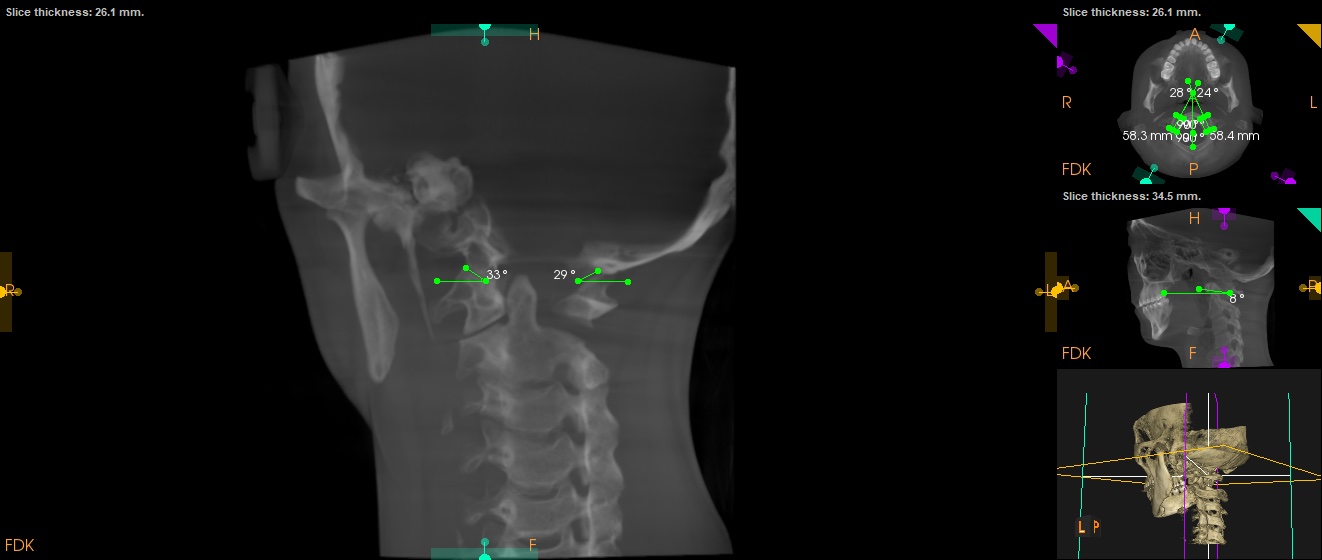
-calcified elongated styloid bilateral -specialist letter says 44mm, vs CT says 37mm
-says on CT “Bilateral stylohyoid calcified ligaments are elongated, measuring approximately 3.7 cm
bilaterally. The distal tips lie within parapharyngeal fat, deep to the level of angle of mandible.
These pass in close relation to the external carotid arteries and internal jugular veins bilaterally
without compression.”
The specialist that I have seen says that most of these symptoms could not be caused by the elongated styloid (only the localised pain in the styloid region and skulll base and throat pain). I am worried that I may have some kind of vascular compression though or at least should be checked for it- but he says I do not need imaging for the vascular structures. Feeling lost and stuck!
@goldenretriever welcome to the foru. Dorry to hear about your symptoms. I have similar to you. I think elongated styloids can definitely cause all your symptoms.
I suspect the brain fog, ear ringing, memory issues, and visual snow are all due to IJV compression. The dizziness may be that or carotid compression or vagus nerve compression. Hard to say without seeing your imaging. Do you happen to have your imaging on hand? We can help take a look!
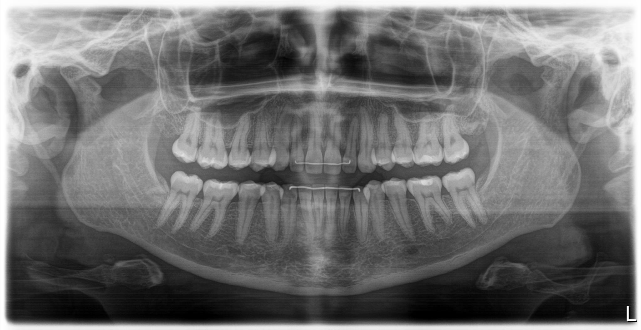
thanks @TML ! sorry to hear you are going through similar symptoms too! Heres my panoramic x-ray and CT scans that the chiropractor took. I havent got the CT imaging taken by the head and neck surgeon but hopefully they would be able to send this to me also. I saw my rheumatologist today who ordered a neck MRI/A of the vascular structures -I am unsure whether this is the correct imaging though to check for vascular compression?
@goldenretriever see attached your imaging. Both of your styloids appear elongated. One appears to be one long piece, while on the other side you have a gap where the styloid looks like a normal length but then a cm or so lower the stylohyoid ligament looks calcified.
Did the CT with the surgeon involve contrast? It doesn’t seem that the chiro CT had it. If you get one with contrast I’ll be able to help you find vascular compression!
I will say, even without contrast, I can tell your right styloid is closer to your C1 than your left styloid. Which means I suspect your right IJV and potentially vagus nerve is being compressed. Won’t know for sure without contrast though.
thanks so much @TML . Unfortunately the head and neck surgeon I saw told me I didnt need CT with contrast imaging so I am unsure how I can get a specialist to order it for me. I asked my rheumatologist for imaging today and he said that it was best to see the vascular structures on MRI Angiography…. not sure if the MRI Angiography images will be any use? I have been told by other users that I need CT venogram? Is that different to CT with contrast? Maybe someone from New Zealand knows who I need to see to get this kind of imaging ordered
Hi good you’ve joined us! I agree with @TML that you could possibly have vascular compression from some of your symptoms…
A CT with contrast would be better to show the styloids & any compression; an MRA might show compression of the blood vessels but not necessarily what’s causing the compression as it doesn’t always show the styloids very well. Plus the contrast medium with an MRI is gadolinium , which can have some side effects (we’ve had discussions about that if you want to search for that, it’s worth reading up on it for an informed decision).
A CTA/ CTV is a CT with contrast timed to show the arterial phase & venous phase, although there is a more detailed/ invasive scan also referred to as a CT venogram with pressure gradient manometry & checks flow/ blood pressures in different areas, that’s not always necessary…
If you can, you could have a look for some research papers in the research paper category which explain IJV compression symptoms & the best scan to show this- there are quite a few about ES causing IJV compression- so hopefully one or two would have useful info for you, & then you could show this to your doctor?
Although we know of a few doctors in NZ who are aware of ES, I don’t know if they’re experienced with vascular ES- unfortunately there’s so much ignorance around it that sometimes doctors don’t believe it can cause the symptoms we know it does. I had bilateral IJV compression, despite being told by my local consultant telling me the styloids couldn’t do that, but luckily because of the info on here, I was able to get a referral to a more knowledgeable doctor, have the testing done & surgery with him.
@goldenretriever - Welcome to our forum! Your symptoms definitely are those seen w/ styloid elongation/stylohyoid ligament calcification & vascular compression. As @TML said it’s possible you have both jugular & carotid compression happening depending on your head position.
The advantage of a CTA/CTV (besides not using the gadolinium based contrast) is those can be done “dynamically” i.e. w/ your head turned in different directions which can help show what’s being compressed & what’s causing the compression. An MRA/V can’t be done dynamically & as @Jules noted, because MRIs predominantly show soft tissues, they can obscure the bones that are causing or contributing to the problem. Is it possible for you to request that the rheumatologist change the order for your scan from an MRI/MRA to a CTA & CTV?
Here’s a link to a good post about gadolinium contrast, but I have to say not everyone gets a bad reaction to it. It’s just wise to be informed of possible side effects if you’re going to receive it. CT scan contrast is iodine based so it flushes out of the body more readily.
I hope this information is helpful. We’ve only had a few members from NZ & some have needed to go to AU to have their surgeries especially when vascular compression is involved.
The head and neck surgeon telling you that you don’t need contrast is silly. Do you have a family physician? Could ask them for a CT with contrast. Honestly it’s hard to say which type of CT to get. If you get a CTA you risk missing the IJVs, if you get a CTV you risk missing the carotids. Sometimes just a simple CT with contrast where they slowly leak the contrast into you can be the best chances for seeing everything. My CTA missed one of my IJVs. I also got a CT with contrast done but I don’t have access to it so not sure if it captured everything.
Thank you! I am glad you guys have brought up gadolinium - I would otherwise not have had any idea to weigh up the possible side effects with the benefits of the scan. It sounds like it is not the best scan for diagnosing vascular eagles anyway.
Great ideas- thank you! Currently working on trying to get a CTA/CTV with contrast timed (and finding a Dr who will listen!)