Very new member here and I’m currently in the process of being investigated for CCI/AAI and IJV compression.
Short story, I’ve had migraines from childhood, autoimmune issues, now diagnosed with hEDS, POTS and MCAS, and have thoracic outlet syndrome as well.
I’m having a STACK of symptoms and have been down many many rabbit holes of information gathering (thanks late-diagnosed ADHD ) and have ended up here.
I’m in Australia, and have an appointment with a neurosurgeon next week who “apparently” is well versed in EDS, IJV compression and CCI, so I’m hoping I won’t be dismissed.
I’d love it if peeps could take a look at my scans and let me know what you guys see.
I’m asking him to rule out (or in) the following:
CCI / AAI
Chiari Malformation
Internal jugular vein compression
Jugular vein aneurism
and Eagle syndrome of course…
If folks could let me know what is obvious to you, I’ll feel more prepared.
THANKS!!!
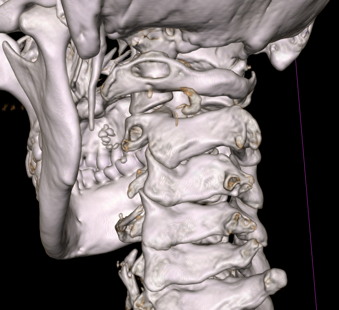
Your styloids are pretty long, I’m sorry that I’m not able to label any of the images though… It looks like they’re also quite close to the C1 process, so in the second image you can see compression of the IJV . You can also see what looks like collateral veins which are common with IJV compression. The left side isn’t so clear , you can see that area as well, the IJV looks smaller so could be the non-dominant IJV. Compression of the more dominant IJV can have a more significant effect.
I don’t know about spotting CCI or Chiari on images, so can’t comment on those, but your styloids are definitely elongated & look quite pointy, and it does look like you have IJV compression on the right. Hope this helps!
Thanks so much for your input @Jules, I appreciate your response.
Yes, from my laypersons research this is what I see too, to me it also looks like the right styloid is more angled than the left, which could also be adding to what I also see as compression of the IJV.
I always find it interesting reading the radiology reports. I never trust them as they never cover everything. I also don’t trust Doctors who solely rely on the reports, and don’t look at the scans (rare for surgeons, but I’ve seen others).
This is an extract, the rest is fine…
“The jugular veins have normal appearances, draining into the subclavian veins, and these into the superior vena cava. There is normal enhancement and no filling defect.
No obvious cervical venous abnormality.
Other findings:
There is a tortuous right internal carotid artery with a retropharyngeal course, causing bulging of the posterior wall of the oropharynx.
There is asymmetry of the palatine tonsils, which are slightly larger on the left with partial effacement of the left palatoglossal fold.”
@htren -
We’ve had members upload videos before but I don’t know how they did it. They’re usually just videos of their skulls rotating around an axis sometimes 360º & sometimes 180º. Is that what you’re trying to upload?
I also don’t know anything about detecting CCI/AAI, or Chiari in imaging so I’m sorry I can’t help with that either. I can tell you that your C1 is quite irregularly shaped w/ the left side being more unusual than the right side & your C2 vertebra is also quite different than most others I’ve seen. I don’t know if those facts are contributing at all to the symptoms you have. You’re also correct that your right styloid is quite a bit more angled inward than the left one.
Though C1 isn’t always perfectly symmetrical, I’ve never seen one as asymmetrical as yours is. As I said, this may be irrelevant, but it may also be contributing to your symptoms.
I also looked at the C1 and C2 and wondered what the heck…!! Perhaps something went weird rendering the 3D images…? Or maybe they’re just screwed
And Yes, it was just the 3D rotation video I was attempting to upload.
I do have migraines, head, shoulder and neck pain, raging tinnitus, dysautonomia, weird nerve stuff, and more, so fingers crossed these scans may be the start of an explanation, past “just Hypermobile”.
Good spot from you & @Isaiah_40_31 , they don’t look right! Worth asking about… we’ve had members with unusual vertebrae before, like Arcuate Foramina Bridge , but these are often dismissed as congenital deformities and don’t cause symptoms…
I saw that when I looked at your imaging last night, @htren. Your whole C1 is so different than any I’ve ever seen, I don’t know if it’s a problem or not. It could be contributing to CCI/hypermobility since it’s not a normal shape/connection between C1 & C2. I don’t know if there’s anything that could be done about it w/o causing additional problems in your neck because your body has grown & adapted to your odd upper cervical spine. You could see a physiatrist or orthopedic doctor who specializes in cervical spine issues for an opinion.
Migraines, tinnitus & dysautonomia go along w/ IJV compression. In cases of IJV compression up near the skull base, the vagus nerve, which is a big player in dysautonomic symptoms, is often squashed alongside the IJV as they closely share space within the carotid sheath there. Neck & shoulder pain are commonly the result of spinal accessory nerve dysfunction being caused by elongated styloids/calcified s-h ligaments. Once the styloid(s) are significantly shortened, those symptoms & others caused by irritated nerves, often go away.
Thanks again for your insights!
I’m weirdly looking forward to my neurosurgeon appt on Thursday so see what he thinks.
I’ll be seeing Dr Rao (I’m in Australia).
@htren - You can look forward to your appt on Thursday for another reason, too! Now you have some idea about what’s going on with your neck so you can have a more informed discussion. Maybe even give him some information he doesn’t have!
It almost looks like a portion on the right is broken off? At C1. It looks very strange. (the image looking straight at back of skull) The little circle on the left is missing on the right unless imaging is off.
htren, you did great job on this imaging. Just an FYI, my daughter has a host of your same DX. She was DX with ADHD caused by POTS. That meaning if you didnt have POTS, you didnt have true ADHD. There is hope….