So I recently had a CT scan with and without contrast. I received the results today and wanted to know what your thoughts are on them. It says my styloids are less than one inch long and that all major arteries and veins and grossly patent( meaning not obstructed). They said there is mild calcification of both styloids though. What does this mean? I pasted below what the report said.
TECHNIQUE: Following the administration of intravenous contrast, axial CT images of the neck were obtained. Coronal and sagittal reformatted images were reviewed. COMPARISON: None. FINDINGS: Pharynx/larynx: The nasopharynx, oropharynx, hypopharynx, and larynx are patent without asymmetric soft tissue. The parapharyngeal spaces are clear. The trachea and cervical esophagus are unremarkable. Oral cavity: The oral tongue and floor of mouth are intact. Glands: The parotid and submandibular salivary glands are unremarkable. No calcifications are seen along the courses of the major salivary ducts. The thyroid gland is unremarkable. Cervical soft tissues: There are small lymph nodes in right and left neck that are not enlarged by size criteria. No soft tissue masses are identified in the neck. The major cervical arteries and veins are grossly patent. The bilateral mastoid styloid processes measure less than 1 cm in length. There is mild focal calcifications of the bilateral stylohyoid ligaments (series 6 image 193 and 202). Other: No acute abnormality is identified in the visualized inferior brain. The skull base is intact. Tiny mucous retention cyst in the left maxillary sinus. The sella is intact. The visualized upper lungs are without significant abnormality. The contents of the spinal canal are incompletely assessed. The prevertebral soft tissues are unremarkable. No acute osseous abnormality is identified. IMPRESSION: The styloid processes are not elongated. There is mild focal calcification of the stylohyoid ligaments. FINAL REPORT THE ATTENDING RADIOLOGIST INTERPRETED THIS STUDY WITH THE RESIDEN
@Ihurt it means that there are a couple spots where your stylohyoid ligament is calcified. So your styloids are normal length, but the ligament connect them to your hyoid bone has pockets of calcification. See image attached showing the anatomy.
Depending on where exactly the pockets of calcification are, they could be brushing against nerves or vascular structures causing some grief. Hard to know where yours are calcified specifically without seeing the imaging.
Thank you, this makes sense. I sadly live in Illinois and there are not many neurosurgeons who are well versed in ES here. I think there is one at university of Illinois ( Dr. Konstantin Slavin). Not sure how in depth his knowledge is on this matter though. Not sure what my next step should be in who to have look at my scan to see if these mild calcification are contributing to my symptoms or not.
It’s curious that your styloids were noted as being less than 1 cm long as a normal styloid is 2.54 cm or 1 inch. 1 cm is the length styloids are cut back to when elongated as that is very close to the skull base. I wonder if the radiologist meant they are less than 1" vs less than 1 cm. If so, they could still be causing trouble as normal length styloids if they are very thick, angled, curved, twisted, knobby, or pointed can cause the same types of symptoms as longer styloids.
I agree with everthing @TML said about the calcifications on your stylohyoid ligaments. You can request a disc of your CT scan, & if they didn’t include any 3D images, you can convert the CT slices to 3D using RadiAnt Viewer (radiant viewer.com) for PCs or Bee Dicom Viewer App for Macs. Seeing your scan in 3D will also help you better understand what’s going on in your neck plus you can upload it here so we can also give you an opinion. Radiologists are often not very familiar w/ ES & vascular compression so misdiagnose those in scan reports. That happened to me.
I agree with @TML & @Isaiah_40_31 ; we’ve had members whose styloid processes aren’t elongated but have had calcifications on their ligaments which can cause pain, sometimes these can even be like grains of rice all along the ligaments … if you can get the imaging & upload it here we can have a look, & if the doctor in your area isn’t helpful you could always send the imaging to other doctors on the list, some do video or phone consults…
Thank you, I will have to see if I can covert my CT to 3D. The PA who ordered the scan said she doesn’t believe my symptoms are from my styloids. This is a head and neck surgeons office ( ENT) here in Chicago and I’m not certain if they are very well versed in this. I mean she is listed in the files and knows about ES, but not sure how much. They attached the pics of my scan on my patient portal through NilRead but its not 3D.
I know a friend of mine has a virtual appointment with Dr. Peter Nakjai in Arizona. He would not even give her an appointment until she had a CTV! Luckily she was able to get her doctor to order one for her. I don’t think many specialists ( neurosurgeons) will give you an appointment unless you already have a CTV, is that correct? I just have the CT with contrast.
Some doctors are okay without it, not everyone has vascular compression, & if symptoms don’t indicate vascular ES then it’s another procedure to go through if you’ve already had a CT…
It should do; although it does depend on the position sometimes; if people get symptoms with their head in certain positions then sometimes a CT can be done like that, but not many radiologists do that… A CTV is a CT with contrast timed for the venous phase, a CTA is a CT timed for the arteries… some doctors like to do testing for pressure gradients across the veins to look for compression, (cerebral venography?) ,this is more invasive & is fair enough if they’re looking for other compressions, but isn’t something you’d want to do if you don’t have symptoms of compression.
@Ihurt I think what could be a good first step is to download your original CT imaging to your computer, then download Radientviewer, then open your CT imaging in radientviewer and convert the imaging to 3D in radientviewer. Then take some screenshots ans post it here. Once we can take a look we can add some notes to your screenshot that you can use to advocate for yourself to doctors. Seems to be one way to get a foot in the door when radiologists keep dropping the ball.
Okay, so I was able to convert my CT to 3D. Here are a few images. I do not have too much of a clue what I am looking at. I think I can see the styloid. My report again said that my styloids were less than 1 cm in length. There was mild focal calcifications of the bilateral Styohyoid ligaments. What are your thoughts?
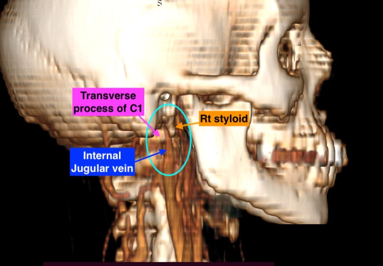
@Ihurt - Your styloids are naturally remarkably short, however, your right IJV looks compressed between the right styloid & the transverse process of C1 to me. Your left IJV is dominant & looks ok.
It also looks like the right greater horn of your hyoid bone may be bumping into your carotid artery though I can’t tell whether it’s the ICA or ECA in the image. I couldn’t see the where the left greater horn ends as your left IJV is in the way. I could also be wrong about the hyoid as the angle of the picture makes it hard to tell for sure if it’s contacting the carotid.
In the picture below you can see how tiny the space is between your right styloid & the TP of C1 - that can indicate IJV compression you can also see that the IJV is much narrower at the top than down where I labeled it.
I think it would be good for you to get an opinion from one of the doctors who does vascular ES surgery. There are a couple who also do surgery to shorten the greater horns of the hyoid if those are contributing to your vascular or other symptoms.
Wow thank you so much for pointing these things out. It would explain why I am having some of the symptoms I am having. Well, here is the bad thing, I can’t get an appointment with a vascular specialist out of my state for two reasons. One, my insurance won’t cover anyone out of state. Second, the really good vascular surgeons listed here who deal with this will not even talk to you unless you have a CTV done and sent to them first. My insurance won’t cover a CTV since mine is considered normal to the doctor I saw. What a conundrum. I mean I know Osborne will accept a virtual appointment, but he is not a vascular surgeon. However he is well versed in this. Ugh, what a conundrum. I also was wondering, on my CT report is stated that all major arteries and veins were patent( meaning open and not obstructed). How can they tell that on a CT scan?
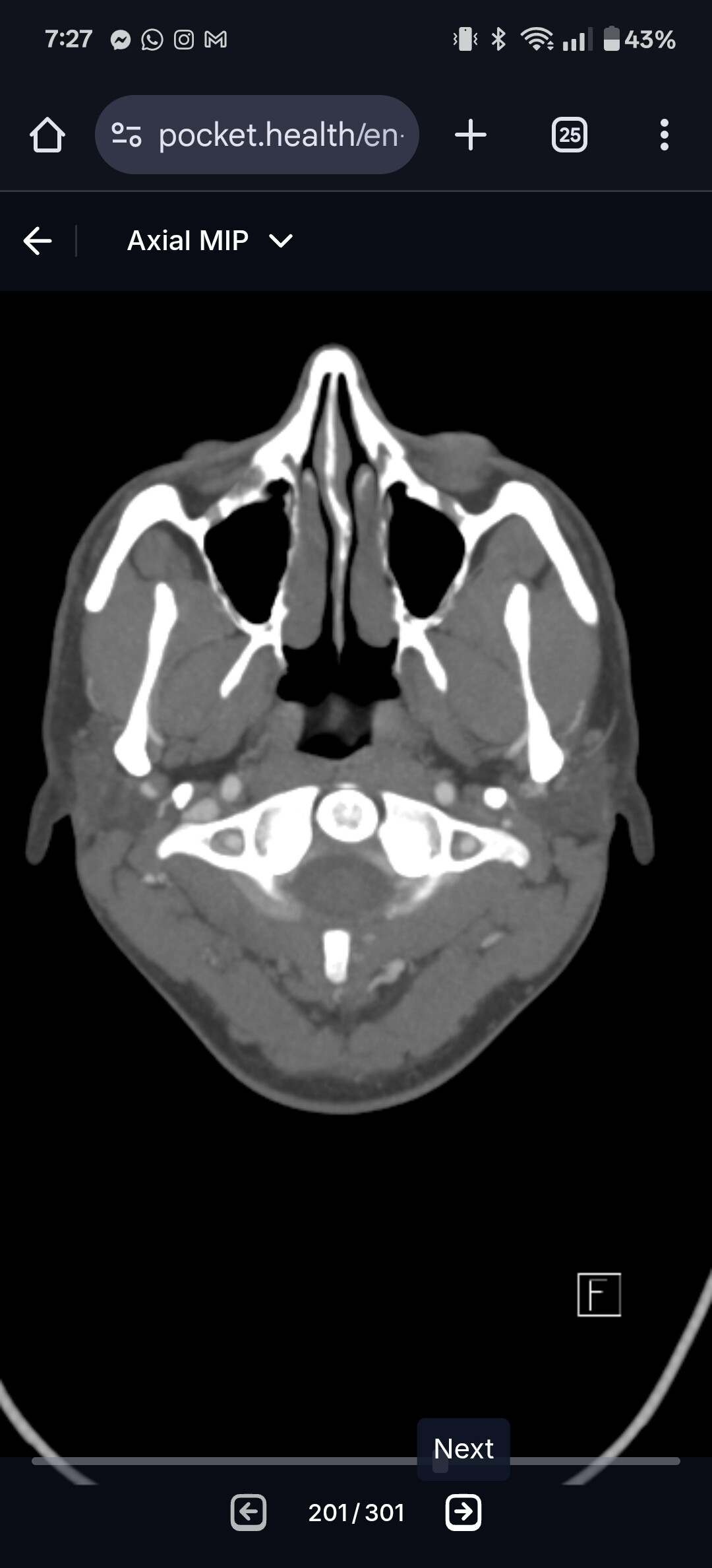
@Ihurt can you go to the axial view of C1 and upload a screenshot of it? I attached my own imaging to help you know what to look for. It can help us see the IJV compression better!
@Ihurt radiologists really drop the ball when it comes to vascular compression in the neck. It’s almost like if they don’t see thrombosis or aneurysm then everything is good. So basically you had contrast in all your vascular structures and they didn’t see any cut open so they think you’re all good. Additionally, radiologists only seem to care about ICAs, and so the ECAs and IJVs are often overlooked. And even when IJV compression is found, they’ll often say it’s nit significant or that it’s a natural tapering point in the IJV. Frustrating to say the least.
There are a few discussions about how to appeal with insurance companies, but it is difficult for you given that you’re being fobbed off with being told there’s no issue… Also if you were to have surgery, because your styloids are so short, & the compression is still potentially there, they would need to be taken back completely to skull base to remove that compression, & some surgeons do leave a little stub of styloid, probably a similar length to what yours are… It would be interesting to get Dr Osbourne’s take on this if you can do a consult with him… I didn’t think that all the doctors insist on other imaging other than what you’ve had already for an initial consultation, as your CT shows the blood vessels pretty well; have you checked with Dr Costantino’s office in NY? I might be wrong- there is confusion with the ‘CTV’ - as a CT venogram, a CT with contrast which shows the veins is what you’ve had done. More detailed imaging & testing of pressures across the veins is a more invasive test which not every radiology dept would do, also called a cerebral angiogram/ venogram …
‘CT cerebral venography (also known as a CTV head or CT venogram) is a contrast-enhanced examination with an acquisition delay providing an accurate detailed depiction of the cerebral venous system.’
Thank you for the reply. I definitely could get a virtual consult with Osborne. However, he is not a vascular surgeon and doesn’t deal with compressions from what I’ve read from others on here. He charges $250 for a video consult. Constantino will not even give you a virtual anappointment unless you have had a CTV. A friend of mine has an appointment with him and he insisted on a CTV and MRI of the head before he will give her a video consult. Same with Dr. Nakaji. Do youthink Dr.Osborne would be able to give more insight into this?
He’s not a vascular surgeon as you say, & doesn’t do C1 shaves so if the C1 process is causing more compression then that might not be helpful, but he has given members good info & a diagnosis when they’ve struggled with getting that, so it’s up to you
Thank you so much for the response. I definitely will upload a screenshot of my C1 for you to look at when I get home a little later. This whole thing is very frustrating with trying to get help from specialists when there aren’t many around.