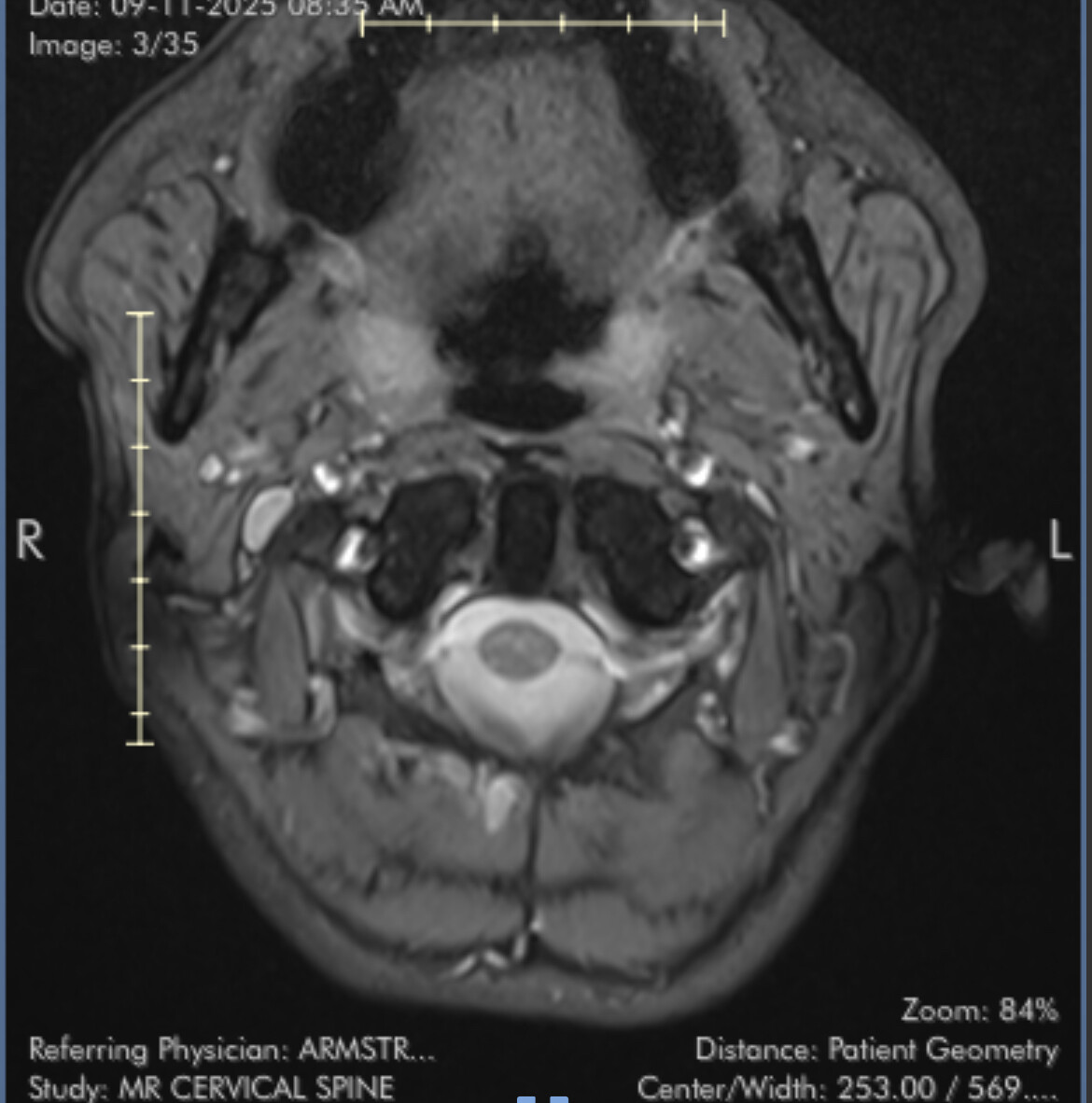
Recently visited my ENT due to ongoing pain on the left side of my neck and throat. The pain worsens with swallowing and yawning and often feels like I have something stuck at the back of my throat on the left (my left tonsil also protrudes but unsure if this is related). I’ve also had other circulatory issues and pulsatile tinnitus when laying down. When showing my ENT the pain location he mentioned Eagles Syndrome and is currently consulting with a colleague as to next step. I had an MRI a couple of months back and wondered if anyone could look at the images? I’m aware that MRI isn’t great and I’ll need a CT but thought it was worthwhile sharing with everyone in this group as it seems very knowledgable!
Additionally I’ve had some very weird symptoms that I thought were unrelated, but have seen there could be a link to the vagus nerve. It’s crazy home impactful this condition can be. I’ve been struggling so much recently
I’ve just learned that I cannot upload images until I’m a more established member Would still appreciate advice on how to navigate this process and reassurance. I’ve done what everyone says not to and googled, so am scared of vascular compression!
@BikerG - Your account should have updated as soon as you posted, but it failed to do so. I’ve upped your account so you can now post images. I look forward to seeing your MRI but am not good at knowing what I’m looking at unless the images are 3D. I’m glad your ENT is at least familiar w/ ES as so many still are not.
Your symptoms do sound suspiciously like they could be related to an elongated styloid process causing internal jugular vein compression. This diagnosis is becoming more common than the non-vascular version of ES on our forum.
The vagus nerve is one of the nerves most frequently irritated by elongated styloids & in cases where there is IJV compression between the styloid & C1 vertebra, the vagus nerve also gets trapped at that location which makes it “mad” which causes more vagal symptoms.
Hi & hopefully you can try uploading images now, just make sure there’s not any personal info on them for your privacy. We’re not very experienced at looking at MRI images, but will help if we can!
When you get a CT, make sure it’s ordered to show from the skull base to the hyoid bone, & that they look at the size, width and angle if possible. And have a CT with contrast too so you can see if there’s any vascular involvement. If you can get copies yourself that’s helpful, as occasionally they get mislaid/ not sent on time if you get referred to another doctor!
This past year has been progressively challenging and I’m trying not to feel hopeless. The vagus nerve related symptoms are very scary and have had a real impact on my life.
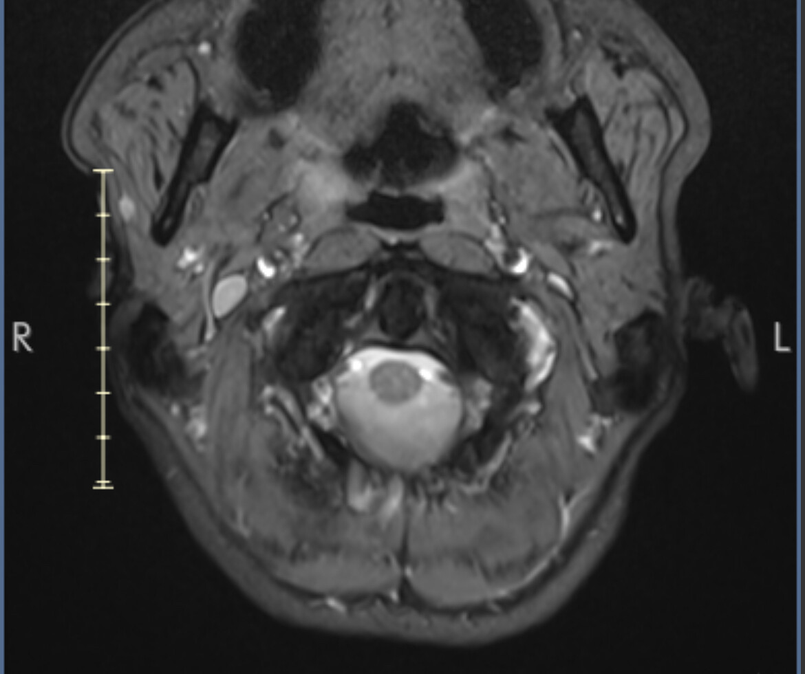
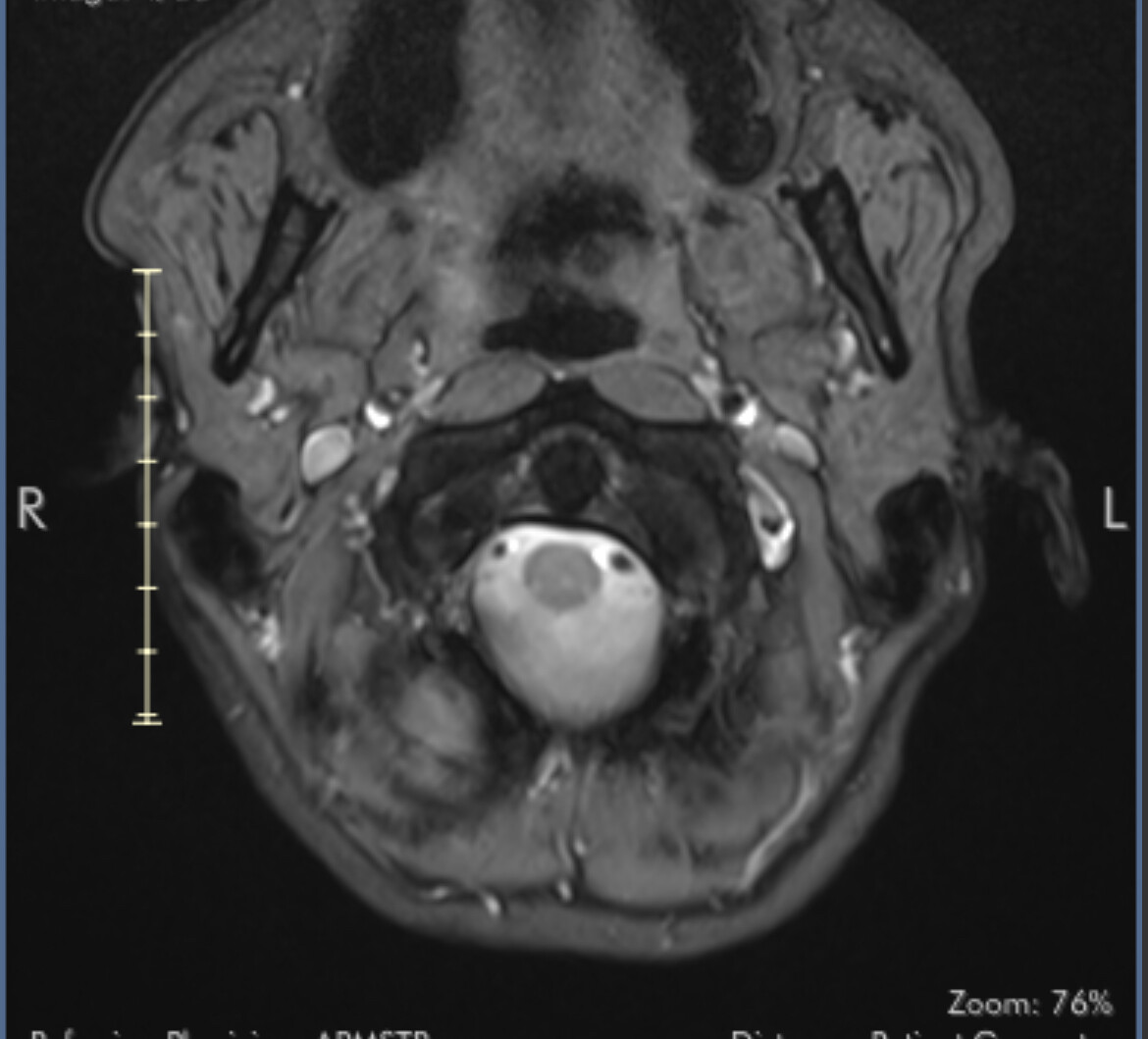
I’ve uploaded some images now. I know they aren’t ideal but I’ll hopefully get my hands on the CT angiogram I had at A&E soon.
Totally understand. Hopefully I’ll be able to get my CT images soon
My ENT thinks Eagles and, ever since he mentioned it last week, I’ve been so worried that I have vascular compression and something bad is going to happen. I’m sure you see people have this concern a lot. I’ve been trying to find resources to put my mind at ease that it isn’t going to suddenly cause a severe vascular event but information is really lacking!
It is a common concern with VES, but it is really, really rare for anything bad to happen. Carotid compression can in extremely rare circumstances cause a stroke or an aneurysm. IJV compression (which it sounds like you could possibly have with head pressure and pulsatile tinnitus) is even less likely to cause a severe vascular event as you say. With compression there is always the very slight possibility that a clot could form where blood is backed up, but I can’t recall anyone having that. There can be cognitive effects with IJV compression if it causes intracranial hypertension, there have been one or two discussions about this. I would try not to be concerned about what could possibly happen, although I know it’s easier said than done
@BikerG - Unfortunately, I’m in the same boat as @Jules & nothing in the MRI images you posted looks familiar to me. I do look forward to seeing your CT images when you’re able to get them.
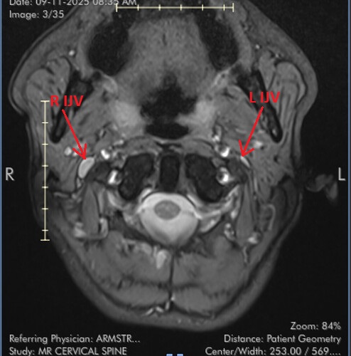
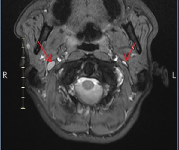
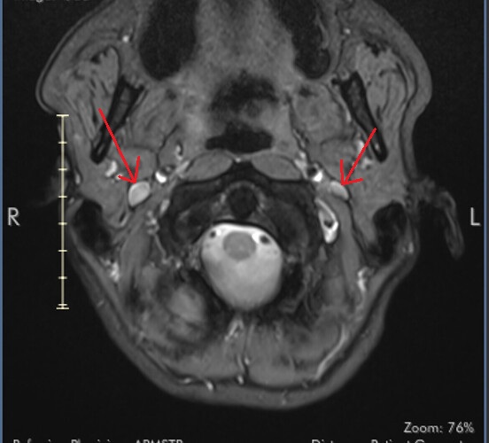
I marked the images for you and marked the IJV.
So you can see how the IJV passes by the transverse processes of C1 which are compressing. In this position the right IJV does not have severe compression, but in Figure 2 you can see that the left IJV is almost invisible, and in the other images there is significant narrowing.
How much this affects your problems should be assessed by comparing your symptoms and the disease.
On MRI, it is very difficult to see the styloid processes.
Thanks for that @tesla001 ! I did presume that those were the IJVs, but then had a wobble of confidence as they seemed to be the same density as the vertebral column so I didn’t want to say something wrong