Got a first X ray with calcified styloid process, need ur help

@junonbss - OOPS! I had just read your other posts that mention your imaging is tomorrow but then I saw the pictures you posted & immediately forgot. :zany_face:

Another option for Macs to create 3D images from CT slices is Bee Dicom Viewer App. I did read in your description of tomorrow’s imaging that it said - " Reconstruction in the three planes of space." which may mean that you’ll get 3D images already done for you. I hope that’s the case as it will save you time & possibly some frustration.

2 Likes

Hihi don’t worry !! I use HOROS for Mac and it’s really great and free. I got others views from an 2017 MRI : I think the problem is really clear !

Are we ok ? It’s belong C1 ? Sorry got so many differents images

1 Like

Your styloids are very scary looking @junonbss! I’m sure getting them resected close to your skull base will make a HUGE difference in how you feel!!

2 Likes

@junonbss @Isaiah_40_31 is not exaggerating stating your styloids are scary. Wow! I will be praying your CT and appointment tomorrow lead to a resolution for you, surgery in the very near future!

3 Likes

I take your prays with me :folded_hands:t3::sun_with_face:

3 Likes

Wow, they are very long! And quite angled too! Will be interesting to see if there are any changes on the new images…

1 Like

Hi everyone!

I wanted to give you an update after what turned out to be a very frustrating day……..

This morning, I went to my CT scan appointment, only to find out that it had been cancelled because the “wrong appointment type” had apparently been selected. What is especially frustrating is that I had called the imaging center yesterday to confirm everything, and nobody mentioned any issue. As a result, the scan has now been postponed to July 29th.

Later today, I had my appointment with the maxillofacial surgeon. Unfortunately, she told me that she does not personally manage Eagle Syndrome patients and will not be the surgeon treating me.

However, after reviewing my case, she felt that more detailed imaging was needed. She requested:

  • a dedicated CT angiogram (angio-CT) to better evaluate possible vascular compression,
  • an MRI to assess surrounding structures and potential compression.

I was fortunate enough to obtain an MRI appointment for July 28th, so at least I will have both examinations completed by the end of the month.

What worried me is that she seemed quite concerned when looking at my imaging and symptoms. She also referred me to Pitié-Salpêtrière Hospital in Paris, which is a major tertiary referral center, but I am afraid the waiting times may be very long. At this point, I honestly don’t know who I should turn to or which specialist in France has the most experience with Eagle Syndrome, especially if vascular involvement is suspected.

Another thing that frightened me during the consultation was the discussion about surgery. I already have a right recurrent laryngeal nerve paralysis from previous thyroid cancer surgery. My left vocal cord is the only functioning one, so she emphasized that if I ever need surgery in this area, protecting the remaining nerve is absolutely critical. Hearing that made me quite anxious about the possibility of another neck surgery.

For now, I am trying to stay focused on getting the MRI on July 28th and the CT angiogram on July 29th, and hopefully these exams will provide clearer answers.

If anyone knows of surgeons or centers in France (or nearby European countries) with significant experience in Eagle Syndrome, especially complex cases involving possible vascular compression, I would be very grateful for any recommendations … I know it’s really complex in France

Thank you all for your support :face_exhaling::sun_with_face:

3 Likes

@junonbss - I’m sorry for the cancellation today, but it’s good the maxillofacial doctor ordered even better imaging than you would have gotten today.

Make sure that CT angiogram also includes the veins in your neck. Often angiograms only look at arteries but we’ve had many more members with internal jugular vein compresssion than compression of the carotid arteries. It would be a shame for your to get the CTA & not have the IJVs be visible.

It’s hard to tell how close the styloid are to your C1/C2 vertebrae in the images you posted. If you have IJV compression, that’s the area where bony compression is most likely to occur. Compression from soft tissues can be found in various places along the IJVs.

The vagus nerve is the nerve which innervates your vocal cords is the nerve which could possibly be damaged by a neck surgery, however, it’s protected by the carotid sheath in which the internal carotid artery & internal jugular veins both also “live”. Any very experienced skull base surgeon knows that & should be careful when working in the area near the skull base.

The closest doctor to you who does styloidectomies & IJV decompression surgeries is Mr. Axon in England. Next would be Dr. Aghayev in Turkey. We don’t know of any doctors in France who do ES surgeries. Both of these doctors are very experienced with the type of surgery you would need.

•Mr Axon, Addenbrooks Hospital, Cambridge (Otolaryngologist/ Skull base surgeon, very experienced with ES surgery, special interest in Pulsatile Tinnitus. Also does private work at Spire Lea hospital, Cambridge, for Spire Health care. Mr Patrick Axon - ENT Surgeon (Ear, Nose & Throat) | Ear, Nose & Throat Surgery | Spire Cambridge Lea Hospital

•Dr. Kamran Aghayev - https://kamranaghayev.com
Link to set up a consult: Get Expert Neurosurgeon Consultation & Second Opinion Online

2 Likes

I’m sorry that you’ve been messed about with your imaging appointment, that’s so frustrating! But as @Isaiah_40_31 says, to avoid another mistake definitely make sure that the veins are scanned too- sometimes this is called a CTV, as it seems more likely in your case that you have IJV compression…
It’s unfortunate that there’s no knowledgeable vascular ES doctors in France, many countries are the same. You could try looking for a head and neck neurovascular surgeon in France, perhaps there’s one in the Paris hospital you’ve been referred to?

1 Like

Thank you all so much for your advice and for taking the time to always help me :butterfly:

I have now requested an appointment at Pitié-Salpêtrière Hospital in Paris, and my maxillofacial surgeon has also ordered both an MRI and a CT angiogram.

The CT angiogram is specifically intended to evaluate any vascular compression (including the internal jugular veins if possible), and the MRI is to assess possible nerve involvement and provide additional information before any surgical decision is made.

For now, I think the best approach is to first consult several surgeons in France and see what each of them recommends. I want to carefully compare their opinions before considering treatment abroad.

Thank you again for your kindness, your experience, and for pointing me toward specialists such as Mr. Patrick Axon and Dr. Kamran Aghayev. It really means a lot to have your support during this difficult journey!!! :folded_hands:t3:

4 Likes

Hi everyone,
I recently had both a contrast-enhanced CT scan and an MRI for my suspected Eagle Syndrome.
Unfortunately, I’m struggling to interpret the MRI images myself, and I was quite disappointed with the radiology reports. They don’t really address the specific questions my surgeon asked. Despite the explicit request to evaluate possible carotid or jugular compression as well as cranial nerve involvement, the reports simply conclude that there is no obvious problem, without providing any detailed analysis.
Because of this, I have started reviewing the CT images myself while I wait to see a surgeon with real experience in Eagle Syndrome. My goal is to better understand the vascular anatomy and whether there could be any vascular involvement.
According to the radiologist, both styloid processes measure 4.4 cm (44 mm), are symmetrical, and show no obvious abnormality or vascular compression.
I’ve attached quite a lot of images (I apologize for that) but I thought it would be better to include more rather than too little so you can see the anatomy from different angles.
If anyone notices anything, has suggestions, comments, or recommendations on how to better evaluate these images, I would be incredibly grateful !
Thank you so much in advance :pink_heart:

1 Like
1 Like
2 Likes

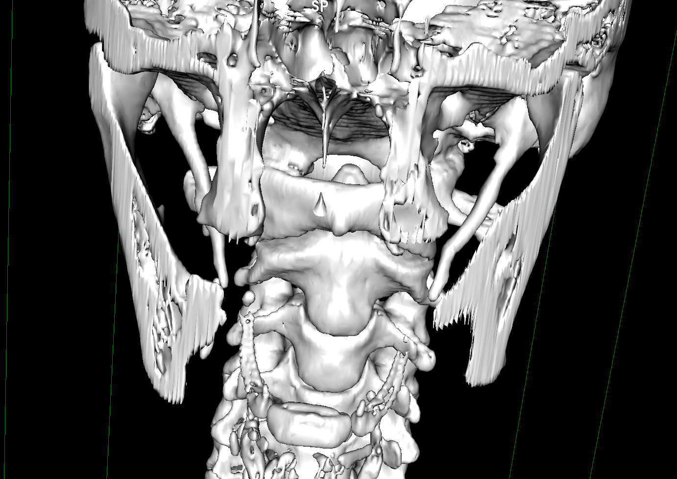
@junonbss - You posted some very good pictures. Could you post one that’s from the front? There are several from each side & the back but not a full front image. That would also be helpful. Your styloids are quite thick at the skull base & appear to be angled inward somewhat significantly which could be putting them in contact w/ your internal or external carotid arteries. From the collateral veins you have on the back of your neck on the right, it appears there’s at least some IJV compression occurring on that side, thus the development of the collaterals. On the left the collateral formation is far less.

I’ve annotated what I see in a few of the pictures you’ve posted. Some are self explanatory & others I have put comments under:


Mild collateral formation on the left thus possible mild IJV compression but also possible carotid compression/irritation by the styloid


Significant collateral formation on the right side which could indicate more serious IJV compression on the right. It also appears your right transverse foramen may not be fully closed as it is on the left. This is more visible in the axial image you posted:

In each of the two pics below, there is a narrow, “fuzzy” (out of focus) area on the IJVs which appears to be compression but I can’t tell what’s causing it:

In the picture below, it looks like the right greater horn of your hyoid might be in contact with your external carotid artery. There wasn’t a comparable picture of the left greater horn, so I can’t comment on the left side. Irritation or compression of the carotids can cause some similar & some different symptoms than IJV compression causes.

I don’t know if any of what I’ve pointed out is significant, but the info is at least worth taking to your ES consult to use for asking questions & getting a better understanding of what the doctor sees & thinks is relevant.

3 Likes

Looks like there could also be compression further down in the mid-neck. Im just catching up on your story but I can share I have many of the same symptoms you do. Ive tried most all modalities and getting botox in the temples and jaw and occipital area regularly (every three months) is about all that has helped. I have advanced osteoarthritis in my left jaw and it gives me fits all the time. I too had a worsening when I had a lower molar extracted. It put me in the emergency room in extreme pain 2 days later. I was down for 2 weeks. I told the oral surgeon I had bad TMJ and to use caution but I think he did not listen. I believe my jaw was opened too wide and I had some bad bruising on my face. My regular TMJ doc said it looked like an "aggressive " extraction. During this time, I developed serious vertigo and I haven’t been normal since.

I can say that over the years, regular massage including cranial-sacral therapy has really helped me. My shoulders lock up too. Dry needling has been good too but I love the cranial sacral work. It is very gentle work but so powerful. You a look up providers in your area at the Upledger website.

I would definitely push to find a doctor who is experienced in Eagles and does look at vascular compression and possible C1 shaving. It needs to be an ENT with experience in skull based surgery. Any doc you see that doesn’t know about this will never get you anywhere except frustrated.

3 Likes

I agree with @Isaiah_40_31 , that there does seem to be some compression of the IJV between the C1 process and the styloid , especially on the left, but then in some images it looks like it’s not being caused by bony compression, and in the axial image it doesn’t look like there’s much compression! But there also looks like there’s compression lower down as @Snapple2020 says, around C5ish, so that could be caused by soft tissue, SCM muscle maybe?

The styloids do look like they’re possibly in contact with your carotid arteries and the hyoid bone processes are quite long and close to that too…

You could get your images evaluated by somebody else, some members have used online radiologists, or perhaps have a consultation with Dr Aghayev, or Kjetil Larsen at MSK Neurology so you have a clearer picture to take to your doctors?

3 Likes

Hi @junonbss, I live in France so understand the difficulties of getting diagnosed. Radiological reports are often depressingly lacking.

Unfortunately you will very quickly realise after a few consultations that it is difficult to find a surgeon that has alot of experience in styloidectomies and correctly removing them. The second difficulty will be getting any useful assessment of your internal jugular veins by a radiologist or surgeon. If you haven’t got symptoms suggesting completely thrombosed internal jugular veins you won’t be taken seriously and even if it was the case you will be given a medical solution which may or may not work :frowning: . Fortunately for you this doesn’t appear to be the case.

However you could try getting in contact with Dr Stephanie Lenck a neuroradiologist at Salpetrière Paris. She is doing research on the glymphatic system and should be well aware of the importance of correct venous drainage in the brain and neck. She was recommended by Dr Hui, a prominent neuroradiologist in the field of cerebral venous congestion in the US to another member on this forum @CristianSerious.

If I was you, I would also seriously think about sending your CT scan with contrast to Dr Kamran Aghayev in DICOM format via WETransfer after initially contacting them via Whatsapp with your symptoms. He doesn’t charge to initially send you his oral video assessment. This oral video assessment will be nothing like any of the radiologicaI reports you have probably received in France as he actually correlates your symptoms with your scan findings. I did this for both my sons who are 25 and 27 years old. You can then have an online consultation with him where he goes into more detail to explain the CT scan and its 3D rendering. He will also explain any eventual surgical procedures that he recommends. This consultation with Dr Aghayev would be useful before your intended consultations with surgeons in France.

For my part both my sons were operated on by Dr Aghayev, both had bilateral styloidectomies and surgical decompression of the internal jugular veins. My first son (24 years old when he had the operation) had severe bilateral compression of his internal jugular veins with severely varicosed collateral veins which explained his headaches and head pressure. This required removing a part of the transverse process of C1 on both sides as well as myectomy of 3 muscles in the neck and partial myectomy of a 4th muscle to effectlvely open up the jugular veins. My second son has just come back from his operation in Istanbul. He had total obstruction of his right internal jugular vein and severe stenosis of his left internal jugular vein. He required bilateral resection of of the transverse process of C1. His recuperation from the operation was amazingly quick. He was able to move his neck within 24 hours and was eating normally in the same amount of time. This rapidity of convalescence is just a reflexion of the level of experience Dr Aghayev has acquired now with operating on Eagle patients and vascular Eagle patients. Keep in mind that you will not find this level of surgical expertise or experience in France even if you do eventually find someone that accepts to operate on you.

Best of luck. Don’t hesitate to contact me if you need any further details.

5 Likes

I’m not a doctor. But still… You don’t have compression of the internal jugular veins in the C1 area. You may have compression of the internal jugular veins lower down (all Dicom files are needed). Regarding the nerve, CT scans won’t show anything. In this case, the diagnosis can only be made based on symptoms such as stabbing pain in the eye, face, ear, and shoulder blade. These operations are still experimental, and there is no precise diagnostic procedure. The surgeons who perform these operations can only hypothesize where the problem might be. You can anonymously download all the Dicom files from the disk (without revealing your personal information) using the website dicomlibrary com


Thank you so much for all the research and notes you sent me. I will send you some new, hopefully clearer, images of the hyoid bone and the anteroposterior view.
I have the impression that, looking further down on the axial section, the foramen is indeed closed?
In any case, thank you so much; it helps me a lot to better understand and to move forward with my research! :pink_heart:

3 Likes