Hi everyone! I’m still waiting for my first ES consult but am also wondering if my symptoms are more aligned with hyoid bone syndrome or a combo of both. Both of my stylohyoids are elongated (I think 4.1 cm and 4.3), not sure if anything is visually wrong with my hyoid bone. My worst symptoms are:
-foreign body in throat
-difficulty breathing; it often feels like my throat is tight and constricted so it’s hard to get air down. I also get full laryngeal spasms that cut off my air.
-difficulty speaking; it’s painful, frequent vocal fry, and really hard to breathe
-difficulty swallowing, but this symptom comes and goes
-honorable mentions: sudden severe insomnia, head pressure triggered by movement, throat/neck is externally tender
For anyone here that had both, was it all treated in one surgery or did you have to do multiple? And just curious if anyone has similar symptoms to me?
I believe we’ve had a few members who’ve had both ES & HBS. However, with your comment about head pressure triggered by head movement, I have concerns that you may have compression of your internal jugular vein(s). When the IJV(s) become squashed between the styloid process(es) & transverse process(es) of the C1 vertebra, the vagus nerve is also commonly compressed between those two bones, too, as it runs closely alongside the IJVs where they exit the skull base.
Your symptoms of difficulty speaking, vocal fry, & difficulty breathing all point to vagus nerve irritation or compression. Have you had a CT w/ contrast done? If not, please request that from the ENT whom you consult. A dynamic CT w/ contrast would be even better. That’s a CT that is done w/ your head in several different positions rather than only in neutral. A dynamic CT can show how vascular compression is occurring w/ certain head positions where it might not show up in a neutral head position. Additionally, it will better show whether the greater horns of your hyoid bone are tangling w/ your carotid arteries lower down. Being able to see the great horns of your hyoid can also help make a determination as to whether or not they appear too long (which is mostly what causes symptoms of HBS).
Excess calcification of the thryoid cartilage can also cause vocal changes & swallowing difficulties. That should show up in your CT scan as well if the scan is done from skull base to hyoid bone or thyroid.
It’s important for you to get your CT imaging on a CD before you leave the radiology office. If they’ll give you two copies that’s even better as you’ll have one to keep & one to share w/ a doctor or doctors you decide to have consults with. We’d also be happy to give our opinions on your imaging if you want to post some pics here. Just be sure to remove any personal information before posting them.
Thank you for the detailed response! I’ve had one CT done with contrast at the ER one of the first times my breathing got really bad. It covered my neck down to my upper bronchial tubes; months later a different doctor noticed the elongated stylohyoids when I sent them a copy.
I’m also suspicious of jugular vein compression because of the head pressure. It’s just sounding like my breathing issues aren’t typical of ES and I’m desperate to figure it out.
Have you heard of anyone having treatment for both scenarios in one surgery? Or is that too much trauma to your throat/neck in one sitting?
It’s a tricky one, as the most experienced doctors who treat vascular ES I’ve not heard of doing hyoid surgery as well, maybe others who’ve seen these doctors (Dr Nakaji in AZ, Dr Costantino in NY, Dr Hepworth in CO & Dr Cognetti in PA) can give you some info?
We have had some members who have had breathing issues probably from vagus nerve compression as @Isaiah_40_31 says- the vagus nerve exits the base of the skull with the IJVs so it makes sense that it could be being irritated too if you have IJV compression.
Obviously it’s important to make the right decisions, if you’ve not had a CT with contrast it might be useful to get that done, especially timed to show the venous phase, and that might help you decide which doctors to get a consultation with?
@Jess1 - If you convert your CT images to 3D, or if you already have some 3D images, you’re welcome to post them here for our non-medical opinions. We’ve looked at a lot of CT images over the years & sometimes see things radiologists & doctors miss or don’t think are significant when they really are. If you do post images, please make sure any personal information on them is covered or removed.
To convert your images to 3D you can use radiantviewer.com (for PCs) or Bee Dicom Viewer App (for Macs).
Thank you, I’ll give that a look. I need to get a separate CD reader because my laptop doesn’t have a CD port and that’s how the hospital sent me my images. But I’ll check out that site and see if the images are clear enough to post, thank you!
I was looking at Dr. Nakaji but he won’t see me until I have a venography done and I’ve been struggling to get it ordered; it’s like my current doctors have never been asked to order that test before. I also thought he only treated vascular issues (e.g., if my thyroid cartilage is causing issues with my hyoid bone but not compressing anything, I’d assume he wouldn’t touch it?)
Yes, I’m not sure that he would treat hyoid or thyroid cartilage; you’re in a tricky situation as it sounds like you might have vascular ES, so you could do with getting a CT done with contrast to find out that for sure before you have surgery, but equally you might need that hyoid etc being done too- unfortunately you might not be able to get everything done in one surgery, but it would be good to get the scans done and then get some opinions before making that decision? Some of the doctors do online consults so you might not have to do in person appointments?
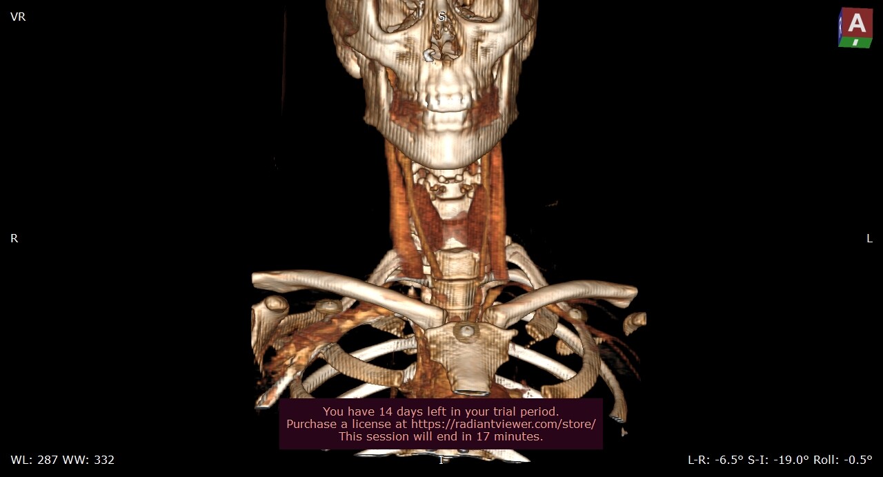
Hi! I just got RadiAnt - what view is most helpful? Unfortunately, viewing from the back of my neck isn’t great as you can see the pillow/platform I was laying on. But the front and side view are legible.
Front & side views are great! I think radiant has a “cutting” tool that may allow you to remove the pillow/platform you see on the back side. To protect your privacy, please make sure to remove any personal information from the pictures you post.
okay here are some screenshots, let me know if any should be more zoomed in or tilted a different way. The one thing I do see that’s weird is bone fragments or calcifications above my thyroid (I can find better screenshots of that if needed). I’m personally struggling to confirm my stylohyoid length, but my orofacial pain specialist said they measured 4.1 and 4.3 cm when they looked at it.
I do have axial view but it cuts off towards the top of the hyoid bone. The CT scan I’m using was taken during an ER visit so it wasn’t the most specific imaging.
@Jess1 - I’ve annotated some of the pictures you posted. The left half of your C1 is unusually shaped in that you have 2 transverse processes w/ one “leg” being shorter than the main one. You also have completely lost the lordotic curve in your neck which is called “military spine”. This is correctible using gentle PT exercises but can take some determination & is a slow process. Loss of the lordotic curve can result in putting the styloids & greater horns of the hyoid closer to nerves & vascular tissues in the neck. That said, it’s been speculated that straightening of the cervical spine when IJV compression is present (as it is for you especially on the left) may be a physical response to try to give more space for the IJV to open up more. In the top image w/o contrast, it looks like the greater horns of your hyoid are pretty close to your spine. I’m not sure if this is significant in your case. In that position, the greater horns can sometimes make contact w/ the common or internal/external carotid arteries, but in the absence of carotid symptoms, it’s likely not a big deal.
Thanks for the thorough response! The annotations are also very helpful . I forgot to mention I was in a car accident 10 years ago so the loss of the lordotic curve is from that. My symptoms started about 1 year ago so I’m unsure what changed a year ago since the shape of my cervical spine isn’t new.
The other weird thing is I was struggling to find and measure my styloid process (the bone was easy enough to find and thanks for confirming via annotation). My orofacial pain specialist measured them and said they’re both a little over 4 cm but I couldn’t find that…but maybe it didn’t carry over well on 3D?
@Jess1 - I forgot to mention that, as you can see, your left styloid is quite thick & club-like where your right one is thinner & more more typical of the styloid shapes me predominantly see on here. Truly length is only one feature of the styloids that can play into whether they’re causing symptoms or not. Their angle of growth, how curved they are, how thick, twisted &/or pointed, they have outgrowths coming off them all can contribute to symptoms even in the absence of excess length.
I’m sorry I’m not terribly techy (my son taught me how to annotate images) so don’t know how to measure the styloids. The lengths that can be measured in a CT scan usually aren’t very accurate anyway because there are tiny spaces between the CT slices so the styloids are often found to be longer than measured in imaging once a surgeon goes in to remove them.
That’s interesting! So given that one of the criteria for surgery is estimated stylohyoid length, what is usually used instead of CT?
Your so did a great job teaching you to do the annotations! They’re really helpful. I measured using the little measurement tool in radiant but since I’m not radiologist, idk if I’m doing it correctly.
Good question! I don’t know if radiologists take that into account or not! I’m not sure that all the ES doctors are that specific with the lengths, or just use them as a general guides, you can see with the scans for most members on here that their styloids reach down to or past the C1 processes, which if the styloids are ‘average’ length of 2.5cms I don’t think they would reach down that far…