Hi my name is Truman, and I’m 22 y/o. I have been struggling with symptoms going all the way back to me at 12 years old; starting with my ears and my jaw. Over the last 10 years I have had a lot of developments especially when it comes to new kinds of pain and the confusion that comes along with it. Finally after struggling for years with cervical issues and concentration among many other things, I decided to quit my job and move in with my mom so we can get the care that I’ve been needing. Earlier this year we saw Dr. Ryan Osborne who is based in LA, where he said that I had lots of indications of eagles syndrome. In April we had the first styloidectomy removed and it seemed have some impact on me right after the surgery, but as time has gone by I have continued to feel worse and worse reverting back to how I felt before I had gotten the styloidectomy. I am going to be seeing the doctor’s at Loma Linda based in CA in a few weeks for further evaluation. What I am curious most about is whether or not the cervical issues, as well as my headaches and difficulty speaking and breathing that I’ve been having are a result of hyoid bone syndrome and would like to hear any feedback from anyone who has dealt with hyoid bone syndrome and if someone has had surgical treatment for it, what their experience was. I have mild disc degeneration and right foraminal narrowing. I struggle to support my head and my neck, and my spine pops throughout the day. It would be great to know if anyone has dealt with this and whether or not they have had success in treating this, and what their lifelong consequences are to having both/ or one of your hyoid bone trimmed.
@TrungoHunk - Welcome to our forum! Were you diagnosed w/ bilateral styloid elongation or stylohyoid ligament calcification? If so, you may need to have the other styloid resected to get full symptoms relief. My symptoms got worse after my first styloid was removed but after my second styloidectomy & a number of months of healing, almost all of them went away.
I’m sorry to say that I don’t think you’ll get any significant help from doctors at Loma Linda. My reason for saying that is I’ve been on this forum for 11 years & no one has mentioned a doctor from that institution having knowledge about ES in all that time. There are many great medical institutions in this country that are completely clueless about Eagle Syndrome & Hyoid Bone Syndrome who should be well versed in both. I would suggest saving your time & $ by getting an appointment w/ Dr. Chhetri at UCLA Medical Center. He is very knowledgeable about HBS & does the surgery for it.
•Dr Dinesh Chhetri, UCLA, Los Angeles (ENT/ Otolaryngologist, Head and Neck Surgeon) Dinesh K. Chhetri, MD - Otolaryngology - Head & Neck Surgery | UCLA Health (Possibly not doing Eagles surgeries any more [1/13/24])
Have you been evaluated for hypermobile Ehlers Danlos Syndrome (hEDS) or Cranio Cervical Instability (CCI) or Atlanto Axial Instability (AAI)? The symptoms you’ve mentioned above could indicate one or more of those diagnoses. Here’s a link that will be helpful re: hEDS evaluation. You can have it done by your PCP using the form in the link. Print 2 copies & fill out one for your own information & give the blank one to your doctor to use to evaluate you.
Cedars Sinai Med Ctr has a pain management center & they deal w/ EDS so that would be another place to look into for your neck.
Here are some other resources that may be helpful for you:
We’ll be happy to look at any imaging you have if you want to share it here as we often see things that radiologists miss or dismiss as not being of consequence when they truly are. Please bear in mind we aren’t doctors so nothing we tell you can be considered diagnostic. It can be used in discussion with your doctor though. If you do post images, please remove any personal info on images you post to protect your privacy.
radiantviewer.com (for PCs) or Bee Dicom Viewer App (for Macs) are useful tools for converting your CT imaging into 3D images which will help you better see what’s going on in your neck.
Hi thanks for replying I appreciate it, and to answer some of your questions I have a bilateral/ elongated stylohyoid, with some calcifications next to and from the styloidectomy process. I also have seen a genetic counselor from Kaiser who did not seem to think I have eds. His conclusion was that I may have some criteria to fit certain disorders I didn’t have enough to qualify for anything. You may be right about the other styloidectomy being necessary to be symptom free but I have other things that make me want to rule out HBS before I have my other styloid taken out. I didn’t detail this in my previous message but I have other neurological symptoms such as visuals, especially when I close my eyes which is accompanied by nausea, I also feel as though I always have a lump on the right side of my throat where the greater horn and the superior Cornu are. I will try tomorrow to get some images so that I can get some feedback from the forum. Thank you
Feeling like you can’t support your head does seem to be a ‘classic’ CCI symptom, so that’s worth maybe looking into… It could be that your hyoid bone processes are touching a carotid artery which could give you visual disturbances, although this can be a symptom of high intracranial pressure too, which can be from the styloid compressing the IJV… certainly worth posting your images to see if we can see anything, & having a good read up of different symptoms to see what matches. Although if you did see an improvement of symptoms with your first surgery, and now they’ve come back, it could be that as @Isaiah_40_31 says you need the other side removing, and also worth considering that you might have some re-growth, especially given your age, so a new CT (with contrast) might be worth getting done if you can (if you’ve not had imaging done recently)?
I’m trying to submit some images I took of my scans but it is saying that I cannot embed media items. Do you know what I can do to get around this?
I’m sorry for the slow reply, @TrungoHunk. I just checked your account & it’s been upgraded so you should be able to upload images into your text box now. Please try again. If you still get the same message, please let me know.

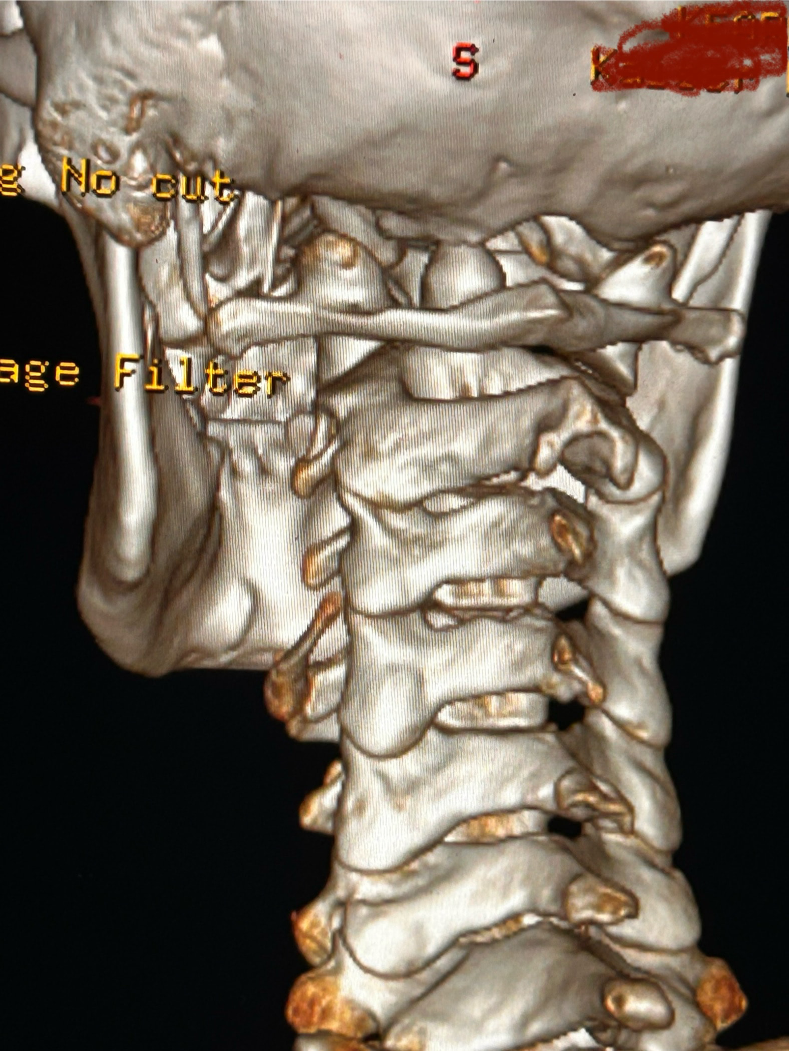


Here are some images of my 3-d ct, thanks for taking an interest it means a lot!
Oh and I almost forgot these were taken before I had my right styloidectomy
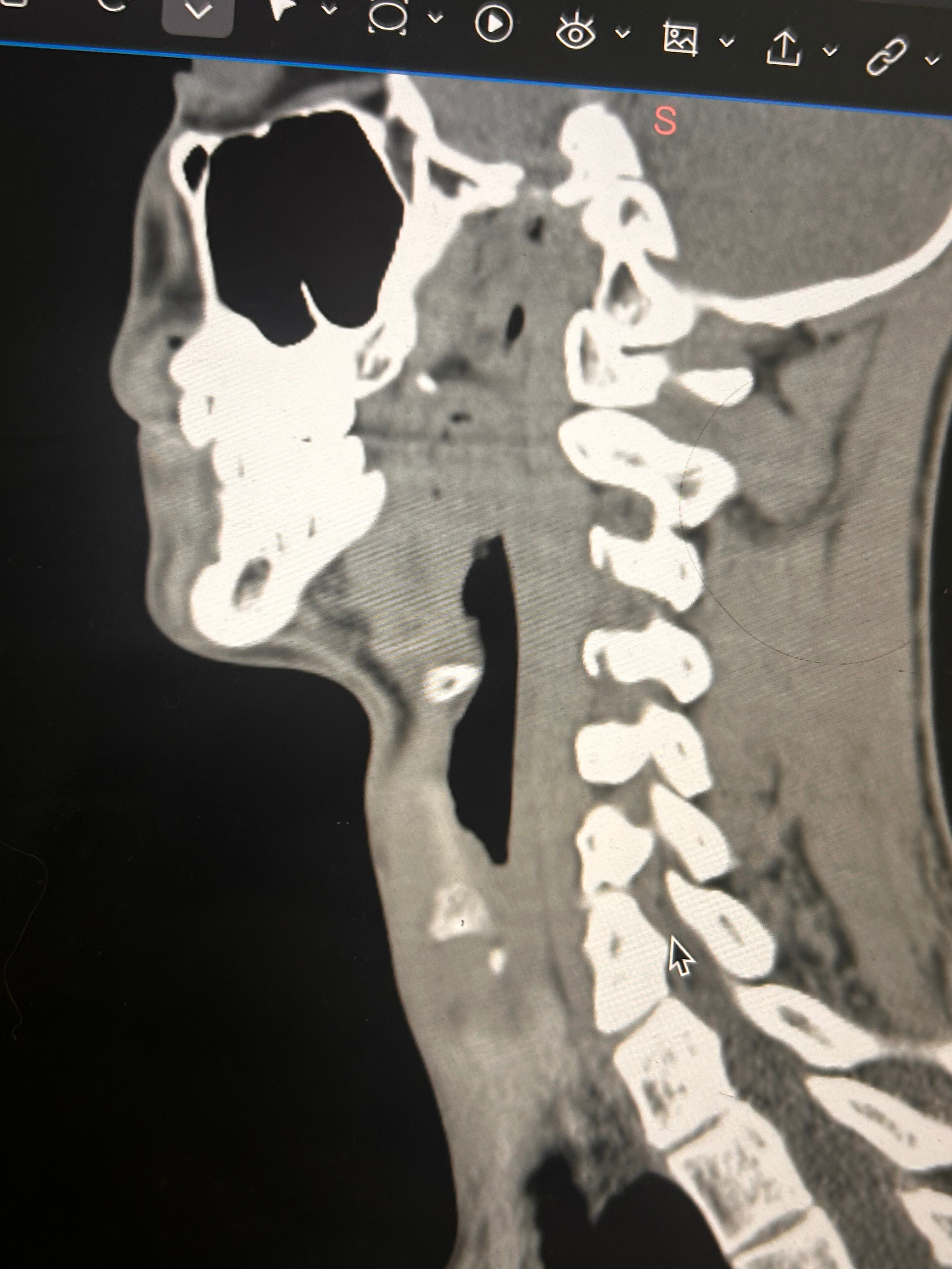
Things of note that I can see in your imaging - You’ve completely lost the natural lordotic curve in your cervical spine. That alone can bring the styloids & hyoid bone into closer proximity to nerves & vascular tissues in the neck though it has been speculated that the neck curve is lost when there’s IJV compression due to ES as the body’s way of trying to create more space for the IJV so it can drain the deoxygenated blood from the brain more efficiently. The lordotic curve can be restored w/ gentle exercises but it takes patience & is a slow process. The following link has helpful information about cervical curve restoration.


In this image of your left styloid, it appears from the angle of the picture that your left styloid is extremely close to the left transverse process of C1 (green circled area). If that’s truly the case, your left IJV is likely being squashed between the two & that means your vagus nerve is most likely being squashed at that point, too. Some of your symptoms could be vagal related. Also due to the angle of the image, it appears the greater horn of your hyoid could be very close to your spine which can cause internal/external carotid artery compression but can also cause symptoms that point to hyoid bone syndrome as you’ve suggested. Unfortunately, it doesn’t appear the CT scan was done w/ contrast as none of your veins or arteries appear in the images you posted & contrast is necessary to see those.
I could give you more precise information if your styloids, hyoid & C1 were shown in the sagittal plane (from the side) vs. from behind as they are. Below is a picture showing the anatomical planes of the human body:

Can I just ask when the images were taken, was it before your surgery with Dr Osborne? And which side styloidectomy did you have?
I’ll just add in as well that in your first image, the hyoid bone processes look pretty thick, more than you’d expect, so could well be part of your issues, but I do agree with @Isaiah_40_31 that your left styloid is very close to the C1 process, the right side looks a little longer than average… Also looking at the 1st & last images (I’m sorry, | can’t label them for you), there’s calcification below your hyoid which I’m presuming is the thyroid cartilage, it lookd pretty chunky compared to most people’s, so this could possibly be causing some issues too?
Yes these are from before my first surgery with Dr. Osborne and I had my right styloidectomy. I have also wondered about the cartilage. Last year I saw a couple of orthopedic doctors, one of whom suggested that I look into the cartilage because of how large it was and how it might be affecting my military neck and my ability to extend my neck.
Thank you so much I really appreciate you labeling things and being as detailed as you are. I think that you could definitely be onto something. I feel as though I have gotten better feedback from you and Jules than I have from my visits to Kaiser. I’m going to try to get a couple images from the Sagittal plane for you to look at. I have other symptoms that I haven’t mentioned just because there are so many like eye and tongue tension, really tense muscles from the top of my head to my shoulders. I also notice especially at night while I am lying down it seems to aggravate whatever is going on, and that is when I most notice the visual disturbances. I also contstantly feel the need to swallow, and sometimes I feel a choking sensation. Thanks again!
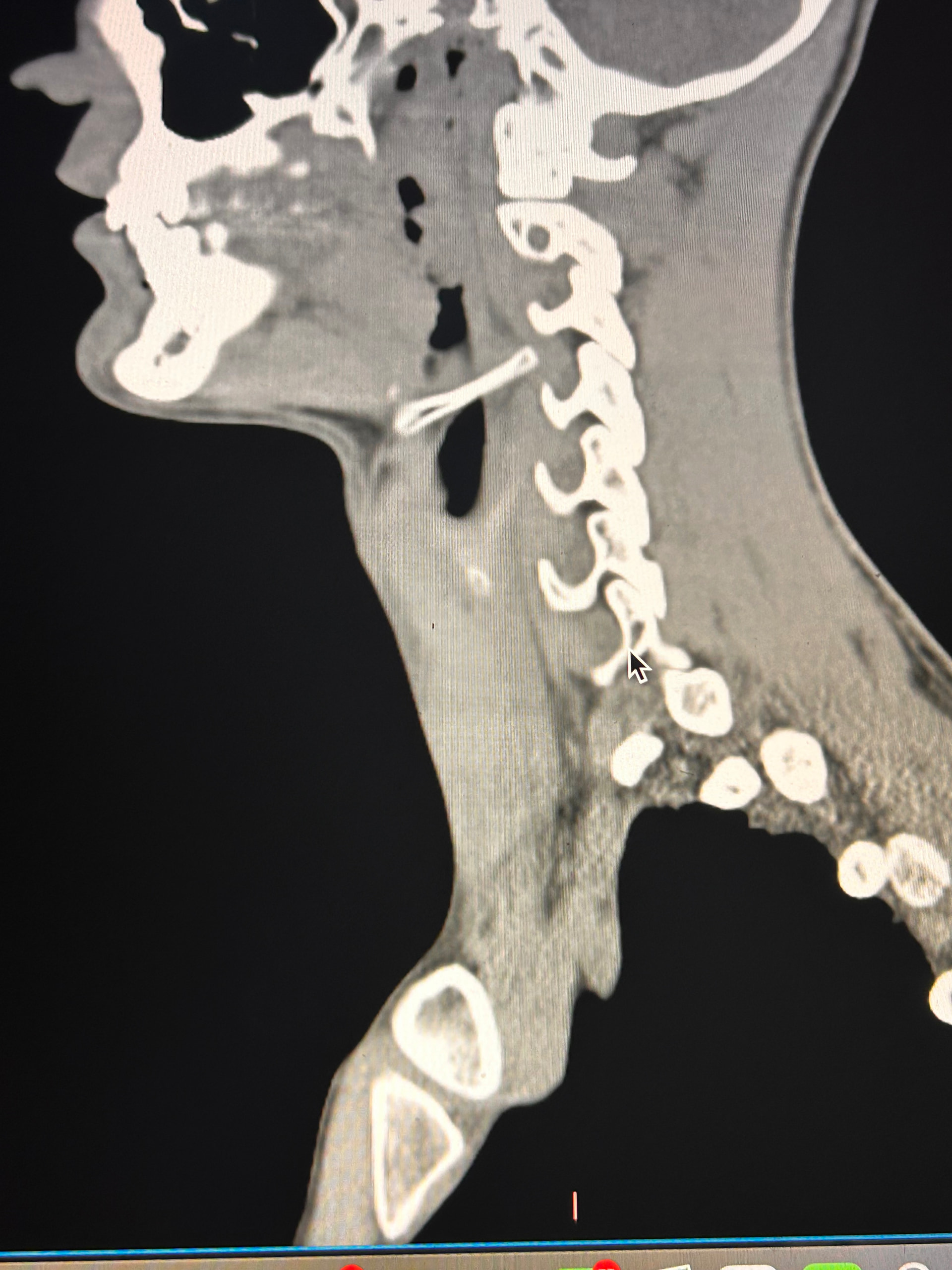

Here are some of my 2-d scans, thanks. The top one is my right greater horn and the bottom is the left
Thank you for the additional images. After reading @Jules observations, I went back over the first 4 images you sent & realized I’d miss some significant things. I’ve annotated more images for you below:

This one shows your right hyoid greater horn from behind.

I totally missed that I could see your whole hyoid bone from the front in this image. Jules is right, your greater horns are very thick from top to bottom. You can also see that your left greater horn is pretty straight, but the right one has an upward curve to it. I’m not sure what the calcification is below your hyoid but it’s most likely thyroid since the thyroid sits just below the hyoid in the neck.

In this image, you can see the differences in the shapes of your hyoid’s greater horns. The right one is more curved but the left one is thicker in width medial to lateral. In this image from the front it looks like both greater horns may be contacting your cervical spine in certain head positions, but again, that may an illusion created by the angle of the image.

In the final two images you sent, the right greater horn is very close to your cervical spine so there’s a good chance it’s causing you some problems as well as possible carotid artery irritation or compression in different head positions. The image of the left one doesn’t show as much of the greater horn so I can’t tell how close to the spine it is. Can you back the left image out a bit so it looks more like the right one?
Do you have worse symptoms on your right side in spite of the styloidectomy?
I’m sorry to tell you this, but Kaiser, especially in SoCal, has been totally worthless in helping our members who have Kaiser insurance w/ ES. A few have been able to appeal to see Dr. Jian at the Sacramento Kaiser, but it required much jumping through hoops for them to get there.
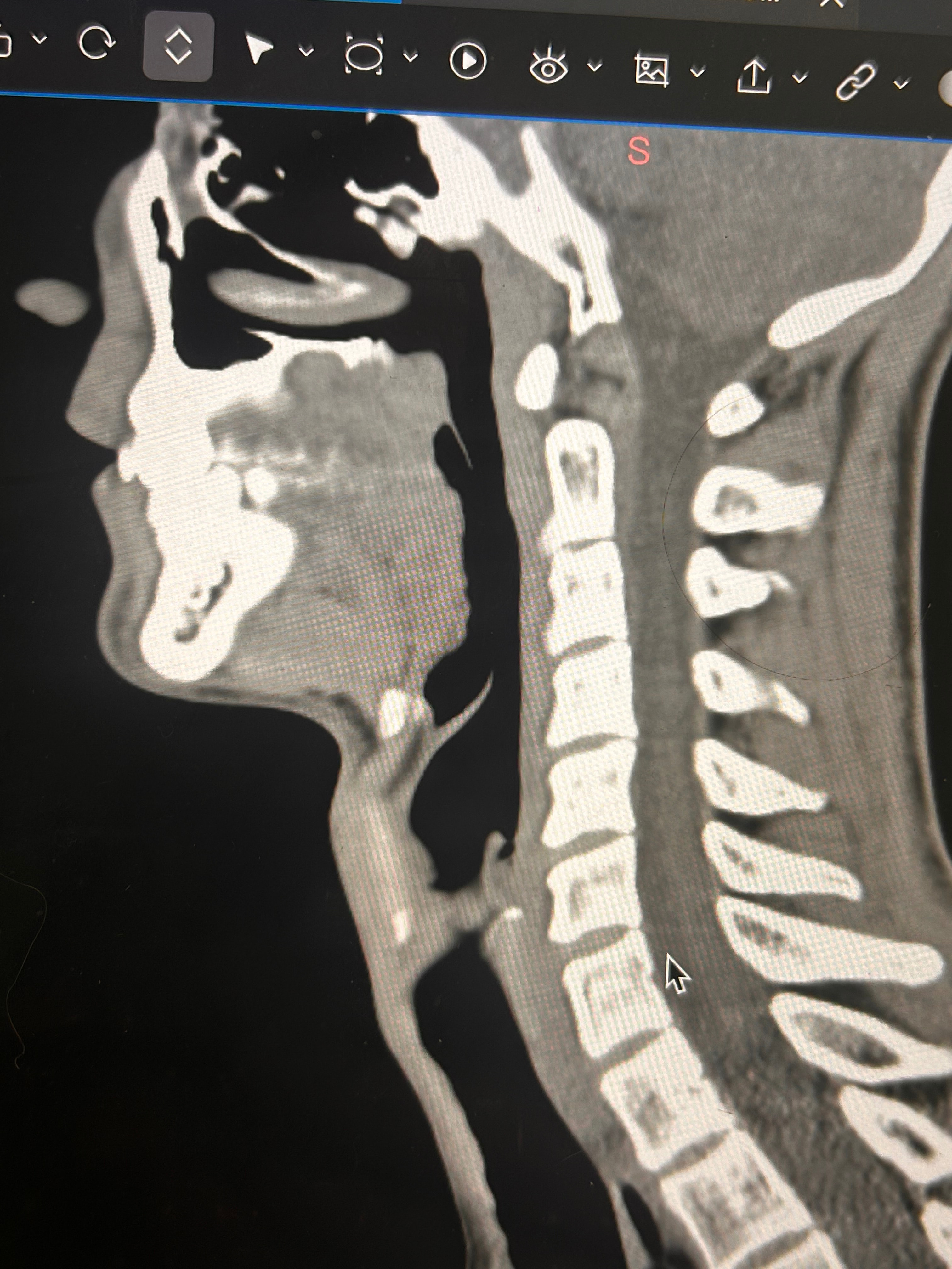
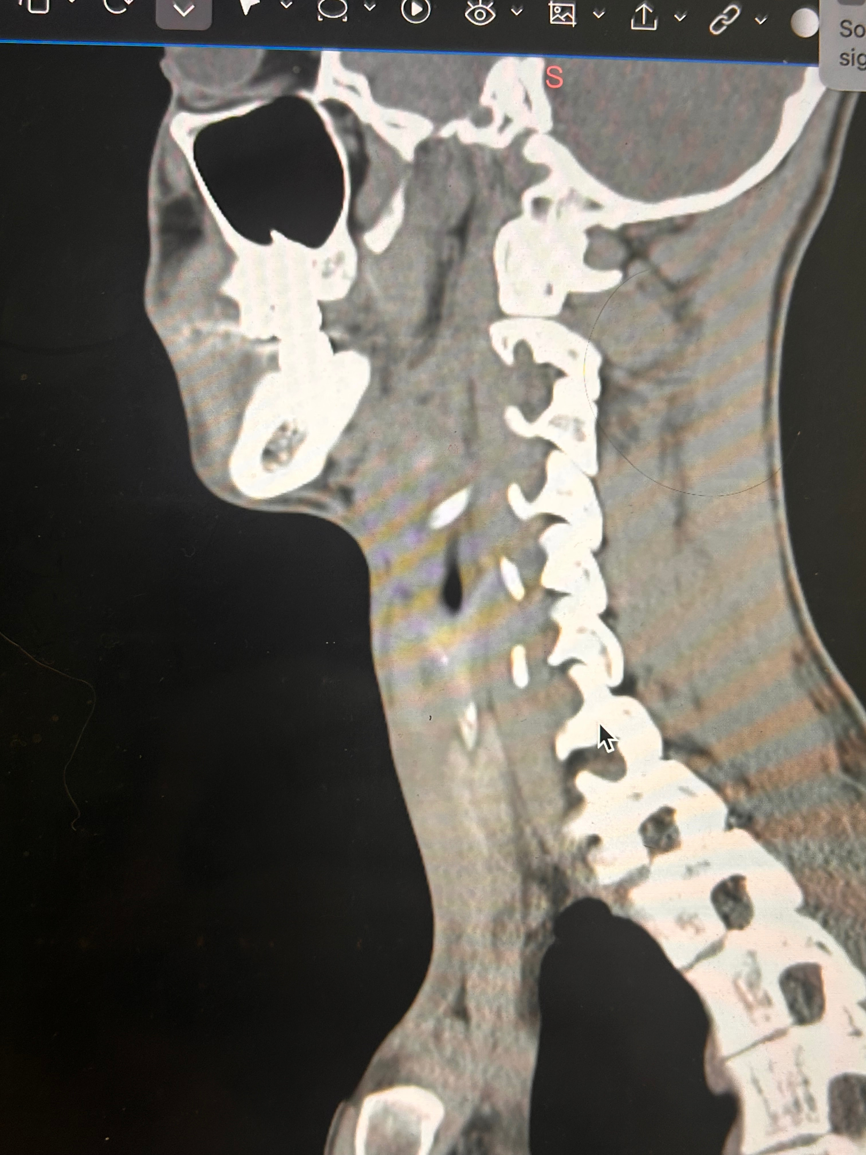


I was trying to get an image of the whole greater horn on the left but I could only get small glimpses of it as I went through the slides. Maybe because of its shape. As far as pain on the right that is where I usually notice issues, especially the lump I feel whenever I swallow, although the left can also get very tight and painful at times.
Yes, it doesn’t show well on those images…