Imaging on Dicom. Would love any thoughts

Hi all, I have been looking at my imaging on Radiant and finally got it all loaded to Dicom. I have been seen by Dr Hepworth and he sees bilateral compression at C1 and styloid along with stenosed Right Transverse sinus. The radiology report reads though that there is also compression at T1ish on the Right side and someone very helpfully called out that it looks like the carotid sheath might be too small? and compression of carotid at C2?
If anyone would like/be willing to have a look at my imaging I would be greatly appreciative. I have a virtual apt with Dr H this week (on the 19th) and want to ask him any questions that might help. I want to be sure that we know of every possible issue before making a final decision on what to tacle first if that makes sense… (for example, someone said that the bulging disc in my neck looks like it might be impacting the spinal cord…?)
So here we are:

My CTA/V: https://dicomlibrary.com?study=1.3.6.1.4.1.44316.6.102.1.2023091405417618.397024556928338295155

And MRIs (there are a few all done in a very short period as my neuro asked for additional imaging):

Brain and Cervical: https://dicomlibrary.com?study=1.3.6.1.4.1.44316.6.102.1.2023091323941923.986719746311488772051

Head and Skull base: https://dicomlibrary.com?study=1.3.6.1.4.1.44316.6.102.1.202309132001868.4625217053213954095821

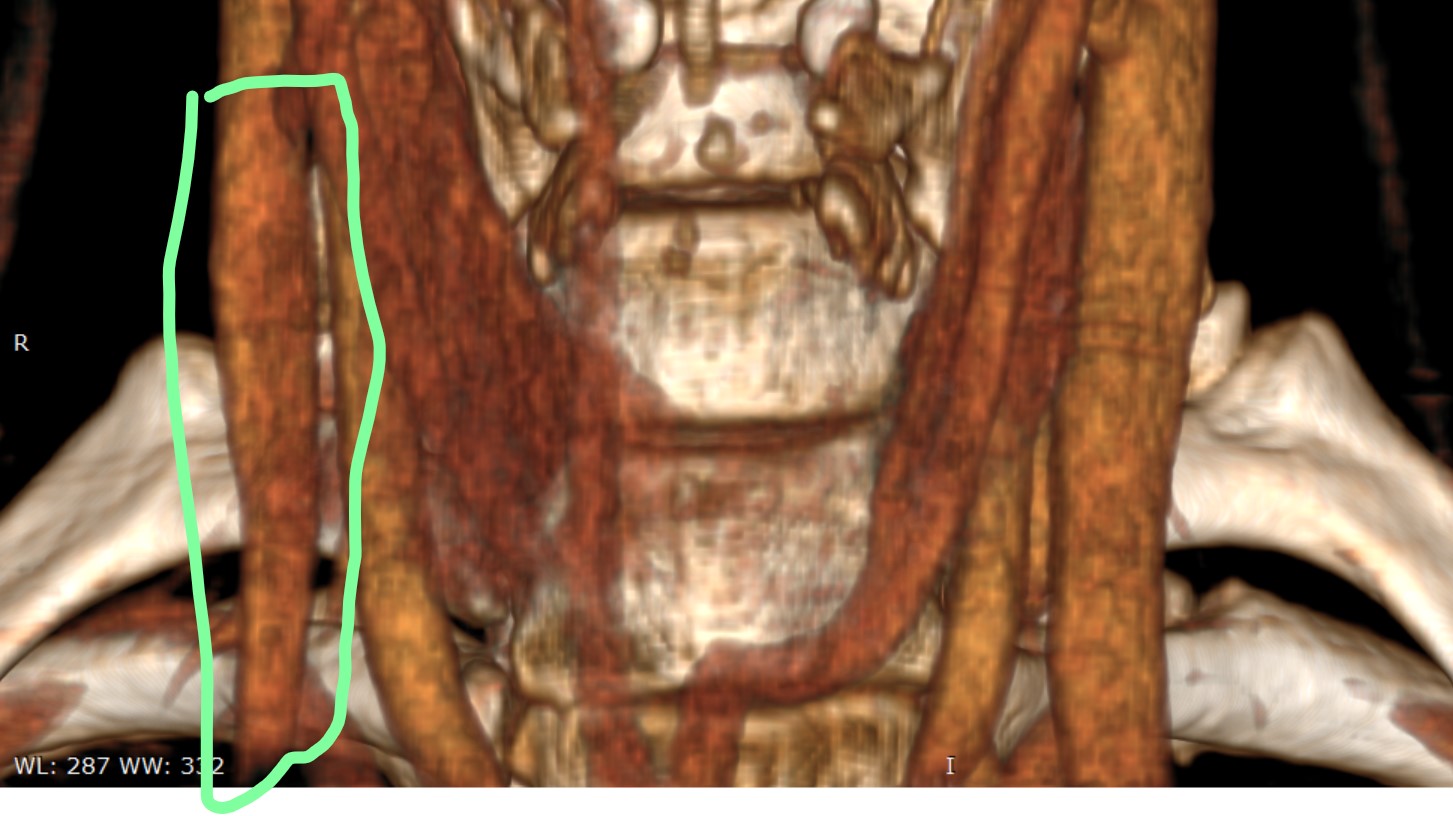
Specific to look at Jugulars: https://dicomlibrary.com?study=1.3.6.1.4.1.44316.6.102.1.2023091318421350.442605827735541973519

Thank you so much for any thoughts! We will be driving the next couple of days with large pockets of no connectivity at all, but I will check in whenever I can :slight_smile:

2 Likes

There are others that can delve much better than me into this but something I noticed is that your deep cervical veins are extremely pronounced on your CTV. Likely due to your stenosis at C1. Was this done supine or were you standing?


1 Like

Thank you very much for looking and for saying that. If it’s the same thing, I did forget to add that the radiologist mentioned large condylar and suboccipital veins. Do you think that that’s what they are?

The imaging was all taken supine.

Wow… Is that large almost horse shoe thing your arrow is pointing to in the second photo a vein???

Yes, that is a vein. Usually those are more pronounced when you’re standing as the jugulars naturally collapse, but yours are very large supine. I’m sure others can chime in on their thoughts but it’s something that stood out to me.

1 Like

Wow! I don’t know how they should look but am guessing not like that. And then to your point, they would get larger upright… They may indeed be contributing to back of neck pain and dizziness…


I wanted to add a few snaggit pics from the 3D created in Radiant in case that helps anyone… Again, there is clear L side dominance (with stenosis coming from the transverse sinus on the R) but with significant pinching between styloid and C1. There is also compression lower in the R jugular that the radiologist called out but I have no idea what structures or muscles are there that could be causing it… I think I can see it where the vein slims down in the green circled area…

@akc,

I can definitely see the C-1/styloid compression on the left IJV, but I don’t see compression in the green circled area you referred to on the right IJV. Sometimes the compression lower down can be caused by the internal carotid artery, muscle or scar tissue.

I’m sorry not to be more helpful. I wish I was better at reading these scans.

You have been extremely helpful, Isaiah and I thank you ao much. You’ve pointed out things and guessed at causes where I am clueless (despite hours and hours on google trying to see and understand what “normal” looks like to compare).
So from the bottom of my heart, I thank you.

1 Like

Thank you, you’re very sweet, @akc! I aspire to @KoolDude’s level of understanding but have a long way to go to even get close!!

I will cross my fingers that @KoolDude notices this post and has the brain-body bandwidth to have a peak too then :slight_smile:

@akc Sorry I was super busy lately with school open and my kids being small still means tons of work. Also, I have to work and babysit my symptoms but I did notice that you already talked to Dr. H and he did diagnose you with bilateral compression of the Jugular veins by Styloid and C1. So I thought you have a case going on and onto the next phase for surgery. Since yours were so clear from imaging and doctor noticed it, I thought they were enough to proceed with surgery.

I did quickly take a look at your CTV images and you have a textbook Vascular Eagle Syndrome with IJV compressions by C1 and Styloid. Your left IJV is the dominant one and is severely compressed by C1 and Styloid and would need a styloid removal and shaving of the C1 to fully decompress it. The right side is smaller and connects to a smaller transverse sinus so opening it won’t drain much but you definitely need the left side operation since it is draining most of your brain fluids.

I also noticed you have bit larger Arachnoid granulations in your Transverse sinuses. You only need to worry if the left one gets too large to block the dominant left TS. You just need to monitor that for now.

Here you can see that the C1 & Styloid compression on the left IJV and C1 compression on right IJV.

Left IJV (light blue arrow) compression by C1 (blue arrow) & Styloid (red arrow).


Right IJV (light blue arrow) compression by C1 (blue arrow) & Styloid (red arrow).

Your Collaterals (red circled veins) and Suboccipital Cavernous Sinus (SCS) (light blue arrows) are dilated because of the compression of the IJVs and blood is being rerouted to them as @elijah has pointed out. Once the IJVs are decompressed they shrink back a bit in most cases


You have dominant left transverse sinus (TS) (red arrows) and smaller almost hypoplastic right transverse sinus (red arrow). You also have Arachnoid granulations in your Transverse sinuses ( blue arrows) although smaller ones are nothing to worry about, the bigger ones pose a problem of blocking the sinus. Yours are moderate in size. Since left TS is the dominant, you need to monitor it so it does not get larger to occlude the vein.


That is all I can see in your CTV. I have not looked into your MRIs. I think you need to address your left side C1 & Styloid compression (both not only styloid). Wishing you all the best.

2 Likes