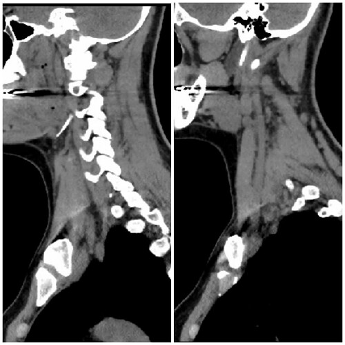
I have done ct scan without contrast. But I don’t know is there possible to see styloids…
I’ve annotated one of your images but am not sure if your right side is the left image & your left side is the right or vice versa. You have one styloid that is visible along w/ C1 but the other is hidden in the very white area near your skull base on the other side (I labeled about where it should be).
On on side (left?) it looks like your styloid may be causing some compression of your internal jugular vein. On the other side (right?), it appears either you have a significant section of calcified stylohyoid ligament or the greater horn of your hyoid is elongated. Either way, it appears you have some vascular compression
on that side as well. Usually when compression occurs down that low, it’s the internal or external carotid artery that is getting poked.
The axial image you posted is good but I can’t see much of significance because you didn’t get contrast, however, some of your larger veins/arteries appear to be visible in the sagittal images you posted.
I’m sorry this is likely not terribly helpful.
Thank you for your long description. ![]()
![]()
![]()
It is very helpful! I really hope doctors will see that my symptoms are caused because of my styloids and will fix it.
How do you learn so much?
It’s a bit strange because on your axial images, it looks like there’s a reasonable gap between the styloids & the C1 processes - I can’t label your images as @Isaiah_40_31 has I’m afraid as I’m not very tecchy, but if you look at this image:
the styloids are the white dots , so there does look a reasonable space, but you would need a CT with contrast to show any compression of blood vessels.
In this image, the styloid looks really chunky though at the top, which can cause compression:
I hope that you’re able to persuade the doctors to look into an ES diagnosis & if you could get a CT with contrast that would be really helpful- one with your head in the position which causes the worst symptoms would be great (a dynamic CT), but not many places do this…
On your other post you mentioned that you’re allergic to contrast, that’s a pain! Would that be to iodine? CTs use iodine for contrast, MRIs use gadolinium?
@Jete without contrast it’s hard to tell for sure but I’ve put red line/dots around what I think is your IJVs. Your left (right side of image) looks to be more compressed than your right.
I am alergic to many things, medication, foods, laundry detergent. Everything. ![]()
That makes sense because on angiography they say: “Well-developed right posterior communicating artery. The left posterior communicating artery is not visible.”


