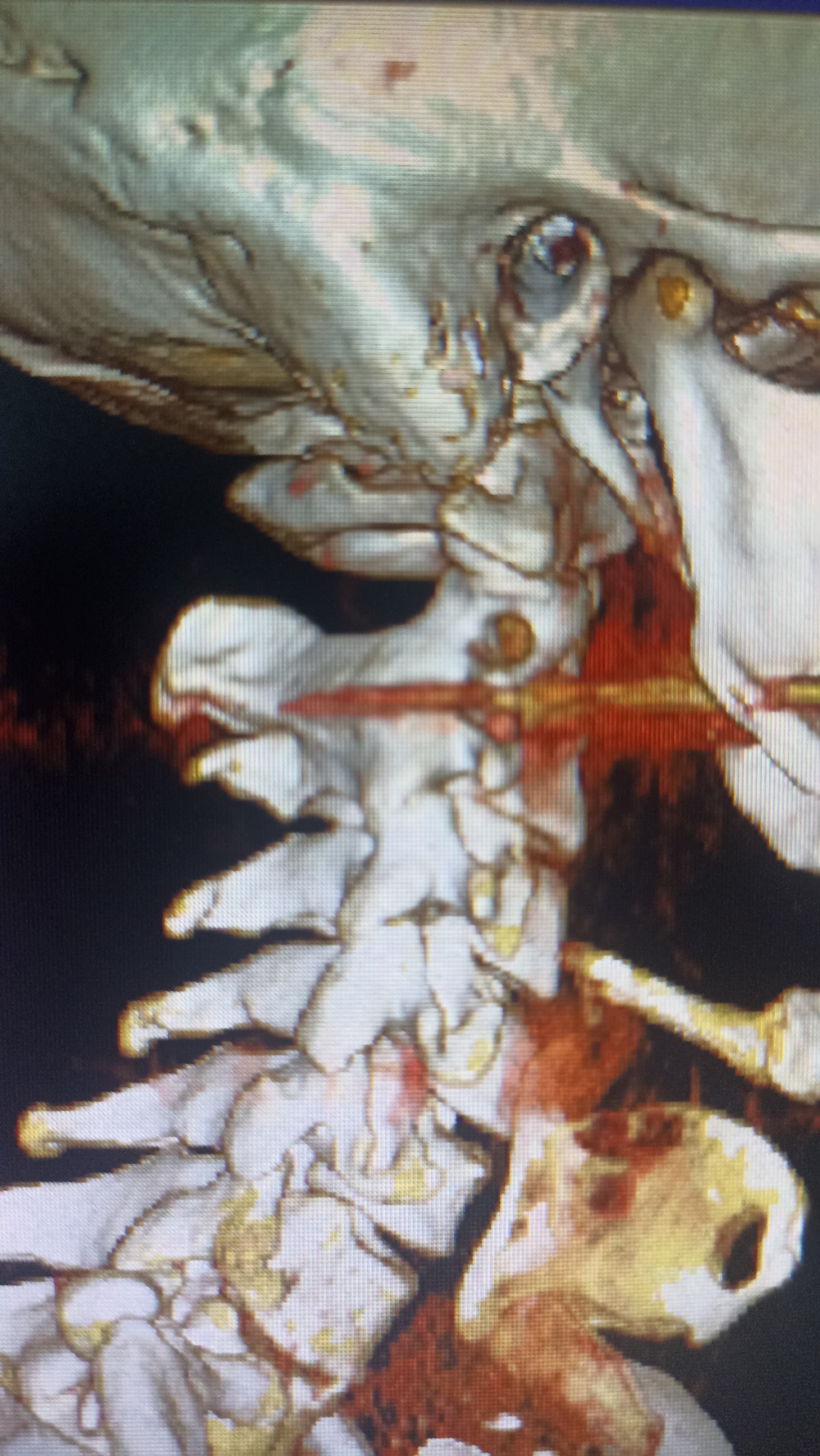
First time having a go at this and I can’t see the veins properly or ligaments one styloid looks to be at a complete different angle to the other and going behind the jaw
Can anyone help or advise me please on best way to do rhr scans or have I got the wrong scan here
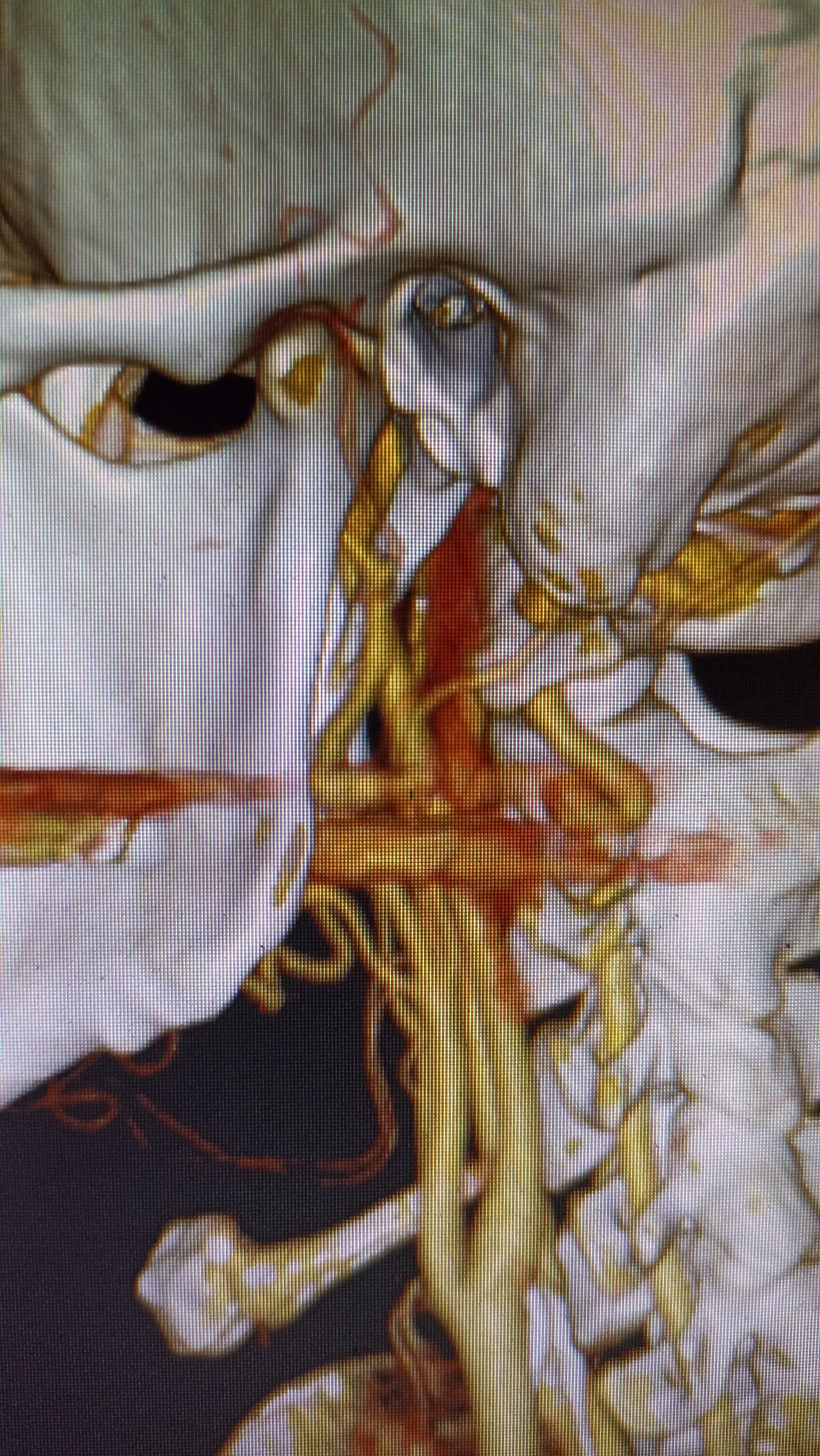
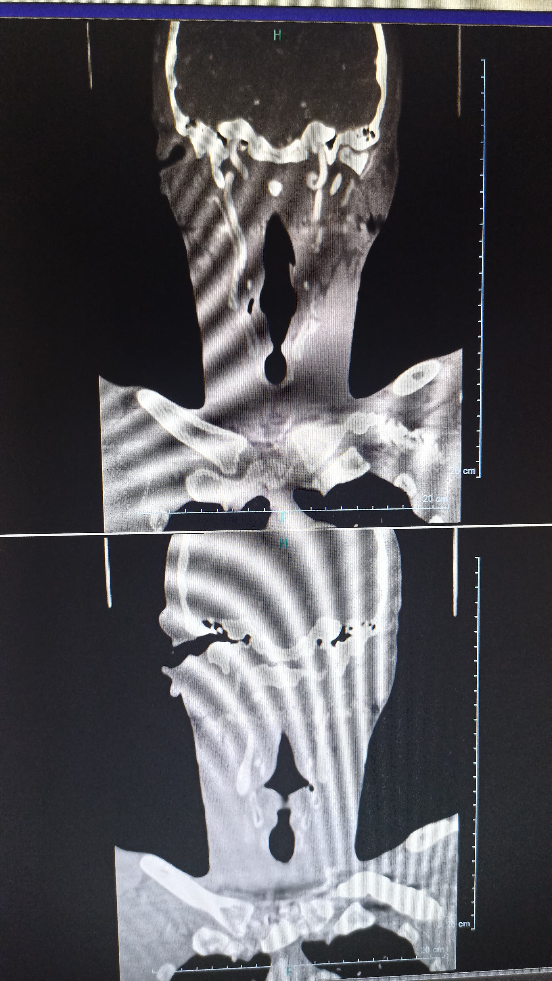
I did comment on your other post as well… you can’t see the veins at all as this is a regular CT, not the contrast one. The left side especially is very angled, they’re both quite wide at the top so could be compressing nerves at the skull base. The left side looks like there’s a decent space between the styloid & C1 space so less likely to cause IJV compression, I can’t tell with the right. You can only see the stylo-hyoid ligaments if they’re calcified, yours aren’t, so that’s good. The hyoid bone processes do look quite long though, that can sometimes cause issues.
I can’t help with your scan images but there is some good news: you appear to have no stylohyoid ligament calcification coming off your hyoid bone & your neck has a good lordotic curve (many of our member have perfectly straight cervical spines). Having that curve helps prevent shoulder & neck issues beyond what you may have now.
Regarding your styloids, both look thick at the top & very angled. The tip of the right one looks very pointed. I can’t see the tip of the left one. They don’t look super long but the angle & thickness could definitely be plying into your symptoms.
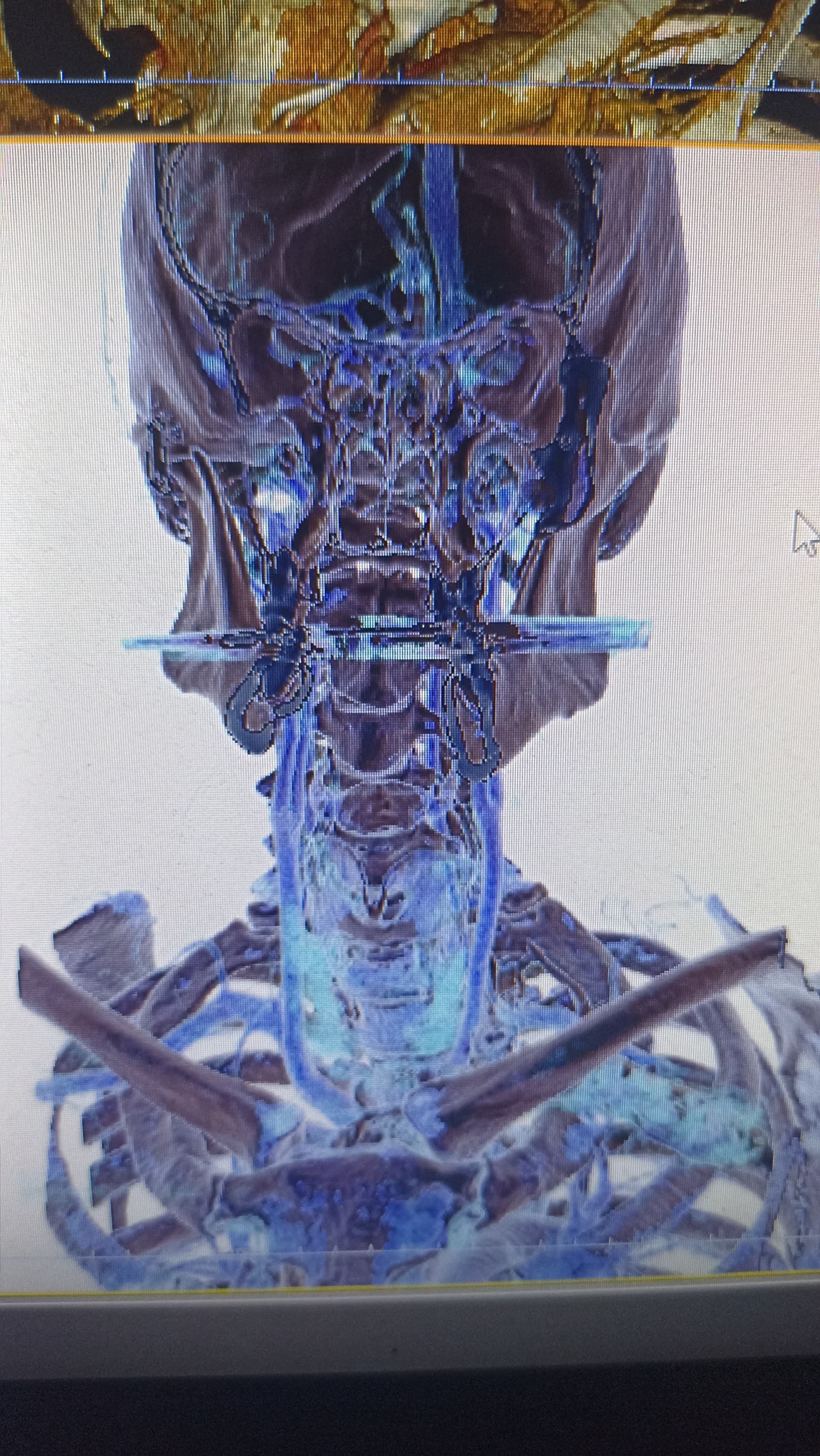
Once your veins are visible, there may be more obvious problems.
The veins are visible in your second set of images but not the first. Sorry I didn’t see the vascular images when I wrote my post.
The left IJV does not looked compressed & neither does the right but the IJV isn’t totally visible on the right so I don’t know for sure. Because of the angles of your styloids, it’s possible you have internal carotid artery (ICA) compression but I can’t see evidence of that either.
It really seems the styloid thickness & steep angle of styloid growth are most likely what the problem is. Please don’t take what I’ve said as diagnostic. It’s just my opinion.
Hopefully others with more knowledge will comment, I can’t see any obvious compression, but sometimes just the styloid touching an artery can be enough to irritate it & cause symptoms, I may be wrong but the hyoid on the left side looks quite close to the carotid bifurcation, where the carotid artery divides into internal & external arteries, this is just a guess though…
You can try @vdm, @Dobbs, @GCD, & I know there are others, but they’re escaping my mind right now. I’m leaving one person off the list as he’s recovering from surgery at the moment.