Hi, I am new here and like many, had never heard of Eagle’s Syndrome. I have been having problems for many years. In 2012 I developed GPN and it took 3 years of various specialists to get that properly diagnosed. I developed swallowing problems a couple of years ago and the last year has been nightmarish on the journey to figure out why.
I have ankylosing spondylitis and I thought maybe I had arthritis in my ear, so I didn’t have much hope. A very clever ENT figured it out and sent me to a surgeon and I was supposed to have my left styloid taken out last Wednesday but insurance approval did not happen until the day after the surgery was to take place.
Thanks to a good friend who was concerned and asked a lot of questions, I now have many questions myself before I try again.
I am grateful for this group as I have learned so much in a short amount of time. I have never seen such in depth research from so many in one place. I feel like I somehow won a lottery.
My main concern at this point is that it appears that muscles ( at least I think it is muscles) are enlarged on my left side. I ‘m figuring this would make it difficult to get to the styloid and also increase the risk as the jugular vein is right underneath. Has anyone had this experience?
@Pain-in-the-Ear sorry to hear about the insurance piece. Really frustrating.
Which muscles in particular? I’m guess the SCM? Sometimes people have to get muscles removed (i e., posterior digastric) if they are interfering with blood flow. Surgeons seem to do a decent job of avoiding muscle damage when performing styloidectomy as far as I know. Cranial nerves are the biggest worry, more so than muscles.
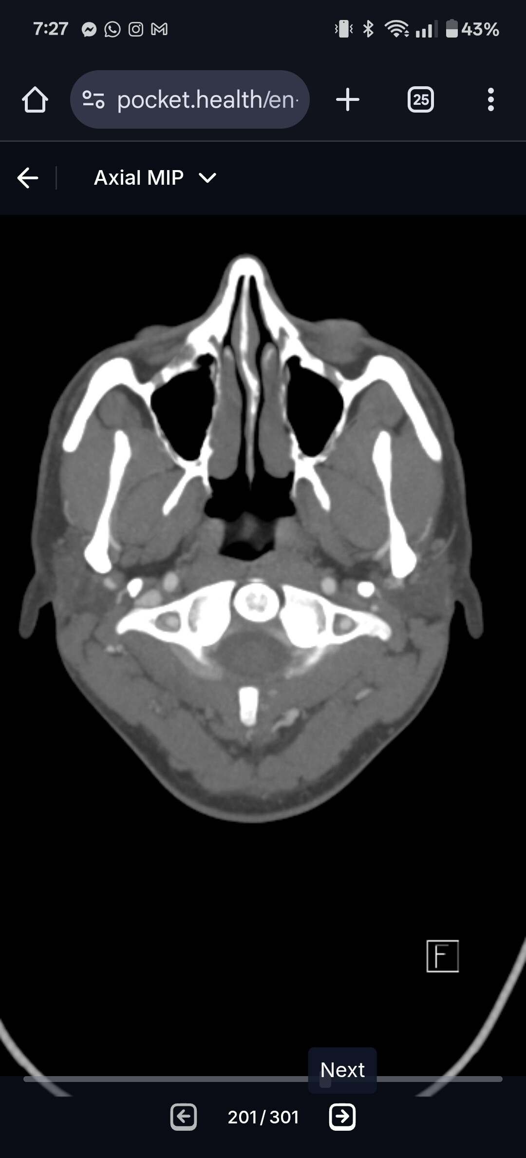
If you post some images if your CT (assuming you got one done) I can help you spot the compression points.
I find the axial view the best. If you go to C1 I can show you the landmarks. I’ve attached my own imaging to help you know where to look.
Thank you for the information. It was an x-ray I was looking at and I do believe one of the muscles was the digastric muscle. I do have several CT’s and an MRI. I will try to locate an like the one you posted .
@Pain-in-the-Ear - You can make your CT (& maybe MRI) images into 3D using either radiantviewer.com (for PCs) or Bee Dicom Viewer App (for Macs).
There are very few surgeons who actually remove muscle during surgery, however, some will “debulk” a muscle that is putting pressure on a vascular tissue such as the internal carotid artery or internal jugular vein. The digastric muscle is under your chin. The SCM is a major muscle in your neck. Both the SCM & posterior digastric muscles can be problems in cases of vascular ES though your symptoms have not sounded like you have that.
SCM:
Digastric:
We’ve had several members w/ enlarged SCMs due to irritation to the spinal accessory nerve by an elongated styloid, & none have had a problem w/ the IJV during their surgeries. Doctors who do ES surgeries on a regular basis know where the underlying nerves & blood vessels are so their incisions are made carefully & the neck or throat is opened very gradually. For the external approach (AKA transcervical), electrodes are placed on the face, shoulder, tongue, & soft palate to monitor the nerves that could be injured during surgery. These alert the surgeon when (s)he is getting to close to one of the monitored nerves so the surgical approach can be changed to preserve the nerve.
I recently was able to observe an ES surgery in person, & the carotid arteries & jugular veins are much deeper below the surface of the neck than you’d think thus the risk of injury to them is very minimal to non-existent unless decompression surgery is required. Even then the approach is very delicately handled to keep them safe.
I have absolutely no idea what I am doing. I tried to find a match on one of my scans that looked similar to your–it’s not exact, but as close as I could get. I have an appointment tomorrow with an orthodontist who is supposed to be really good at analyzing these structures and I hope to get more information.
I am really concerned myself about the nerves as that 9th cranial nerve on my left side has really caused me problems. I just don’t want to end up worse than I am, and while I desperately want relief, I am kind of paralyzed with fear of exacerbating a really bad problem.
Thank you Isaiah–it is the digastric muscle–both bellies on my left side that are enlarged. From the information you gave me, I can now see why I am having such problems with swallowing and choking.
That gives me some peace knowing that the nerves are monitored like that–that was one of my questions–thank you.
@Pain-in-the-Ear when you say “several” CTs, was there a variety of CT types? Like were they all the same method, or was one a CTA, a CTV, a CT with contrast vs without?
Just wondering because this CT image you posted it’s hard to make out the arteries and veins.
A CTA or CTV would be great. The image angle was close to perfect though!
Omgoodness. I think I somehow how two CT scans of my neck- without contrast, one mri of my neck, one of my brain, a ct of my ears, and yes, I had a CTA. I have them all on a jump drive at work, so I will find the CTA tomorrow morning.
We’ve had some discussions about enlarged muscles, so you could search the past discussions for info about that. I think Dr Costantino removes some of the digastric muscle when needed, mentioned in this discussion:
Omgoodness! Thanks! I am seeing an orthodontist today and I hope for some clarity. I have not idea why one side is so enlarged but it is the side I am having difficulties with the GPN.
Uneven elongation of the styloids is pretty standard. It’s rare that a person has equal length styloids on both sides.
Don’t be disappointed if the orthodontist isn’t very helpful or gives negative input about surgery helping you. Orthodontists usually don’t know much about ES.
Ok, my nephew and I managed to make some 3D images. I don’t know what I’m looking for exactly, but there is something definitely messed with my left side which is the side I’m having trouble with and it looks as though my styli is is broken both at the top and at the hyoid bone. Not sure, it is my best guess for not knowing what I’m doing. Any help - any input is appreciated.
@Pain-in-the-Ear - If you could also provide some sagittal plane (side profile) pictures for the images where your IJV is visible it would be helpful. Your jaw is blocking the part of the IJV & potentially carotids that we need to see as well as your hyoid bone.
Your right styloid is very long & looks to be very close to the transverse process of your C1 vertebra bone. Your left side isn’t broken. The left styloid looks to be normal length, but you have a significant chunk of stylohyoid ligament calcification between your styloid & the left lesser horn of your hyoid bone. If your nephew could include left & right sagittal images that show your styloids & hyoid bone plus the jugular veins & carotid arteries that would be great.
He did an excellent job of helping you w/ the 3D images.
@Pain-in-the-Ear - Please make sure your nephew takes your name & age off the images/link to the images before they get posted on here (forgot to mention this earlier). Our forum is visible to the public so this is necessary to protect your privacy. I’ve removed what I could easily, but there may still be some images where your full name/age are present.
I’ve annotated several pictures, but there still isn’t one that really shows what’s happening on your left side as none of the sagittal images were straight from the side so I can’t see the styloid or calcified section of ligament except in the pics which you posted earlier.
I’m still not clear about what is possibly causing your symptoms. Your right styloid is somewhat long & looks very close to C1 on the right side which could be causing IJV compression, but what I can see of your left IJV, it looks ok.
On the left the greater horn of your hyoid looks close to your external carotid artery, but I can’t tell if it’s close enough to be bothering the artery. You have military neck i.e. you’ve lost the natural lordotic curve in your cervical spine which brings the styloids & hyoid bone closer to local vascular tissues & nerves in your neck, increasing the risk of contact w/ those things when you move your head.
It would be interesting for you to have another consult w/ Dr. Hernandez to at least get a better understanding of what he sees in your CT scan which may be causing your symptoms & what his surgical approach would be in your case. Just having that information could give you some relief from worry/fear.
I’ve labeled the relevant arteries & veins + hyoid bone in this image. I noticed that your internal jugular vein may be compressed in the lower section. You can see how it kind of disappears. That could be from your SCM as you noted you had muscle inflammation. If it’s more your digastric, that would explain your swallowing problems.
I’m sorry I can’t be more helpful & don’t want you to draw any serious conclusions from what I’ve pointed out because I’m not a doctor & am truly unclear in your case what the contributors to your symptoms could be apart from your elongated right styloid & the calcified section of your s-h ligament on the left.
Hi - thank you so much for taking the time to mark my images and give me your thoughts. I am just not tech savvy, but trying to learn, thank you also for trying to protect my privacy.
I am just trying to gather as much information as I can so I can ask the questions that I need to.
I’m kind of stuck in a purple haze with no clear direction.
You asked about my symptoms- I may have things that are symptoms that I am not aware of because I didn’t connect them to the awful pain in my ear. Like I park my car and I feel like the car is still moving. That might be one, but I’ve never associated with the pain. In fact, I didn’t associate the swallowing and choking issues with the pain, because I’ve had the pain for so long. Or maybe my left eye spontaneously watering the past year- that might be a symptom too- I just don’t know.
I am going to keep pushing for more information.
What scares me more than anything is I went to have my wisdom teeth removed and ended up in severe pain and damage that has lasted a lifetime. I don’t want to jump into something and end up worse than I am, because this is pretty bad That is my fear.
Nevertheless, I now have a bit of hope too and I’m very grateful for all that I have learned.
@Pain-in-the-Ear when you have the feeling you are still moving after stopping a motion event like driving a car, getting off a boat, or even taking an elevator, it can be similar to mal de débarquement syndrome. I struggle with the same symptom.
A recent research paper showed that IJV compression in the neck can cause it, and that decompression surgery such as a styloidectomy can treat it.