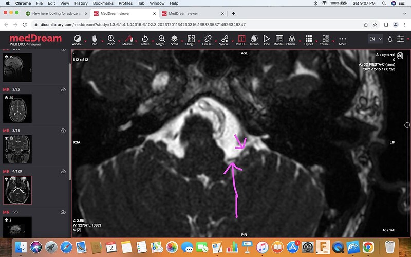
So the MRI you shared with us initially is called a FIESTA MRI. It is a hi-resolution scan that is ideal for evaluating the cranial nerves. So my only concern is what I have identified as a Brainstem Compression is wrong and perhaps a radiologist needs to evaluate the cranial nerves too; as to not put all our eggs in one basket. I think its up to you, but you could call them and see if you can re-upload your MRI, or you could just go with the one you have uploaded as it will show the vertebral artery/ brainstem concern most likely.
It seems really odd that a brainstem compression would have been missed on 3 different studies. I’m really doubting my ability to read scans. Again, I could be leading you down the wrong path. I really hope you won’t miss this cash you are spending too much…
It does seem odd if it’s been missed on several scans but highly plausible imo because I’m living with this I know something is wrong.
I think initially they were looking for tumors or cancer’s and possibly other things. It wasn’t until recently the vascular possibilities became apparent and the d-dimer test was done and came back positive.
Hoping it will show the possible compression like the other did. If not I will have to see what the report says and then decide if it’s worth getting the Fiesta MRI reported on or not?
This MRI certainly demonstrates our brainstem concerns. So if they have seen your questions regarding the vertebral arteries and brainstems you should at least get some answers.
Oh that’s good to hear, does it show any nerve compression on this scan?
This is the vestibular compression but as you can see the nerve it isn’t as clear or precise as on the FIESTA. Perhaps they will note the artery’s proximity to the vestibular nerve because you asked. But again, this is a common finding so often they don’t even comment on it. If you google “vestibular paroxysmia” you can read up on it. But this wouldn’t cause d-dimer results I don’t think.
Thanks for confirming. Yes that’s definitely causing some symptoms the nerve compression which funnily enough came around excactly the same time as the other symptoms.
Been having a look at a few medical articles and particularly scans showing brainstem compression and comparing some of the images you highlighted and my scans and its pretty clear this is happening on my scans unfortunately. Now pair that with my crazy list of horrible symptoms and then compare my symptoms to the symptoms of brainstem compression and it just cements it even more.
I’m now thinking really how has this been missed on several occasions and on several different scans and it’s quite worrying. Could there be any possible explanation or reason for this or is it just really bad luck on several occasions, would there be any reason for someone to see this but not put it in the report.
The only reasons I can come up with for the brain stem compression being missed is that 1) The radiologists weren’t looking for it so it was overlooked, 2) whoever read your scan deemed something else to be more of a problem so the brain stem compression took a back seat & wasn’t mentioned. Regardless, if it had been observed, it should have been noted.
I found some articles today suggesting brain stem compressions due to vertebral arteries are sometimes incidental findings and don’t cause symptoms!!! But that wouldn’t be for the radiologist to decide. Their job is to report on the findings and consider your symptoms, but ultimately you need to be diagnosed in a clinical setting. But, of course, yourself and a Neurologist need to know about this, otherwise its hopeless.
I don’t know if you have ever seen your radiology reports but I would start calling the doctors that ordered these scans and ask them to email you a copy of the report. Or call the institutions that performed these scans and ask them for the report. I have a feeling a radiologist would have commented on this but the report wasn’t sent, or it was put in your file without anyone reading it.
The other possibility is I’m completely wrong and what we are looking at is normal…
One more item which might explain throat pain:
The big arrow is pointing to your PICA artery in both images.
The small arrow is pointing to your “glossopharyngeal bundle”. So much like the vestibular nerve, you might have a neurovascular compression of the glossopharyngeal nerve. Glossopharyngeal nerve pain is very specific, so I’m not sure if it applies to you… And again, arteries and nerves are often seen in close proximity so this isn’t a diagnosis, but if you have the sort of pain that matches the criteria then perhaps it could be considered.
I think all my symptoms are due to compression of veins on certain things but why and what’s happened or happening to cause it?
I have so many more symptoms I haven’t mentioned that I wasn’t sure if I was going mad or not and how they were all possibly related because I have a list as long as your arm since all this started. I honestly thought I was dying when this suddenly hit me one day and I couldn’t stand or keep my balance or even talk properly or understand words correctly for a brief period. Assumed a stroke and so did AnE but I’m assuming no obvious signs on the CT scan they did at the time.
Walk with a little limp sometimes, and it feels like I’m walking drunk and can’t walk properly at times
Burning in top of left thigh randomly happens and stays for a long time
Hip pain
Leg pain
Problems speaking at times if I’ve done too much or talked for a bit
Facial and eye pain and burning of face sensation
Struggle to push to go to the toilet like I used to be able too and I get a build of pressure in head or neck if I try and push to hard. Had all this checked as they assumed prostrate and physically it looks okay
High heart rates since this all started too
Terrible Neck pain and don’t like moving my neck as this makes my symptoms much much worse and triggers head pressure that is indescribable this can completely debilitate me
Ent have confirmed hearing loss which it isn’t because it’s the brain noise is so loud I can’t hear over it sometimes
As I understand it only in the UK radiographers and sonographers are allowed to report on scans but this will need clarifying.
I have spent £££ of my own savings on scans and docs appointments and haven’t been able to work for two years but I cannot live like this.
I came on here looking for help because I’m absolutely desperate now and losing hope.
Some of your symptoms do seem similar to when I had bilateral jugular compression- unsteady walking, feeling drunk, head pressure…when you push to go to the loo that will increase head pressure, that was one of the times for me which would set off pulsatile tinnitus…Voice/ speaking issues can be common with ES too, & if the vagus nerve is irritated it can cause heart arrythmia. Face & eye pain or burning can be due to irritation by the trigeminal nerve or facial nerve. Hearing loss seems to be quite common with members too.
I can’t comment on the other issues that @boogs99 has been helping you with!
I will repeat what I’ve said many times - it’s not just the length of the styloids that needs to be taken into consideration. The thickness, how curved & direction of curve, twisted or pointed they are also play into what types of symptoms they can cause EVEN WHEN NOT elongated. Too many doctors look for length alone to define ES. It should be defined by symptoms with the physical features of the styloids being the parameters that are measured not just the length. These physical features also contribute to the possibility of vascular compression. Early on we determined that your styloids look both thick & fairly steeply angled inward. These two features alone are enough to cause many of the symptoms you’re experiencing.
Speaking from experience, the burning in your left thigh, leg & hip pain are most likely coming from your lumbar plexus (spinal nerves) & may only be related due to changes in your posture because of the pain in your neck area.
Hi there, I was wandering if you would be so kind as to ask some of your forum experts to look at some more scans I have obtained. The only thing is these scans are of the chest and abdomen and there is some possible anomalies but I’m not 100% sure what I’m looking at.
Thanks
Matt
I certainly wouldn’t have a clue about abdo scans! Not sure if any of our other members would, none of us are medically qualified though…We have had a few members with Thoracic Outlet Syndrome, also May Thurner Syndrome and Nutcracker Syndrome which are compressions of other veins, might be worth looking into if you haven’t heard of those? Is that what you were looking for with your scans?
Hi Jules
Thanks anything would be good and I think it’s possibly vascular anomalies I’m seeing which I think would look the same or similar to vascular issues in the skull region.


