New here looking for advice and opinions

Anyway here is the links to the scans just thought I noticed some very obvious bulging vein areas

https://www.dicomlibrary.com?study=1.3.6.1.4.1.44316.6.102.1.20240329142155226.12454968986199449076

https://www.dicomlibrary.com?requestType=WADO&studyUID=1.3.6.1.4.1.44316.6.102.1.20240329142155226.12454968986199449076&manage=0377170b638f41e125c00a5ebf0b2053&token=9109b3a1c6a92238eaf67592f5e8190b92e5a2f919410d0af9

CT with Contrast upper body
https://www.dicomlibrary.com?study=1.3.6.1.4.1.44316.6.102.3.202403092640804.6643496183113760012151

1 Like

Hi @Sprinter also in UK. Have been quiet on the forum for a bit as been mega busy. Did you ever get any venous phase CT scan? Could help you rule out vascular eagles, or venous thoracic outlet syndrome.

2 Likes

I’ve been missing you, @LimeZest. Was going to check in shortly. Busy is good as that indicates you aren’t down & out. Any updates regarding doctor’s appointment or other relevant ES information?

1 Like

Unfortunately, I’m also in @Jules’ situation. I can ID some things in the neck/skull areas but know nothing in the chest or abdomen. Hopefully, if there are members on our forum who are able to help, they will speak up.

1 Like

Thanks @Isaiah_40_31 will update everyone soon. Nothing hugely special to update but will do so anyway. Just been mega busy with work and life (and was ill with a terrible viral infection for basically all of february).

1 Like

Rough that you’ve been ill, I hope that you’re over it now :hugs:

Yes doing much better now, it was the most ill i’ve been since i had COVID. Alongside the other health issues i just dont recover as quickly.

1 Like

@Sprinter i’ve been looking through your scans. Before i post anything could you summarise your symptoms? I’ve found something quite interesting in them but want to be sure its real as i think you have some artefacts on your scan that line up in the position im looking.

One question i would like to know as well - does turning your head hurt a lot? Do you have quite a short neck and do you think your head is tilted at all to one side?

1 Like

All of this is really similar to my symptoms, I’ve had all of your symptoms since Nov 2021, on top of those I had a sensation of getting too much air through my nose / not being able to feel the air properly through my nose. I’ve now got squeezing on the sides of my head and my head never feels properly attached, it clicks loads and hurts. Over the last couple of years I’ve tried not to worry about it all so much, I’ve not been able to properly exercise because I’ll regret it the day after (I used to run and so loads of exercise classes) but there was a point where everyone just kept saying anxiety and stress and I ended up just getting on with life as terrifying as it was and did paddleboarding, swimming, ice skating. Unfortunately I still have many symptoms, I’m always tensing parts of my body up, it never relaxes as much as I try, I’ve always got something tensed up.

In your report it says mucosal thickening of the ethmoidal and maxillary sinuses, I also have this apparently. I don’t know if that is any significance. I feel like if I could breathe properly this all might go away.

1 Like

Turning my head does hurt and is sort of stopped or restricted by something so I avoid if I can. Neck doesn’t look tilted to look at but feels it could be inside if you get what I mean.

There are definitely lots of artefacts and oddities in my scan all missed by original radiologists but I’m trying to figure out what is exactly going on and what’s causing all the issues because getting help seems impossible without that.

1 Like

Hi all, so @Sprinter and I have been messaging and i have come up with some possible things for him to pursue in terms of further investigation. But with his permission i thought it would be useful to get some feedback, particularly from the people here who have experience reading scans @vdm @KoolDude @Chan.

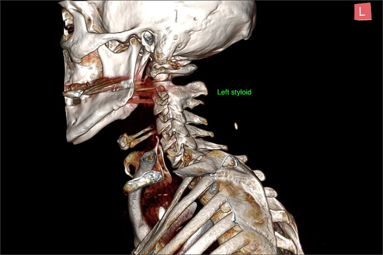
Right so first lets go through the possible eagle stuff. Yes styloids are reasonably long, not huge but quick thick and pointy. They extend below C1 tubercle so therefore have possibility to impinge IJV


In axial view you can see reasonably large interval between styloid and atlas, and you can make out IJVs as this is a venogram (though not the best one i’ve seen). So i really dont think styloids are causing compression.

In sagittal view (side on), i think this is more apparent with some rounding of the IJV over the C1 tubercle.


So for the eagle stuff - i am not convinced styloids are compressing IJV but of course they could still be irritating nerves in the area.

But while studying the scans i came across something that i think is more likely causing symptoms, but could be wrong. Immediately i noticed there is quite a large scoliosis of the spine, having discussed this with @Sprinter he says he doesnt think this is coming from head position during the scan.

After a bit more looking around - his cervical spine just didnt look right which is when i noticed possibly quite an obvious malformation. His C2/3 look completely fused! Now unfortunately this does land almost exactly where the CT artefacts exist due to metal implants in the teeth - but the entire spinous process for C3 looks very small also possible suggesting congenital malformation. But the scan defects here could throw this hypothesis into doubt.


When looking from the front (and chopping part of his head off), you can see there is no disc between C2 and C3. I dont know the cause for sure, but there is a congenital malformation called Klippel Feil syndrome - causing most commonly fusion of C2/3 or C5/6. it does tend to come with a particular appearance like short neck etc. Its not something that can be acquired, but plausible he has had it asymptomatically until a head injury. Or inflammatory response/calcification etc has caused this.

There was also evidence of calcification around where the atlas meets the dens.

Looking at the sagittal view of individual slices these things are still visible.

Now looking at the MRIs you can see a similar disruption of the C2/3 disc space, its not completely calcified here but the MRI was taken many months before the CT. I also did some measurements for craniocervical instability, as my hypothesis is that the fusion of C2/3 has cause adjacent segment disease of C1/2 as is common in Klippel Feil syndrome. This would cause instability in the area, possibly evidence by the calcification around the dens.

Back to to the measurements, i’ve seen people measure the clivoaxial angle (which basically shows how the head is sitting on the neck) in different ways. Either they measure the dura (which i dont like) or they measure dens and clivus (which in my view is correct). Either way, these values should be >150 degrees, but more recent literature is saying that anything over 135 can in principle be normal. These images are very narrow however, both below 125 degrees. Possibly suggesting mechanical damage of the brainstem? But i did notice the clivus itself seems to be malformed.


On the MRI the atlantodental interval looks quite large, it isnt nearly this big on the CT but just pointing it out anyway.

Another common measurement - Grabb-Oakes line is way above the normal 9 mm cut off suggesting the cranium has translated forward over the cervical spine. Other notable things were possibly quite low lying cerebellar tonsils, suggesting a possible chiari malformation? Even in supine they do seem to extend just below the foramen magnum, so could be worse in an upright position.

Lastly the coronal (from the back) MRI view also shows a similar loss of the C2/3 disc to the CT. It also does somewhat show the head tilt and scoliosis from the CT scan also.

So to summarise, i think there could be some serious issues here. I think one of the best doctors to see in the UK about this would be Mr Jake Timothy at Leeds Nuffield to try to get a formal diagnosis, or potentially Dr Iain Smith at Newport Chiropractic.

As always - disclaimer, im not a medical doctor or radiographer and this is not in any shape a diagnosis just someone who is interested to help.
5 Likes

@LimeZest I agree with all your findings in general. The C2-C3 fusion might be congenital, or arthropathy/trauma-induced. Needs to be compared with the images immediately after trauma (if available).
Interestingly, I have facet joint arthropathy at C2/C3. Even started thinking, in my case it might be mild C3 irritation causing a lot of issues (C3 is quite a complex root, including involvement in diaphragm movement as far as I remember).

In @Sprinter’s case I’d think the possible CCI/Chiari/perhaps some spinal cord adhesion pulling down the brain is more likely culprit.

3 Likes

It’s a privilege to (try to) serve :sweat:

Based on @CJsBattle’s and @boogs99 's earlier findings on ‘arterial loops’, I thought I’d see if there’s such a thing as “carotid artery tortuosity” through my search bar. The first hit was the radiopaedia.org page for carotid artery tortuosity:

Classification and symptomatology notwithstanding, pathogenesis is stated to be unknown according to a paper from 2019. There is a connective tissue disease association in the title of that paper.

There was a research paper posted on this forum referencing the recent association of such with Eagle’s. I thought it significant when I saw it on my RSS feed as well, given some coverage in mainstream publications on some degree of relevance of connective tissue disorders to long COVID. I might be remembering incorrectly on that last bit, though.

The KFS finding, which I’ve yet to work through, is quite fascinating. Indeed, as @vdm asserts, I’d wonder at the outset how to rule out arthropathy/trauma-induced. Not sure if @LimeZest’s work above does that implicitly. No comment atm on CCI/Chiari/adhesions :hushed:. I’ve downloaded the first three of the five DICOM studies attached. I will work through them tomorrow morning.

Hope that’s something to go on in the interim.

2 Likes

Thanks you @LimeZest for taking the time to help @Sprinter , way above my knowledge but great to give pointers as to what else to be looking into…thanks as always @vdm for helping too, & to @Chan also. You guys are great :smiley:

1 Like

@vdm yes C3 nerve is certainly involved in diaphragm movement hence can impact breathing. This would of course lead to other muscles having to take the strain of this movement if they can.

@Sprinter has since sent me a second opinion report on an old MRI (c.a 2 years old) so probably close to the injury time. That mentions congenital fusion of C2/3 so I think probably this is more likely than trauma.

1 Like

I’m in awe of what you guys spot in people’s scans & know beyond just seeing the visuals. As @Jules noted, it’s way above my knowledge, but I’m super thankful to have this level of “medical” competence here when we often aren’t finding it in the “true” medical community. Between you, @LimeZest, @vdm, @Chan, @KoolDude, @boogs99, @CJsBattle & others who haven’t been on here as recently, our site has provided information that some of our members might never receive from medical consults & which is allowing them to get the care they need for more complete recovery.

THANK YOU!! from the bottom of my heart, for the time you give to helping your fellow members. :gift_heart: :hugs:

3 Likes

Thank you for saying this @Isaiah_40_31. Of course as you say none of us here are medically trained, but personally i dont think radiography actually requires it, i’ll probably go out on a limb and say it could be a hinderance. Yes you need to know anatomy, but to some degree being able to spot patterns is the most important factor. Another thing which is worth pointing out is that we see lots of unusual scans on this forum, so we look into every detail, but most radiographers see predominantly normal scans, so they have a bias into thinking that the next scan could be completely normal, but for us its the reverse.

2 Likes

I totally agree. Book learning cannot take the place of actual experience & taking interest in the minute details of a given case in order to learn as much as possible & so be able to apply that knowledge going forward. That’s where I see you all excelling.

1 Like

Just to confirm all scans were done after I had a wack on the neck that occurred in November 2021 and I got progressively worse after this quite seriously unwell immediately just after the incident but again I did start having some issues leading up to that but they were manageable so it raises some confusion and speculation regarding the injury. Possibly I had something going on and all it took was a hit on the neck and back area to make things drastically worse and life changing?

I have to thank everyone so far with all my heart as if it wasn’t you for you guys and this forum and everyone that has contributed I wouldn’t have a clue what’s going on and what’s happened and currently still happening to me right now. I can’t show my gratitude enough and I’m truly thankful.

Going forward I’m booking a upright MRI scan and it seems it’s going to be very tricky to get any help with this as the NHS and doctors just don’t want to know.

1 Like

Hi @LimeZest I think between you, @boogs99 and @CJsBattle covered this one so well. I am not sure I can add more value here. What you found coupled with possible Brainstem compression that @boogs99 found could be contributing to his symptoms. I would suggest to seek second opinion on his imaging by providing the radiologist with your findings.

1 Like