New here looking for advice and opinions

@vdm @LimeZest @CJsBattle @boogs99 @KoolDude

Hey everyone, is it just me or does the March 2023 MRI (post-contrast) show right internal jugular vein occlusion? Could this somehow be just artifactual?

Here’s a video of me scrolling through the series while waving the mouse cursor around.

I think the CT venography also shows worse IJV stenosis on the right side than left so would certainly fit with that.

4 Likes

Comparing with the November CTV, it almost seems as though the left side has expanded slightly since March to compensate…

Interesting! I havent got copies of my own scans, but my left IJV has done something similar to compensate for stenosis on the right.

3 Likes

I agree with @LimeZest. Interesting for both of you that you’ve noted some enlargement of your less compressed side over time. It seems collateral veins are usually the body’s answer to stenosed IJVs but certainly extra blood flow through one side would demand a larger vein area if possible so pressure in the vein is reduced.

1 Like

It looks less passable then the left one, but I’m not sure about the cause. In my opinion, the IJVs are so easily compressible that different head position, or muscle tension possibly might cause the compression, temporary or permanent.

1 Like

I looked a bit into the bilateral looping/coiling of the extracranial internal carotid arteries well-visible in both the CT and MRI from 2023. At the outset, I sought to find some big-picture information, so I searched Google Scholar with the keywords “carotid artery tortuosity narrative review”. I downloaded five relevant-sounding papers from just the first page, and worked through the first from last year in India by an anatomist.

Review Papers on ICA Looping

2023 India
Morphological Variations of Cervical Internal Carotid Artery and associated Clinical Significance: Narrative Review
The paper with my highlights (952.7 KB)

2019 Europe
Arterial tortuosity: Novel implications for an old phenotype

2017 China
Current understanding of dolichoarteriopathies of the internal carotid artery: a review

2014 Americas
Clinical implications of internal carotid artery tortuosity, kinking and coiling: a systematic review

2007 Brazil
Tortuosity of the internal carotid artery cervical course: case reports and literature review

Some worthwhile details to mention to a doctor, but nothing quite rising to the level of definitive answers.

For instance:

[Internal carotid artery looping] changes hemodynamic [sic] and blood flow to a greater extent than tortuosity. Coiling of the ICA may reduce the lumen of this artery culminating into turbulent blood flow causing umpteen number of neurological manifestations such as strokes, transient ischemic attacks and amaurosis fugax due to cerebrovascular insufficiency. Cerebral emboli and blockage of artery are usual findings due to tortuosity of the ICA leading to cerebral ischemia.

Or:

[Internal carotid artery looping] may squeeze the pharyngeal wall producing obstructive sleep apnea. Abnormalities in the ICA may create problem [sic] during swallowing and speech and also produce feeling of foreign body in the pharynx. In addition to this, [the loops] also cause oropharyngeal pulsatile mass, longstanding hoarseness and upper respiratory distress.

2 Likes

Hi @Chan , I did take a look at the MRI video and compared it with the CTV video. First, I am not a doctor and although it is hard to conclude from 2 videos as to what is really going, my findings are based on what I see in these 2 videos.

Findings :

  1. Based on these 2 videos, my conclusion is what you see as occlusion is an artifact of the T1 MRI. In T1 MRI, arteries and veins are indistinguishable from signal perspective since it shows the same signal for both of them. This is because, it relies on the volume of blood that is following in them which is about the same. So in this case, the occipital artery moves on top of the compressed but patent right IJV and their signals merge appearing as one and giving the impression that the right IjV has disappeared. The CTV, on the other hand, contains more slices and shows the compression of the IJV by the Styloid to be severe. It does not show any occlusion of the right IJV. CTV is done on venous phase, as result, veins appear more prominent than arteries. It also relies both the contrast and the volume of the blood that is flowing in the veins hence can accurately depict any occlusion as flow void. See the screenshots below for details.

  2. I found what appears to be moderate sized arachnoid granulation in your right transverse sinus. I suggest you keep an on it so if it gets larger, it can potentially occlude the TS.

  3. I found what appears to be the markers for IIH (raised ICP). I found Optic Nerve Sheath Dilation (ONSD) and Meckel Cave dilation. They can be better viewed in T2 MRI sequences as the CSF fluid is bright as opposed to dark in T1. These are both established markers for IIH as well as partial empty Sella which I think you found your self.

Hope these adds to your list of evidence to show that you indeed have the tale-tale signs of IIH, possibly induced by IJV compressions.

Here i see that the IJVs (green arrows) and right one is severely compressed by the Styloid(yellow arrows) mainly and to a lesser degree the C1(blue arrows - though small space appears b/w them) and you can see the occipital artery (red arrow) moving towards the right IJV. Reason I want to show the occipital artery is because it will move on top of the right IJV in the next images merging their signal and would be virtually indistinguishable

Here is the occipital artery (red arrow) just before it moves on top of the right IJV (green arrow). You can see the IJV is patent (open).

image

Here the occipital artery (red arrow) is on top of ( superimposed) the right IJV. The right IJV might be there its signal merged with the artery signal. That is an MRI artifact since the arteries and veins are isointense (same intensity or signal) in T1 MRI. Because they contain roughly similar amount of blood, it would be very hard distinguish based on their signal in T1 MRI.

Here is the CTV version

Here is CTV showing the worst compression of the right IJV(green arrow) by mainly Styloid (yellow arrow) and by the C1(blue arrow) to a mild degree. You can also see the faint signal of the occipital artery (red arrow). It is this artery that is more visible in the MRI when it moves across the IJV they have the same signal intensity, they will be indistinguishable. The CTV is done on venous phase, so the arteries won’t be prominent as the veins unlike the MRI where arteries and veins are Isointense (same signal) so when one transposes onto other, you will only see that. I would argue that the CTV is more accurate since it has more slices (299 slices) compared to the MRI. So since the CTV shows severe compression but not occlusion I do not believe it exists and it is mere MRI artifact.

Here I see Optic Nerve Sheath Dilation (Yellow arrows). Since this is T1, the CSF fluid is dark and you can see the dark lines (CSF fluid) that runs along with the optic nerve. This is sign of raised Inter-cranial Hypertension. It is best seen on T2 MRI as it will show the CSF fluid (the dark lines ) as bright white.

Here I see a moderate size arachnoid granulation (yellow arrow) on the right transverse sinus of questionable significance. You might need to monitor if it grows bigger, it does pose a risky of contributing to increased pressure in the brain.

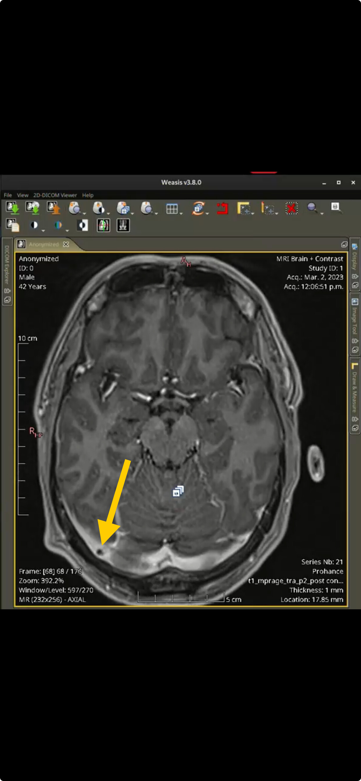
Here is another II marker called enlarged Meckel Cave (yellow arrows). Since this is T1 MRI, CSF fluid will be dark and you can see that they are both enlarged but more pronounced on the right side perhaps coinciding with severity of the compression on that side. Indentation and Transverse Diameter (crossectional diameter) of the Meckel Cave: are considered imaging markers to diagnose Idiopathic Intracranial Hypertension as found on multiple studies.

Source Study : Indentation and Transverse Diameter of the Meckel Cave: Imaging Markers to Diagnose Idiopathic Intracranial Hypertension | American Journal of Neuroradiology

3 Likes

One thing I came to believe is that jugular compression between the C1 lateral mass and the styloid process is actually very both complex and complicated phenomena.

The skull base is supposed to be able to flex forward and backwards on top of the C1, about 30 degrees. Not slide or glide with the point between C1 lat.mass and the SP remaining in stationary, but rotate like on a horizontal hinge.

According to some sources the C0/C1 joint is responsible for as much as 20-50% of the whole neck flexion/extension ROM.

Which means, the distance between the SP which is attached to the skull and C1 lat mass is supposed to vary, leading to dynamic, changing compression levels on the structures in-between, therefore the IJV too.

So, it seems some of the roads lead to impacted biomechanics possibly caused by muscular spasms.

Now whether it’s a self-reinforcing mechanism (impacted SP/SH cause muscle spasms, causing further immobility) is up to debate, at least in my case it was obvious that long SPs/ossified ligaments were causing a lot of resistance (and pain) whenever I moved them outside of their “settled/preferred” position, leading to malignant movement patterns. Removing the styloids helped to break this cycle.

For further information, check this one:

So my conclusion is, the whole concept of the IJV compression between the C1 and SP as a static constant thing might be:
a) accurate in case there is a pathology causing C0/C1 immobility (congenital fusion, arcuate foramen etc), or the angle of SP at “normal” or even “extended” position is so bad that it causes the IJV compression
b) accurate in case it may be caused by stiff and shortened deep neck flexors and overstretched/weak neck extensors, or any other combination of agonist and antagonist muscle imbalance causing improper neck movement
c) inaccurate in cases the muscle spasm is due to the postural or other reasons, and surgical resection doesn’t guarantee any results unless the postural and muscular issues are addressed either before or after the surgery.

Upd:

This one shows how much (approximately) the SP is supposed to move away from the C1 during extension.

And this is the whole cervical spine in action during flexion/extension

6 Likes

@vdm all of what you said are valid. There is big biomechanic component to this disease.

2 Likes

I really appreciate all you guys taking the time and looking at my scans and finding all this. It’s alot to take in and I don’t quite know what to make of it all.

The jugular vein compression does make complete sense to me because of my debilitating symptoms as well as the other issues found as I have terrible head neck pressure and vision issues. What does confuse things for me is that some days I can fell not too bad and others I can be feeling very stroke like is the only way I can describe it, and I’m very unwell and this can coincide with any upper body movements that tense the neck and move the shoulders area as well as neck movements, for example sorting through a few boxes and bending down a bit recently set me off really bad and I couldn’t walk properly or think clearly and the only way to describe it is like I was being taken out the room and couldn’t control my body properly and everything is failing. Other days I can wake up and I’m very bad from the get go then some days I get a fairly ok day with less symptoms affecting me for no reason I can conclude yet.

I guess what questions I have now are for anyone that can help advise is

Where or who do I need to see about all of this and can anything be done to help me. I’ve been struggling so far and have no help despite seeking it any way I can.

Radiologists don’t seem to find anything and the doctors don’t look at my scans they just rely on the radiologist reports put Infront of them.

I have an appointment coming up on the 22nd April at adenbrokes with a Neurosurgeon that I have had really to push for as they even initially turned down my referral stating they had seen my scans and an appointment with them wasn’t needed. It wasn’t until I called them and complained a bit and asked what scans they had excactly seen and who had seen them and also asked if its not a Neurosurgeon I need to see then who is it I need to see, I also quoted a letter written by a general vascular surgeon explaining he advised I need to see a Neurosurgeon that they reinstated a new appointment. So in light of their reluctance to see me initially I am expecting this to be a tough consultation with minds already made up about me without knowing all the facts. So I don’t hold out to much hope.

Again thanks for everyone you’ve been invaluable so far, and I can at least now understand why and what’s happening to me and it makes so much sense where as I was completely without a clue what was happening to me for so long despite all my efforts with doctors.

Any advice on what main points I need try to say and show and get across at my appointment will be appreciated there is alot of information on the thread and many findings and I will no doubt have limited time at my consultation.

1 Like

Wait… @KoolDude, @vdm, I think you guys are mixing up my own 2021 CT-E with @Sprinter’s 2023 CTV!

@Sprinter’s IJV’s aren’t quite as compressed, which is a bit confounding given the IIH markers. Here’s a video I made of @Sprinter’s November-2023 CTV:

Otherwise, wIthout checking the measurements for the IIH markers, I generally found the optic nerves, Meckel’s cave, and sella turcica worth commenting on, as @KoolDude does. The thing about the arachnoid granulations, I don’t even know to look for.

So @vdm, @KoolDude, would you say that the March-2023 MRA shows more right IJV stenosis than the November-2023 CTV? In which case, wouldn’t this be cause for getting one of those dynamic angiogram things?

@Chan My last comments were about this:

And later about @KoolDude’s highlights:

I was referring the CT on the posting below. I did assume it was CTV ( CT Venography) since it clearly showed the venous systems well. If it was CTA ( CT Angiography), then it must have been done in both phases ( venous & arterial) since IJVs and collaterals (which are veins) are clearly seen. Typically, CTA will show arteries clearer than veins.

Well, it is bit difficulty to glean evidence from a video but if your thick black lines along the optical nerve is all CSF, then there is no doubt that the Optic Nerve Sheath is dilated although it is bit difficulty to visualize in T1.

For Meckel Cave, according to the paper I posted "The mean diameters of the Meckel cave on the coronal T2 plane in patients with idiopathic intracranial hypertension were 5.21 ± 1.22 mm on the left side and 5.16 ± 0.90 mm on the right side ". That means the diameter could be as low as 4.26 mm and as high as 6.06 mm on the right side, my guess is, that your right Meckel Cave falls in that range with my visual estimation because it is difficulty to measure it on a video.

T2/Fiesta sequences with more slices than the traditional 27 slice, is good for measuring them.

1 Like

As @vdm is keen to emphasize, some of the relevant structures are very mobile relative to each other. For myself, most of my problems manifest when I sleep, which is when I do not consciously control my neck posture.

Your styloid process does seem awfully close to both the internal jugular veins and the internal carotid arteries, and any bony compression of these structures can, generally speaking, seem to do the sort of things you describe as symptoms. I don’t know that there’s been any explanation in this thread for the IIH markers otherwise. (That feature with the brainstem/pons and the basilar/cerebellar artery perhaps could be viewed as yet another IIH marker, as an intrusion of CSF space into the brainstem.)

Interestingly, the 2023 India paper on ICA tortuosity does mention that “one study relates presence of [variant morphological forms such as coiling, looping, kinking, and tortuosity] to short neck” and this would seem to bring into consideration the findings that @LimeZest mentioned with Klippel-Feil Syndrome.

As @LimeZest offered above, I would perhaps look to set an appointment with this Mr. Jake TImothy at Leeds Nuffield or Dr. Iain Smith at Newport Chiropractic, perhaps they’ve encountered this situation before with the ICA looping. I’d also look to get one of those dynamic angiogram things, or at least ask about it and whether it would be of some diagnostic value in this situation.

If angiogram in certain neck positions shows that either/both the IJVs or ICAs are being impinged on, then I would think surgical options would present themselves accordingly. Or atleast what not to do with your neck. It’d obviously be best if you could find someone to quarterback all this, rather than having to have to fight your way through it all.

I’d hope that others with more experience and knowledge with dynamic angiograms could chime in?

2 Likes

Hi guys apologies I haven’t been on for a while just been very busy for me and with that feeling poorly most of the time because of that. I will hopefully get a around to catching up with the thread over the next few days.

I have just had an new MRI scan done today and wanted to share the images maybe these might help clear up or confirm some of the issues already found and discussed or maybe not?

I haven’t had a chance to look at them yet. There was some Brain, Cervical Spine and neck movement scans taken.

https://www.dicomlibrary.com?study=1.3.6.1.4.1.44316.6.102.1.20240501172312476.56035202466134575026

https://www.dicomlibrary.com?requestType=WADO&studyUID=1.3.6.1.4.1.44316.6.102.1.20240501172312476.56035202466134575026&manage=e2ad6a6f424cde967b55c9cb869368cd&token=2109570791835c757641d90b484dd26447a87871438daa13eb

Hopefully I have uploaded them all correctly if there seems to be any missing please let me know

2 Likes

Did you get a report back? I would say you have some brainstem compression and your c2/c3 looks almost fused together. I’m no expert though.

1 Like