This is my story: June 2024 i had a whiplash accident where i overextended. I had some pain at the base of the skull but not that severely, had some CCI symptoms, i kept on going till the point where i got severe insomnia several weeks later. I am currently sleeping 3 hours a night structuraly with sleeping pills for 1 year now. Got tinitis especially when there is pressure on my neck (laying down). My neck is getting weaker in time and symptoms increase. I suspect that the compression of the IJV are the main cause of the sleeping problem. Or this could be because of compression of nervus vagus.
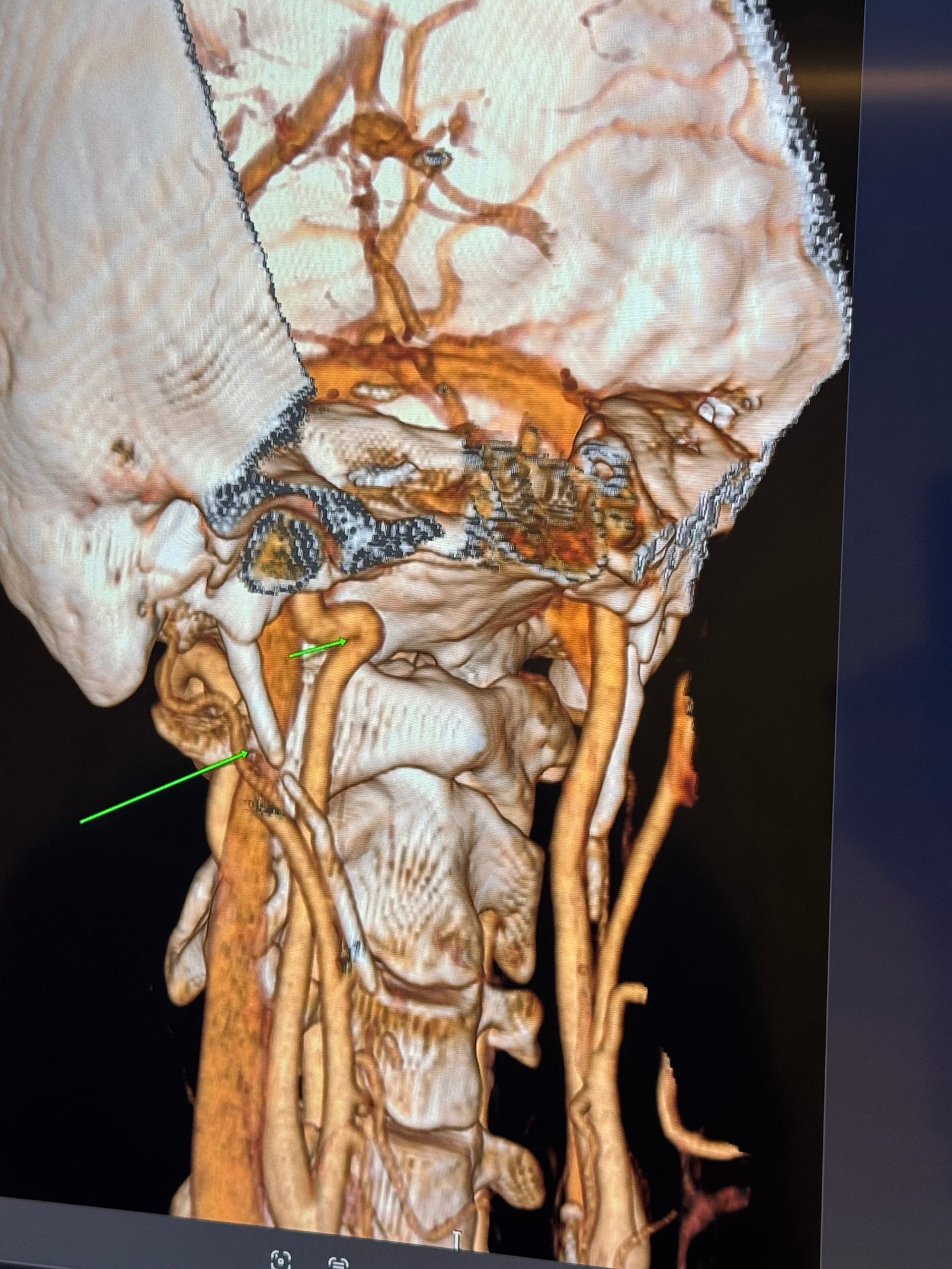
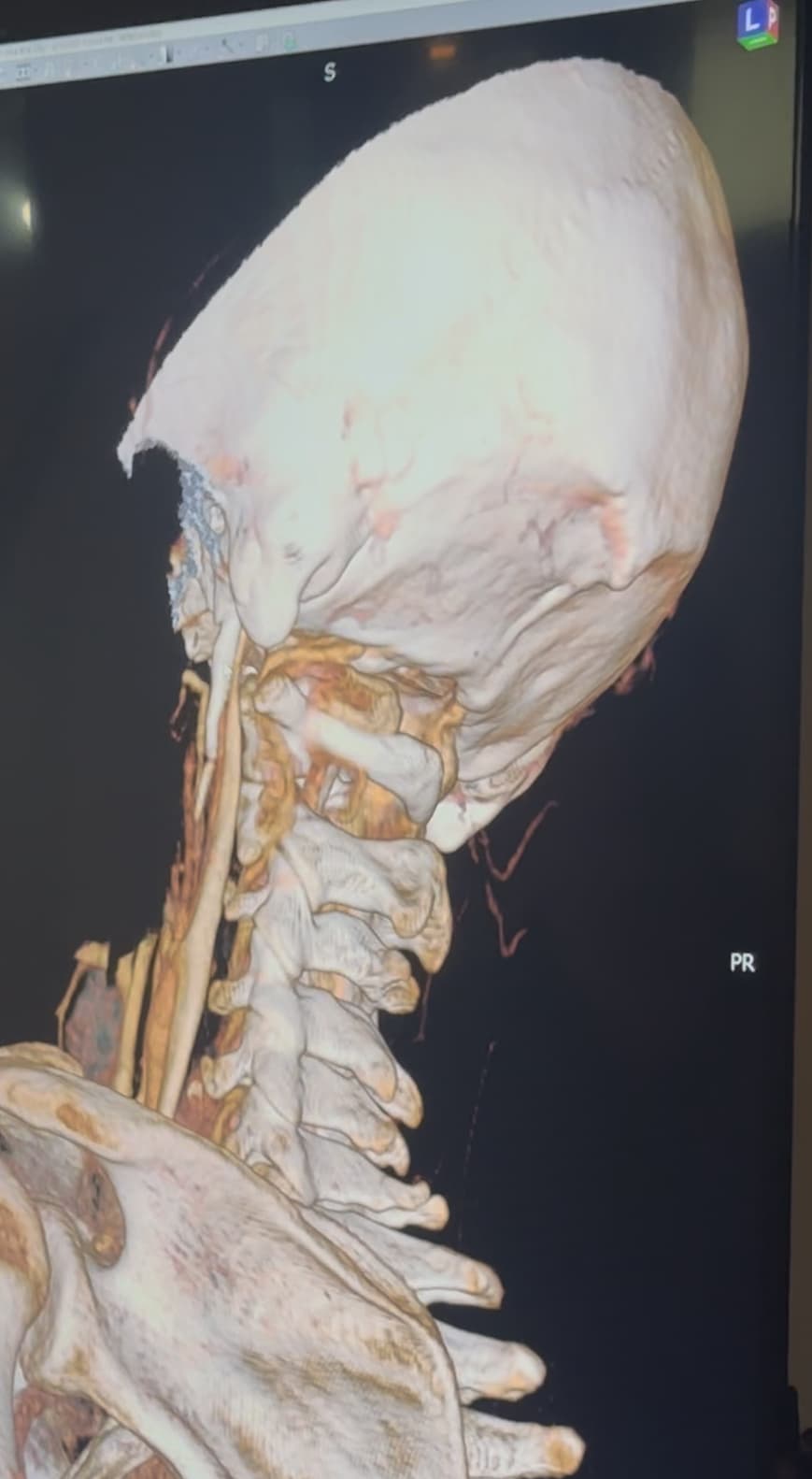
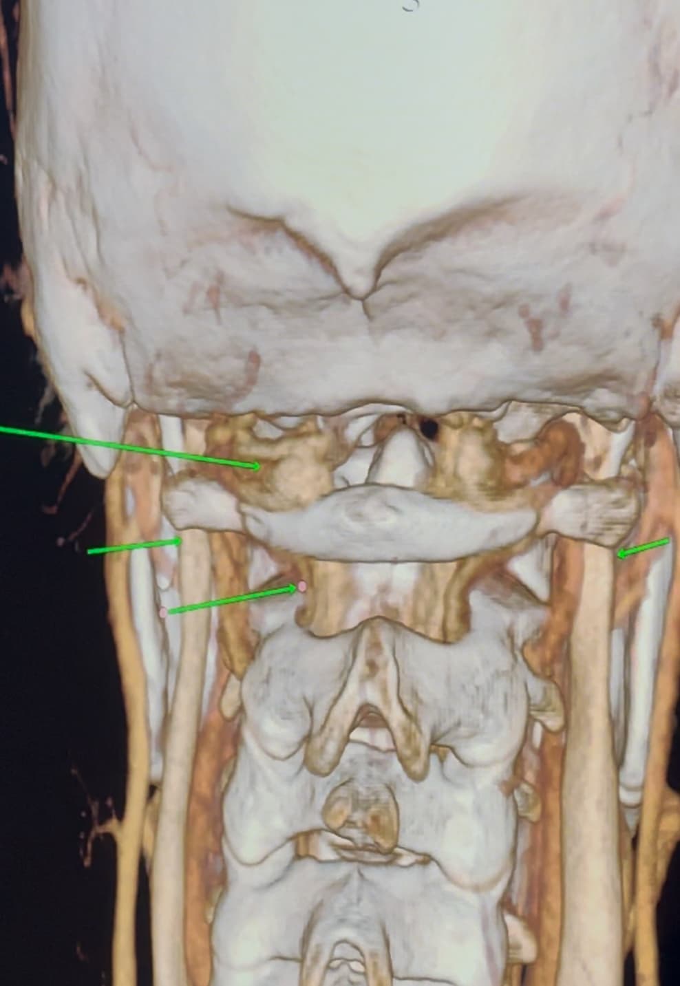
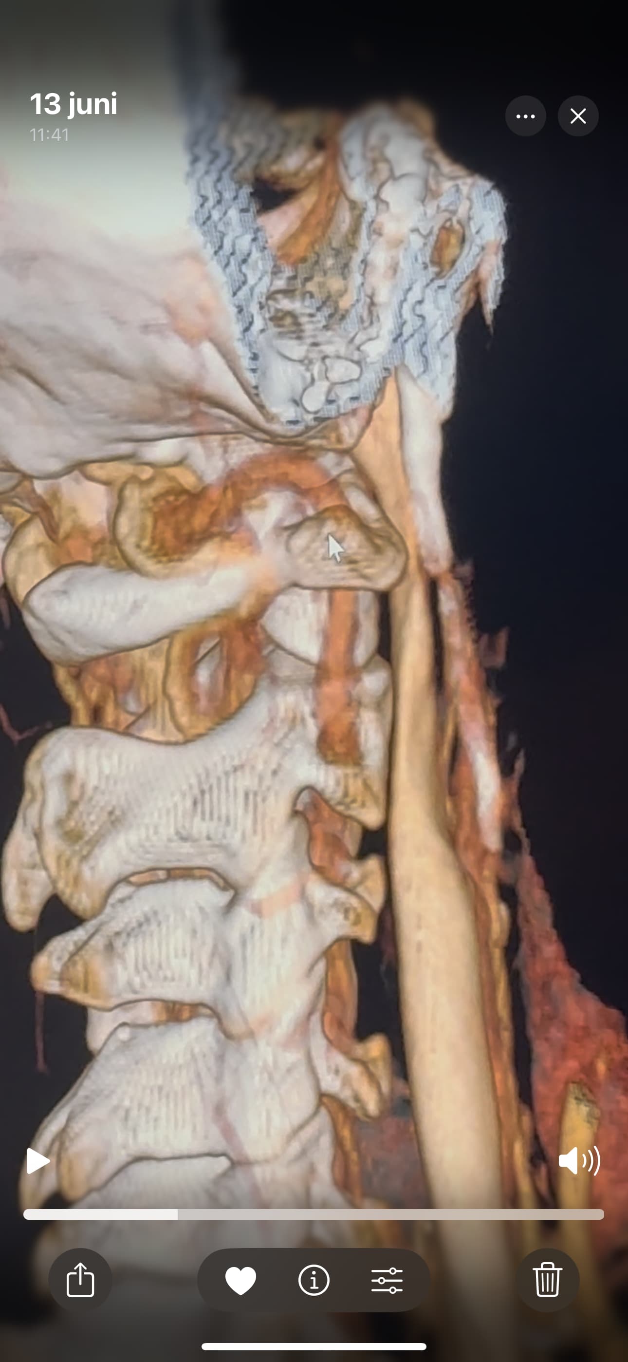
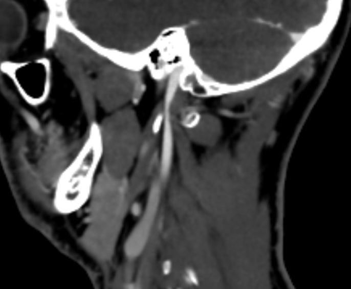
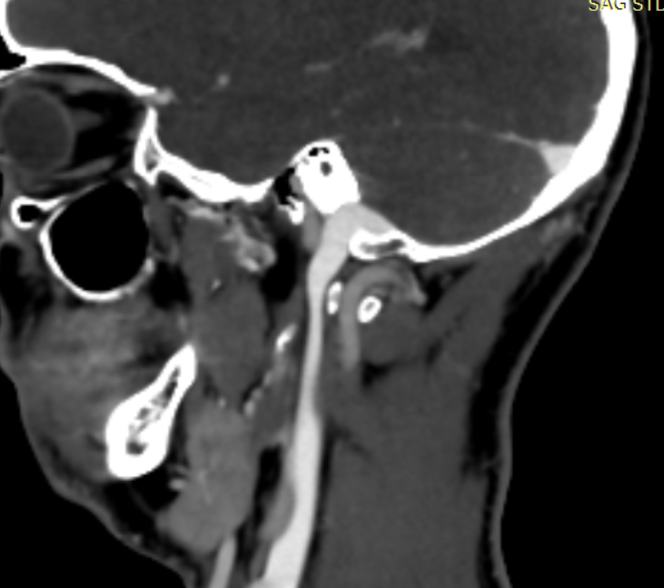
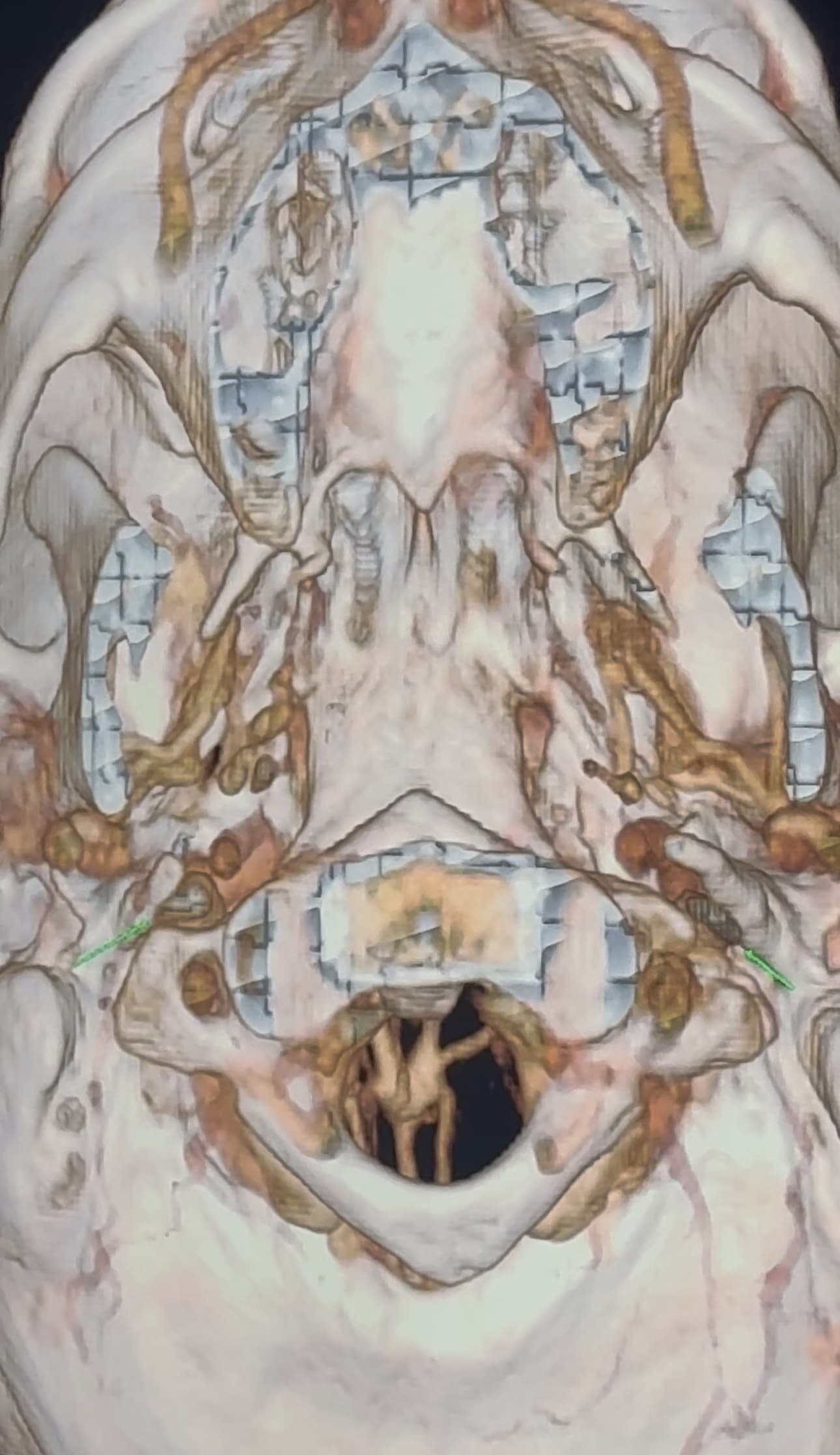
I have a cbct and CVA made wich shows elongated styloids and calcified stylohoids around 5 cm. Space left in neutral position between c1 is 3.5mm. Left IJV compression in laying position but there seems to be mainly some soft tissue compression there. So not due to compression of c1/styloid. The right is my dominant IJV.
They found a kinking carotis artery on the right at the c1 level.
I would be very grateful with any help of somebody more experienced to view the scans.
Hi Rob & welcome! Looking at your CT, & obviously we’re not experts, I would estimate that your styloid & calcified ligament section is longer than 5 cm; on your left in the middle image it goes way past your C2 vertebra! We have found that the measurements from CTs are often wrong as it can be quite hard to measure accurately due to CT imaging being done in slices…
The IJVs do collapse naturally when flat, so it could be due to that, in your 3rd image it looks like the IJV is flattened higher up, so what would be right by the skull base. It could be like that naturally because of being flat for the CT, or it could be that it is being compressed where it emerges from the skull at the jugular foramen - if styloids are quite thick that is possible, & yours do look a little wide at the top… It looks like there could be something pinching the IJV between it & the styloid where you’ve marked too; we’ve had members who’ve had compression from an enlarged SCM muscle, the digastric muscle, omahyoid muscle, as well as other blood vessels & nerves… I’d guess where the compression is that the digastric muscle would be the most likely if it was a muscle, or maybe the stylohyoid muscle, although we don’t hear much about that one!
I don’t know about the kink you’ve marked, we do all have anatomical variations, or whether that’s significant would be something to discuss with your doctor…
Who diagnosed you? Have you been able to see either of the doctors on our list? Prof. dr. Henri Marres - Radboudumc
Dr. Langeveld: A Langeveld - Universiteit Leiden
@Rob12345 - Welcome to our forum! I have nothing to add to what @Jules said, but did want to say WOW! You do have very long styloids. The “kink” in your ICA (internal carotid) isn’t really a kink more of a curve, but I can see based on the image, that it could be causing some slowed or backed up blood flow going into your brain. The IJV compression is curious as it appears to be more than the styloid causing the problem but clearly C1 seems not to be involved. I think Jules has made some logical assumptions regarding other possible compression culprits.
@Rob12345 - I’m having the same problem with your images, they won’t open & note they’re uploading. Please try reloading them & make sure the images are fully uploaded in your post before you hit the “reply” button.
We received the name of several hospitals in The Netherlands that have doctors who are familiar w/ ES. You may also want to check with one of them to get the name of a doctor who can help you. There is another doctor on the list besides the name & contact info @Jules sent you earlier in this discussion thread.
These hospitals in The Netherlands have doctors who know about ES &/or do surgery for it:
1- LUMC in Leiden 2- RadboudUMC in Nijmegen 3- Erasmus UMC in Rotterdam
4- AmsterdamUMC in Amsterdam. This hospital/University has also been doing a lot of research on ES.
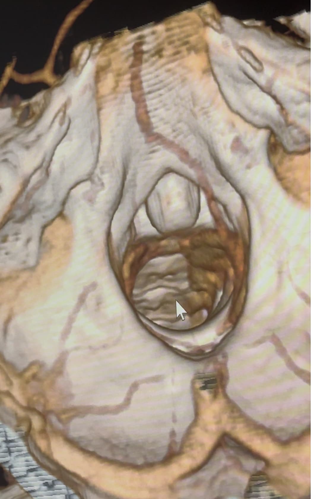
@Rob12345 can you go into the axial view of the original imaging and go to C1? It’s the best view for showing IJV compression by C1 and styloids. I’ve attached my own imaging to help you find C1 in the axial view.
@Rob12345 - I can see you have bilateral IJV compression in the images you’ve posted & it’s possible the middle image in your first set of pictures is pointing to collateral veins. I’m not great at identifying collaterals, but I can say for sure your IJVs are being squashed between your styloids & C1.