Possible Eagle Syndrome or looking in the wrong place?

Hello and thank you first and foremost on sharing in this journey with me!

I’ve been dealing with some pretty bad flare ups of pain and tightness lately in my left jaw/ear area - which have led me to discover Eagle Syndrome (and this wonderful place!).

A little bit about my journey so far…I’ve been seeing doctors non-stop with no success for over 2 years. I’ve been dealing with some symptoms that include: dizziness, lightheadedness, trouble swallowing or catching breath, left shoulder/chest pain, droopy left eye-lid, jaw pain below ear, left jugular distention and tight left veins from left collarbone.

What was the “OK, I need to get looked at” moment was about 4 months ago when I had a Panic Attack. I truly thought I was having a heart attack or a stroke as out of nowhere while working (office job) I couldnt stand, got super light headed and dizzy, heart rate at 130 and vision blurred. I fell to the ground and called 911 who arrived and said it didnt render symptoms to go to the ER.

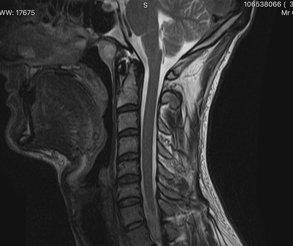
I pushed hard and my doctor ordered a brain MRI W and W/O contrast, CT of Chest W and W/O contrast, and CT spine W contrast. All came back negative for anything serious other than what they called a “Thornwaldt Cyst” on the MRI that they said was benign (except if it gives symptoms of bad breath, post nasal drip, vertigo, or estuation tube discomfort). My doc assured me none of my symptoms matched with a serious condition of Thornwaldt.

I’ve added all of the scans that I could below and would appreciate any and all feedback. I’m not well versed in looking for things on these, but wanted to rule any and all things out before I go to discuss (and obviously share these) with my ENT appointment next week. I apologize if these arent perfect views but they are all I have (and those CT’s and MRI’s are pocket breakers).

My self thoughts are: TMJ, Eagle Syndrome, or TOS (Thoractic Outlet Syndrome). I also am not well versed in the symptoms of Thornwaldt cyst, but possibly could have that looked at more on next ENT visit.

Any help or experiences with symptoms or on these scans would be greatly appreciated! After 2 years of doctors visits (and sounding like a hypo) I can’t find any outlet to help.


Marc

1 Like

@bezm - Welcome to our forum! I’m not well enough versed at reading x-rays to know which of the lines in the area where your styloids “live” might be your left styloid & I don’t want to make a wrong guess. Your ENT may be able to help sort that out. You should take the scans you have of your neck to your appointment with you & you should ask for a referral for a dynamic CT scan with contrast as that will be the best diagnostic tool to start with.

If you spend a lot of time during the day looking down at a computer, try elevating the screen so it’s at eye level as this may help reduce the occurence of the type of symptoms you had the day you went to the ER.

Here are links to a couple of posts our mod @Jules wrote that will help familiarize you w/ symptoms & their causes:

1 Like

@Isaiah_40_31 thank you for the welcome and reading through! Totally understandable - to be honest I’m not entirely sure what I’m looking at either. I do see the left styloid a bit though so It’s the first image (along with the others) I’ll be showing to my ENT doc next week at appointment. Will most certainly ask about dynamic CT too.

I do spend most days looking at a monitor, so will adjust more and see if I can ease tension there.

The posts and welcome message from @Jules has been great. Already noted a few to share with doc asap.

Thank you for taking the time!

2 Likes

@bezm - You’re right! I didn’t zoom in on the first image. You can see your left styloid. I’ve circled it in pink in your image:

It doesn’t look super long but it does look quite thick at the top & dagger-like overall. It’s also pretty angled inward. All those physical features can weigh in on causing symptoms.

1 Like

Yes, I agree that you can see the left styloid on the first image that @Isaiah_40_31 has ringed- I’m not up on the tech to be able to do that with your scans, but you can see it in the other images, & there could potentially be a bit of calcification on the ligament you can see on the second image, but it’s not clear enough to say for certain…it’s potentially long enough to warrant asking the ENT about though, just make sure you’re as prepared as you can be in case they’re dismissive with a research paper or 2. We do also have a section about advocating for yourself on here, might be worth a read before you go…

2 Likes

This was EXACTLY what I was honing in on! I agree with your assessment of it and will bring that up to my doc next week. Thank you for taking a look!

2 Likes

Thank you for looking! I think that’s the best scan to show my doc too next week. That coupled with my symptoms should be enough to warrant more scans and a deeper look. I’ll certainly advocate for myself because this journey has been so debilitating over the last few years that I won’t stop until I can find some relief. Thanks Jules!

2 Likes

Hi All! My ENT appointment went great. The doc truly listened to me and even did some checkups (camera in throat, ruling alot of other things out). She took a look at the scan I provided above that sort of shows the styloid. She confirmed that it did look out abnormal. She ordered a CT with Contrast so I will report back once I can take care of that! Thank you all for the support thus far!

1 Like

That is really great news, @bezm! I’m so glad your doctor listened & checked you thoroughly. The “icing on the cake” is the CT scan getting ordered. We’ll look forward to hearing what you learn from your scan results.

1 Like

That’s really good to hear, I hope that the CT shows what’s going on, well done for for explaining your situation well & advocating for yourself :+1:

2 Likes

Hi all,

I have some updates to share.

Just got results back from my neck CT scan with and w/o contrast with indication to “Rule out Eagle Syndrome”.

Everything was normal/unremarkable except the impression… I’ll copy here:

Impression: Elongated calcified bilateral stylohyoid ligaments could be seen with Eagle Syndrome. The elongation and calcification of bilateral stylohyoid ligaments measuring 41 mm on the right and 39 mm on the left.

I’m not sure which of these images to post because I can’t really see much from them (not sure what to look for). I haven’t heard from my ENT doc who sent me yet, but hoping this is good news. I think I’ve found the culprit…I think I have Eagles…

2 Likes

The fact that ES is mentioned in the radiology report makes it sound diagnostic to me. Are any of the images you have 3D or are they just CT slices? If you only have the slices, you can convert them to 3D using RadiAnt Dicom Viewer - https://www.radiantviewer.com for PC
or
Bee Dicom Viewer for Mac - ‎Bee DICOM Viewer on the Mac App Store

Creating 3D images will help you better see what the slices show when all put together in one image.

1 Like

they are just CT slices. Thank you! Will see what I can do with these and try to provide here.

My doc is referring me to a head and neck surgeon that she referred her last eagle patient to. They are Dr. Leila Mady at Johns Hopkins if anyone has heard of her!

1 Like

wow! This program is insane!

I’ve uploaded the following screenshots after 3d modeling…

Are these my elongated stylohyoids?


1 Like

Good job using the dicom software!! You created some very vivid images. YIKES!! Those are nasty looking daggers you have in your neck @bezm! You should feel much better once they’re gone!!

We have heard of Dr. Mady. Our member @Itsanewdawn is also setting up an appointment to see Dr. Mady soon.

Your right internal jugular vein appears to be getting squashed a bit between your styloid & C1. I can’t tell about the left side because the IJV is distorted. That is something you should definitely bring up w/ Dr. Mady if/when you see her.

2 Likes

Thank you for pointing all that out!!! That’s so comforting to hear another patient will be seeing Dr. Mady. @itsanewdawn I would love to connect!

I’m wondering why if the right was longer than the left (41 vs 39 cm) I’m having more pain/symptoms with my left side vs right? Really no noticeable symptoms on my right…

I’ve posted left screenshots here maybe to tell better:


Thank you for the help!

In the images you just sent, your left styloid looks longer than the right but which styloid is more symptomatic boils down to the angle of growth/elongation, styloid thickness, curve, etc. It also depends which nerves are being aggravated by the elongated styloid. Your left styloid may have more significant contact w/ the nerves creating your symptoms than the one on the right side. This is very common.

2 Likes

Thank you for reviewing and the help tonight, @Isaiah_40_31. So so happy to get this moving along and confirm what I’ve been feeling for a while now.

2 Likes

I’m glad that the CT confirms ES for you, & that you have a referral to Dr Mady. Will be interested to hear how you get on!

2 Likes