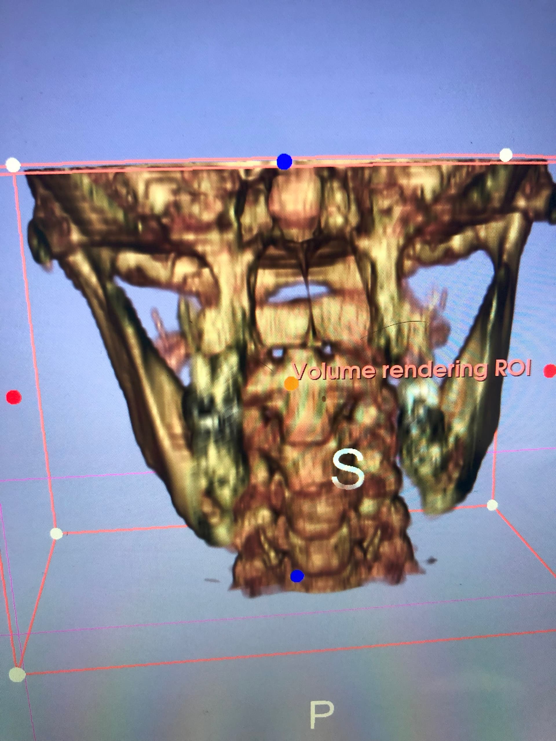
I finally got around to getting a copy of my CT scan (w/o contrast). I converted it to a 3D image and the results look pretty similar to what I saw on an x-ray. Styloids don’t appear to be elongated (can hardly even find them) but you can see calcification on both the right and left side. The right side (where I am experiencing a lot of my symptoms) has a slightly longer calcification than the left side. Left side is shorter but has a few other small pieces.
My question is do those calcifications look big enough to be causing my problems? They don’t look that big to me but I am experiencing a lot of issues that seem typical of eagles. I also have these hard round lumps below both ears with the one on the right being quite a bit bigger. I always though that those were normal human anatomy but according to my husband last night, no they aren’t. The lumps, especially the one on the right lines up right where the calcification is.
I’m still trying to find a doctor to ask about it. I’m thinking of doing a phone consultation but it kind of sounds like both Samji and Conjetti don’t consider only calcification to be a problem unless I’m misreading things here on this forum.
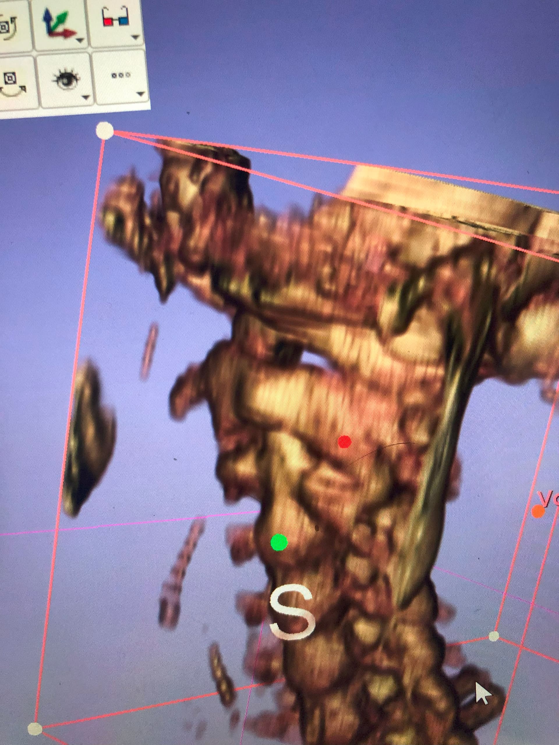
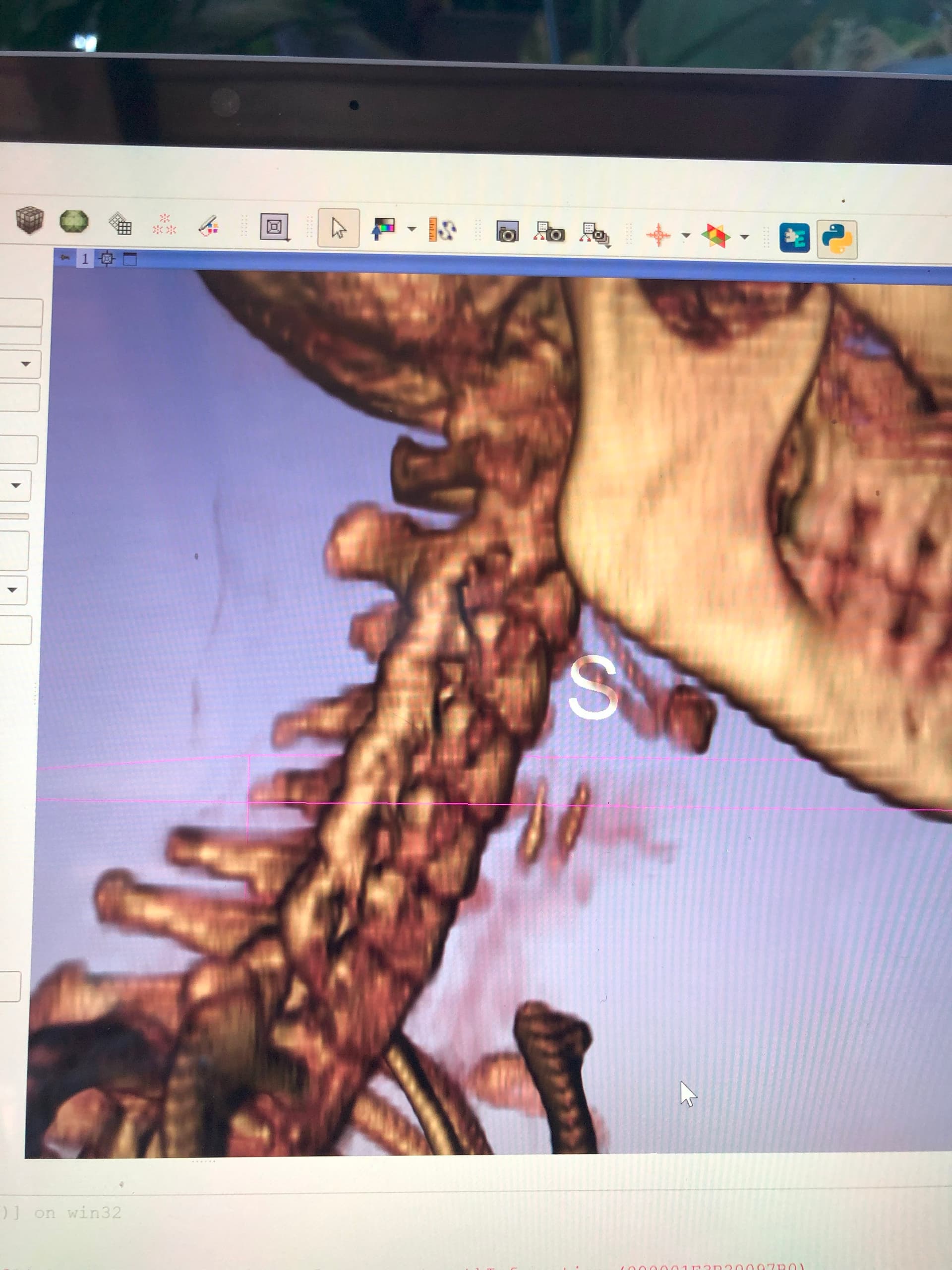
Also for those of you who know the neck anatomy well, what are those two hard bone looking things in the last image below the hyoid bone? Is that additional calcification or is that cartilage or something? It’s also right around one of the spots I’ve been complaining a lot about throat pain.