I have Eagle Syndrome (left elongated calcified stylohyoid ligament), confirmed on CT in 2013 at Mayo Clinic as well as dental x-rays in 2019. Currently, I’m undergoing tests and treatments for a 14-month Long COVID illness, which includes a great deal of cognitive dysfunction as well as other symptoms. The process of evaluation includes ruling out other things. (We already know I have neuroinflammation, based on the clues and labs.) I had a head CT as well as neck and head CTA.
I prepared a request for the radiologist to compare the CT from 2013 to the current scans and measure if the elongated/calcified stylohyoid had grown and if it was causing mass effect on the neighboring blood vessels (such as interior carotid). I took the request, the report from 2013 and the scan from 2013 on a disc. The radiology team asked the radiologist if they could do it, they said yes, so they uploaded my 2013 scan to their system and took my detailed request of what to look for.
I also asked for them to evaluate for possible friability of the blood vessels, considering my dad’s history of having a friable carotid that they could not stitch up after surgery and he died from it. Based on his history and mine, there has been speculation that we have Ehler’s Danlos, which is a connective tissue disease that can cause those types of issues.
The radiology reports for the CT and CTA are back, and everything was found to be normal. BUT…it says they did not compare with other scans and did not do #1 and #2 above. They only evaluated for “amnesia, other” which is the diagnosis the neurologist provided because it is often a broad diagnosis used for cognitive dysfunction.
The good news is they gave me the actual scans on a disc and I can take them to specialists to evaluate for the two things I mentioned above. I don’t have a disc reader at home (I have a MacBook Pro). Any suggestions on that?
Today I had “US TRANSCRANIAL DOPPLER INTRACRANIAL ARTERIES COMPLETE.” They checked the common sites. By the temple. At the neck where the carotids are. At the occipital bone area. And the eyes. I explained that I had Eagle’s Syndrome and asked if they could check multiple positions due to the fact that the calcified/elongated stylohyoid could compress the blood vessels only in certain positions. The tech went to ask the radiologist and the radiologist said that wouldn’t be necessary. He said Eagle syndrome would not make a difference in the scans they took or have a different outcome due to different positions. That doesn’t sound right. Also, the entire doppler ultrasound for all of the positions they checked only took seven minutes. It doesn’t seem they did a very thorough job, for a complete study rather than a basic one. The results are back. It says:
Purpose: Measure blood flow velocity in major intracranial arteries.
Technique described: Real-time ultrasound with color and spectral Doppler.
Reported arteries & velocities (cm/s):
ICA (Internal Carotid Artery): Right 20, Left 19
MCA (Middle Cerebral Artery): Right 43, Left 45
ACA (Anterior Cerebral Artery): Right 34, Left 32
P1 segment (Posterior Cerebral Artery): Right 20, Left 25
@WillisWay - I think you’re focusing on the wrong vascular tissues. With the length of time you’ve had ES &cognitive dysfuntion & now know you have EDS, I believe you probably have internal jugular vein (IJV) compression which is quite common among our members who have hEDS. Even though your father had a problem w/ the friability of his carotid arteries, it may not have transferred to you. IJV compression is notorious for causing brain fog & ES itself can cause neuro- inflammation though I don’t know how that manifests in you. If you’re still inTX, I think Dr. Chan-Leveno diagnoses & treats IJV compression with ES. She would at least be someone to start with to investigate this with. A CTV would also be helpful. That is a CT scan where contrast is injected during the venous phase of the heart beat. They can be hard to get because it seems many radiology offices don’t know how to do them.
I also have a Mac like yours and my husband has an external disc reader that plugs into the USB port so I was able load the scan images off my CD & convert them to 3D. Bee Dicom App works best for Macs. If you can either get ahold of an external disc reader or ask around among your friends & family to see if anyone has one, you may be able to do this yourself.
I completely agree with you. This doctor doesn’t know much about ES as indicated by that statement.
Thank you so much! You have always been so helpful. I tend to put my ES on the back burner due to other health issues going on. (Such as primary immune deficiency and twice-weekly infusions for that.) I was able to function fairly okay until I had COVID 14 months ago and have had Long COVID ever since. I’m guessing that caused a “perfect storm” for neuroinflammation and cognitive dysfunction. Does a patient with Eagle who gets Long COVID notice any correlation? I have greatly elevated TNF-alpha and other inflammatory markers, which were not elevated prior to Long COVID. I am in Speech & Neurological Rehab twice a week for the new challenges. The reason for the CT, CTA, and transcranial doppler has to do with evaluating for my 14-month cognitive dysfunction. I think the ones testing got by with not checking the Eagle Syndrome factor because the ordering diagnostic code was for cognitive impairment. I’m not sure why they accepted my old CT on CD and even uploaded to their system if they weren’t going to use it for comparison! I will check with the recommendations you have given me and report back. Thank you SO MUCH! (Oh! I don’t know for sure that I have EDS, but it has been suggested by some doctors due to my history. I’m considering going to a genetics specialist to test for that and a few other things, since it could be a contributing factor.)
I forgot to state that the webinar addresses long covid and the other systemic inflammation your describing in asscociation with the EDS patient and need to control it as much as possible before surgical treatments. It is a long webinar but should provide you excellant information and resources.
@WillisWay -
We have a few members who’ve had long COVID. We also have members with primary immune deficiency who are on regular IVIG infusions. The fact you have long COVID may be related to your immune deficiency but you likely know that. One member whom I know of who has primary immune definitely had ES w/ IJV compression & after two surgeries (one for each side) some of her worst symptoms subsided to a significant degree.
Any type of viral or bacterial infection ramps up the immune system & causes inflammation in the body thus can exacerbate ES symptoms.
I hope if you see Dr. Slavin, he is familiar with EDS (if you test positive for it) & ES w/ its vascular variations, & can help you.
I’m sorry that you’re dealing with so many issues & that the report wasn’t helpful! Do you notice any different symptoms if you have your head in certain positions- dizziness, fainting etc? It might be helpful to have a read through some of the symptoms of carotid or IJV compression to get an idea of what might correlate with what you’re experiencing, if it’s more than cognitive impairment?
I have gotten the CD with my recent scan opened using Bee DICOM Viewer app. Is there a way I can upload my images to get everyone’s input here? Could someone help me measure the styloid process/stylohyoids? And tell me any concerning facts, such as angle, length, width, calcification, possible compression of nearby vessels/structures, etc.? I’m not very techno-savvy! I’m not sure if it’s too big a file to upload it all?
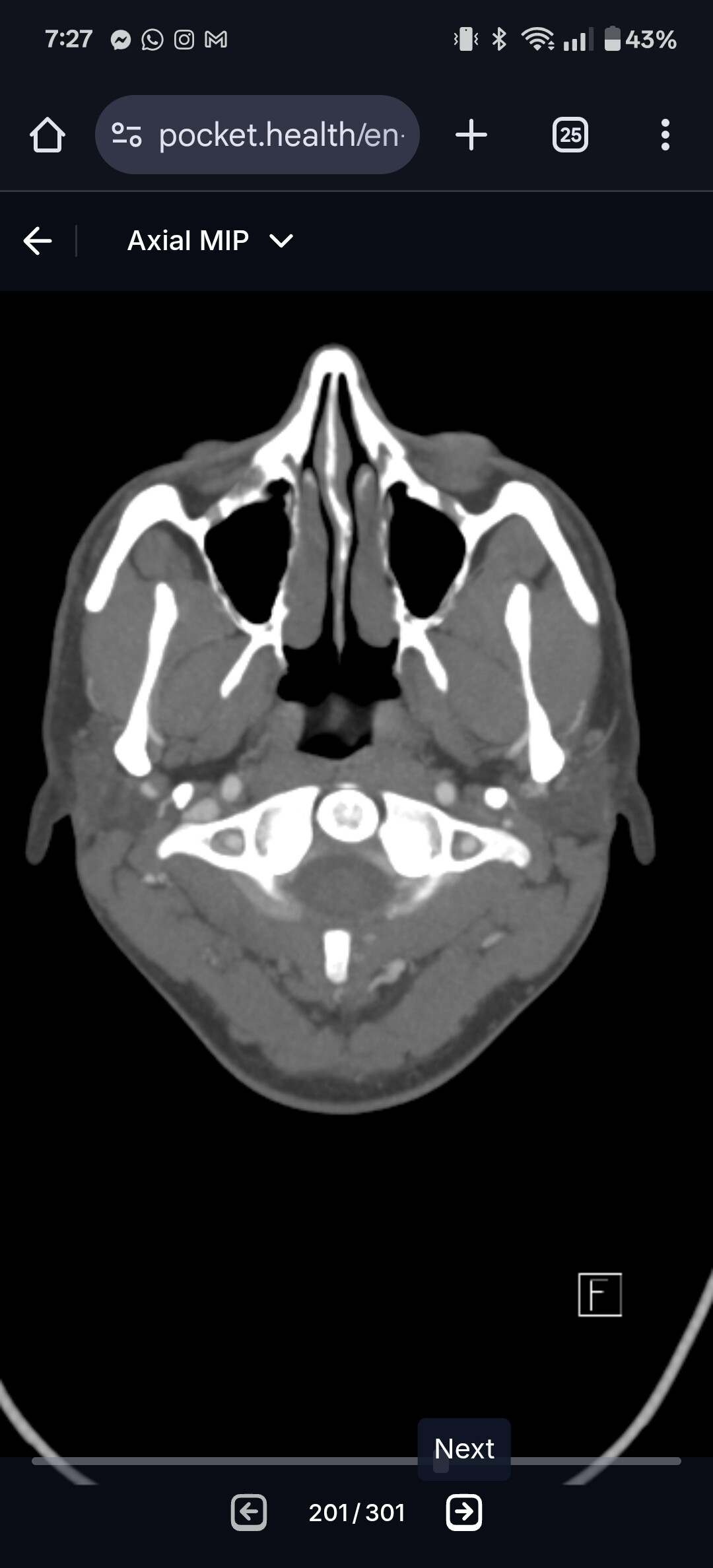
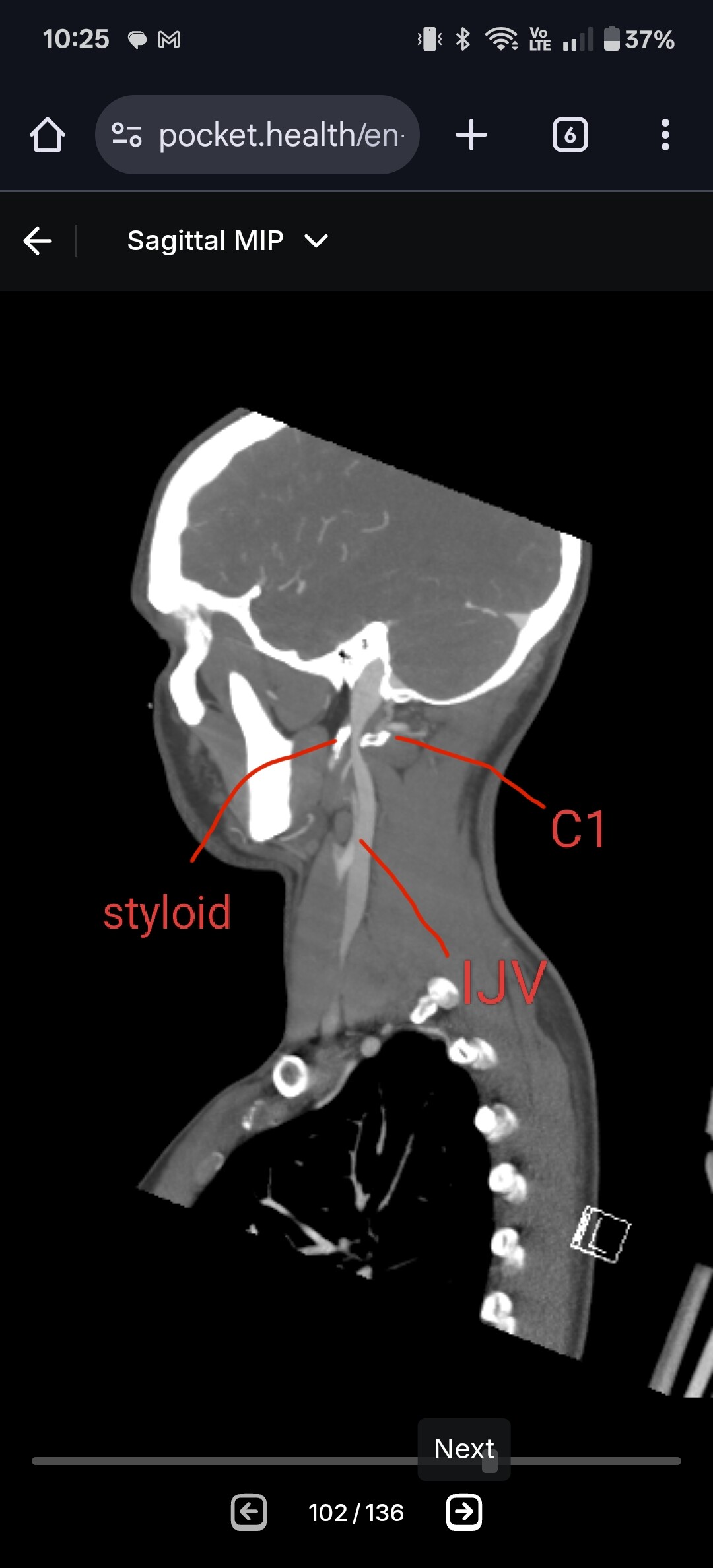
@WillisWay if you go into the axial view of your imaging and go to C1, I can see if your IJVs are being compressed between your styloids and C1. I’ve attached my own imaging to help you know what to look for. It’s the top vertebrae and it has wings with a white circle in the middle.
I have the following options on my scan. I don’t see axial view. I have: 3D, Sagittal, Coronal, Bone Neck, CTA neck. Will any of those work? Also, they don’t have them labeled with C1, best I can tell. They have image numbers, like if we were loading cell phone images.
@WillisWay interesting that there isn’t an axial view. And yes, the numbers is all we have (there shouldn’t be anything labeled as C1 - I was just referring to it as a landmark). Sagittal view can be helpful if there is contrast in your IJVs. See attached my imaging and see if you can make it match.
Feel free to upload the 3D, bone neck & CTA neck, we can still have a look & try & give you pointers- I’m a fellow not at all techno savvy person, so can’t give you any pointers on that though!
I’ve used AI to narrow down the images so you don’t have to sort through hundreds of them. Thank you for reviewing my imaging for Eagle Syndrome. To make navigation easier, I’ve organized each image set and highlighted the slices that best demonstrate the following:
Styloid length (from skull base to tip)
Width/diameter (cross-sectional thickness)
Contact with C1 (atlas) (areas of closest approach/possible compression)
Reference sheet is attached below (filenames provided).
Symptoms prompting evaluation
Persistent neck pain, stiffness, and soreness
Headaches and facial/jaw pain
Sensation of fullness and pressure in throat/neck (like swallowing glass when I sing)
Dizziness and episodes suggestive of vascular involvement
History of ankylosing spondylitis and other inflammatory conditions (noted for context)
Cognitive dysfunction since Long COVID
Imaging Summary
Bone Neck series: clearest for bony landmarks and styloid–C1 spatial relationship
CTA series: shows vascular course relative to styloid and C1
3D reconstructions: useful for overall orientation, angulation, and relative length
I hope this helps streamline your review. Let me know if you need something more. I also have zip files of all the content from the CTA.
Sincerely,
Kathy Willis
It will only let me load five images at a time, so I will do the 3D Reconstruction first. It might be enough for you to do what you need to do to find length, width/diameter and contact/angulation. I will add some more in another message.
Since these images look nothing like what I’ve seen here before, I feel like I’m way off! I have about 300 images to sort through so I had IA help. But they might have selected the wrong ones to share with you. It’s hard because I can only upload five images at a time. I also made zip files, and two of them are small enough that my email will let me send those, but the other is too large even zipped. Should I try sending zip files? I don’t want to keep clogging up the inbox!
@WillisWay - I’ve deleted the images you put up because they contained your name, birthdate, etc. Please be careful to cover up or delete your personal information on any images you post to the forum because the posts here are visible to the general public.
Also, you can have a private conversation & send your imaging to @TML directly by clicking on his screen name as it appears above one of his posts. By doing that you won’t need to delete your personal details if you don’t choose to.