Abstract
The internal jugular vein (IJV) is of utmost importance during various surgical and endovascular approaches, including central access. It descends through the parapharyngeal space, carotid triangle, and sternocleidomastoid region. The anatomical variables of the IJV are mainly related to its calibre and dominance, number of venous channels (i.e., duplications and fenestrations), and compression sites. Specific compressions of the IJV are not exclusively due to the jugular nutcracker between the styloid process (SP) of the temporal bone and the C1 transverse process, which, in turn, should not be granted the eponym of Eagle. The possible morphologies of the SP and ossified stylohyoid chain are discussed here. Additionally, the digastric and sternocleidomastoid muscles, the hyoid, and the distorted carotid arteries may compress the IJV, thereby raising intracranial pressure. Here, a case is documented with a long inferior petrosal sinus adjacent to the IJV, both compressed into the C1-styloid nutcracker, which is an absolute novelty. Multiple compression sites of the IJV are supported here with original evidence. All anatomical variables of the IJV are relevant, as they may lead to stenoses or interfere with IJV cannulation. In rare cases of IJV agenesis, multiple compression sites on the opposite side may significantly alter bilateral cerebral drainage. Different methods may be used to decompress a stenotic IJV, including styloidectomy. In conclusion, the anatomical variables of the IJV should be acknowledged by practitioners and documented on a case-by-case basis.
My Highlights
2.2. The Internal Jugular Vein’s Valves
The IJV valves make a buffer zone between the large central veins and the cerebral venous system [41]. The valves are generally located about 0.5 cm above the union of the subclavian vein and the IJV, in 96.8% of the general population [41]. The IJV valves prevent the backflow of venous blood and backwards venous pressure into the cerebral venous system during conditions where the central venous pressure or intrathoracic pressure is increased, such as chest compression during external cardiopulmonary resuscitation, severe or repetitive cough, and straining [41]. Without competent IJV valves, a sustained or prolonged retrograde-transmitted venous pressure via IJVs might impair cerebral venous drainage and determine neurological deficits, such as encephalopathy, after cardiopulmonary resuscitation [41]. It was previously reported that the valves are usually bicuspid, sometimes unicuspid, and rarely tricuspid, with inconsistent cusp orientation raising questions about their functional efficacy, particularly in limiting competence to foetal life [42].
2.4. The “Tunnel” of the Internal Jugular Vein at the Level of the Transverse Process of the Atlas
At C1, the IJV descends through a veritable vertical tunnel bordered by different anatomical structures, each of these having the potential to compress the IJV. The tunnel of the IJV is limited: posteriorly, by the C1 transverse process, through which courses the vertebral artery, anteriorly, by the SP, medially, by the ICA, antero-medially, by the stylopharyngeus and styloglossus, laterally, by the posterior belly of the digastric muscle, the SCM, and, eventually, the mastoid process, and anterolaterally, by the deep lobe of the parotid gland and the external carotid artery (ECA) (Figure 1).
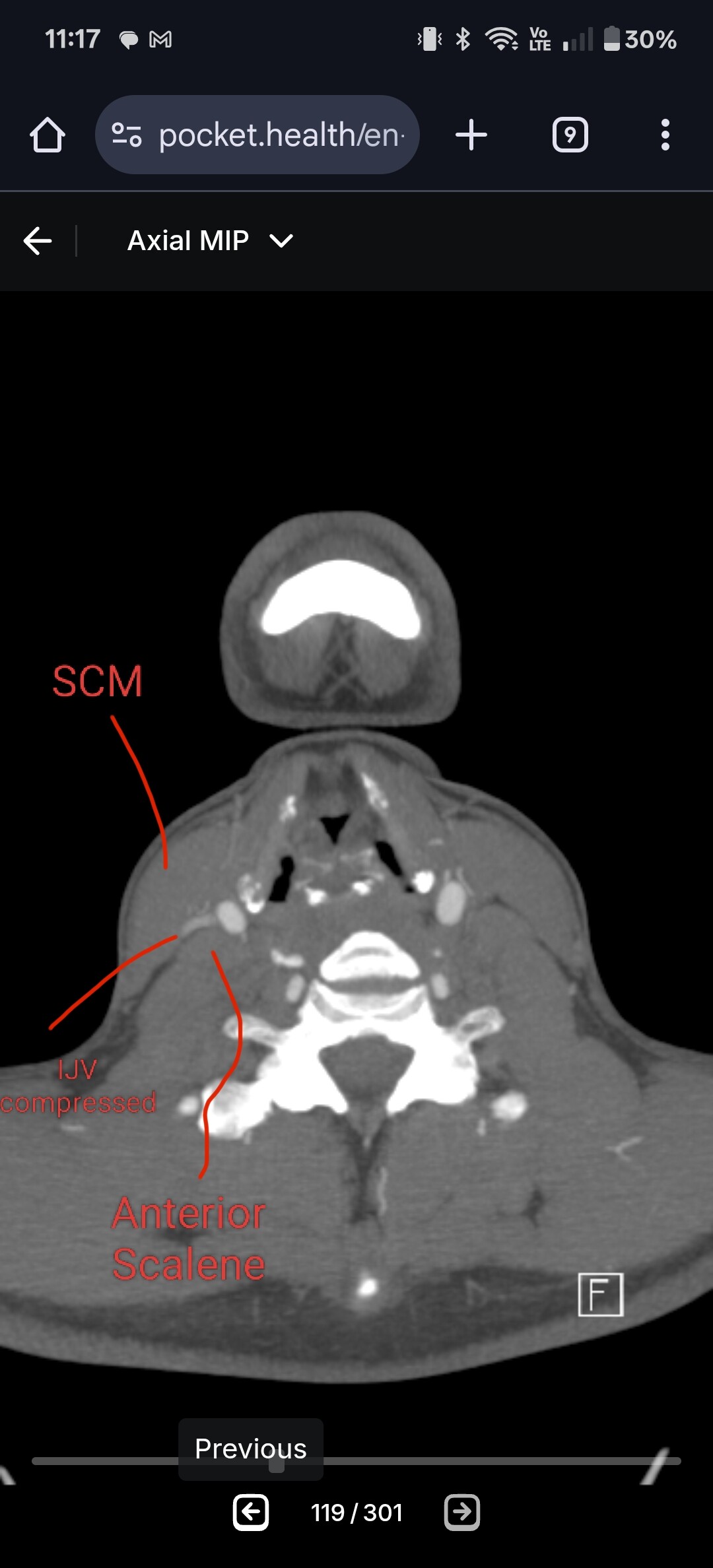
7. Compressions of the Internal Jugular Vein
…
On the other hand, external compression of the IJVs is an effective method for increasing intracranial blood volume and brain volume in animals and healthy humans [155]. It has been reported that, on assuming an upright posture, cerebral venous drainage is distributed away from the IJVs to the deep cervical veins/plexus [155]. Such intentional IJV compression (e.g., with a collar) prevents brain injuries by increasing the intracranial blood volume and reducing brain movement during trauma [30,31,152–154]. IJV stenosis is associated with several neurological disorders, including idiopathic intracranial hypertension (pseudotumor cerebri) and pulsatile tinnitus [156,157]. Such collars on the IJV may offer acute symptom relief for patients with venous pulsatile tinnitus [158]. In cases of extreme bony compression causing IJV stenosis, surgical decompression might be necessary [156].
A Long Inferior Petrosal Sinus May Also Enter into the Nutcracker
The IPS is a paired dural venous sinus in the posterior cranial fossa that drains the
cavernous sinus into the jugular bulb. It receives an inflow from the auditory structures
and the brainstem [179,180]. Endovascular access to the IPS has diagnostic and therapeutic
utility for diverse conditions involving the cavernous sinus and sellar regions [179]. The
IPS can be used for the embolisation of the cavernous dural arteriovenous fistulas or
venous plexuses of the skull base [181]. Bilateral IPS sampling is an essential means
for the diagnosis and differential diagnosis of pituitary microadenomas [181]. A long
extracranial IPS courses along the IJV to empty into it at a lower level [181]. This extracranial
extension of the IPS may be regarded as an accessory IJV [182]. It was demonstrated that
a long or aberrant IPS may also be used for transvenous embolisation for endovascular
management [183].
8. Clinical Anatomy
…
Cerebral venous outflow from the brain is not fully understood [191]. A simple
compression of the IJV may be benign, while stenosis of the IJV can be pathological.
Evidence is emerging that the presence of surrounding venous collaterals and white matter
hyperintensities may assist in distinguishing whether an IJV compression is benign or
pathological [191].
8.3. Thrombosis of the Internal Jugular Vein
IJV thrombosis is an infrequent condition that comprises 1.5% of deep venous thromboses overall and 45.3% of upper limb deep vein thromboses [128]. The leading risk factors include central line placement, cancer, and ovarian hyperstimulation syndrome [128]. Pokeerbux et al. (2020) reported the initial case of IJV thrombosis potentially attributed to venous entrapment between the SP and C1 transverse process [128].
IJV thrombosis may lead to pulmonary embolism [203]. In approximately 20% of cases of pulmonary embolism, the source of emboli cannot be identified, and it may be speculated that IJV thrombosis in a stylo-jugular syndrome may be responsible for some of these cases [203].
Guan et al. (2021) reported a case of cerebral venous sinus thrombosis in the lateral sinus in a patient with bilateral compression of the IJVs due to an overgrown left lateral mass of the atlas, as well as arteriosclerosis and expansion of the right ICA [135].
9. Conclusions
The jugular nutcracker can also involve and compress an elongated inferior petrosal sinus (IPS), which serves as an accessory IJV. This represents a discovery of critical importance for precise preoperative anatomical assessment.
The IJV may display multiple pathways, including fenestrations and duplications across different segments. Its size and bilateral symmetry vary unpredictably. Styloid process geometry differs between patients, meaning the SP may not always compress the IJV. Other nearby anatomical structures may be responsible for IJV compression instead.
Therefore, thorough IJV documentation is essential before any surgical or interventional procedures. Clinicians have to distinguish carefully between proven findings and hypothetical associations.
PS: Still alive if anyone’s wondering ![]() Apologies for lost emails (never get a domain with NetFirms or its parent company).
Apologies for lost emails (never get a domain with NetFirms or its parent company).