Hello all! I somehow got logged out of my account and wasn’t receiving notifications!!
I’d like to let everyone know I’m doing great, able to move around the house and mostly eat anything. I cannot use the left side of my tongue which does cause a lot of issues when speaking and thus my speech is off. I’ve started getting my nerves back and that shit HURTS lol real bad.
@Snapple2020 they didn’t give me anything for swelling but I’ve been taking ibuprofen and Tylenol with the 5mg oxy but i haven’t taken those in awhile now!
Here are some more pictures of the process, I’m also on Facebook if you’re on those Eagle Syndrome groups you’ve probably seen me share on there!
Thank you all so much for being so amazing and helping me through this process!! 
@Jules I definitely have been getting that itchy feeling as well and the whoosh!
these guys were apparently 7cm long 
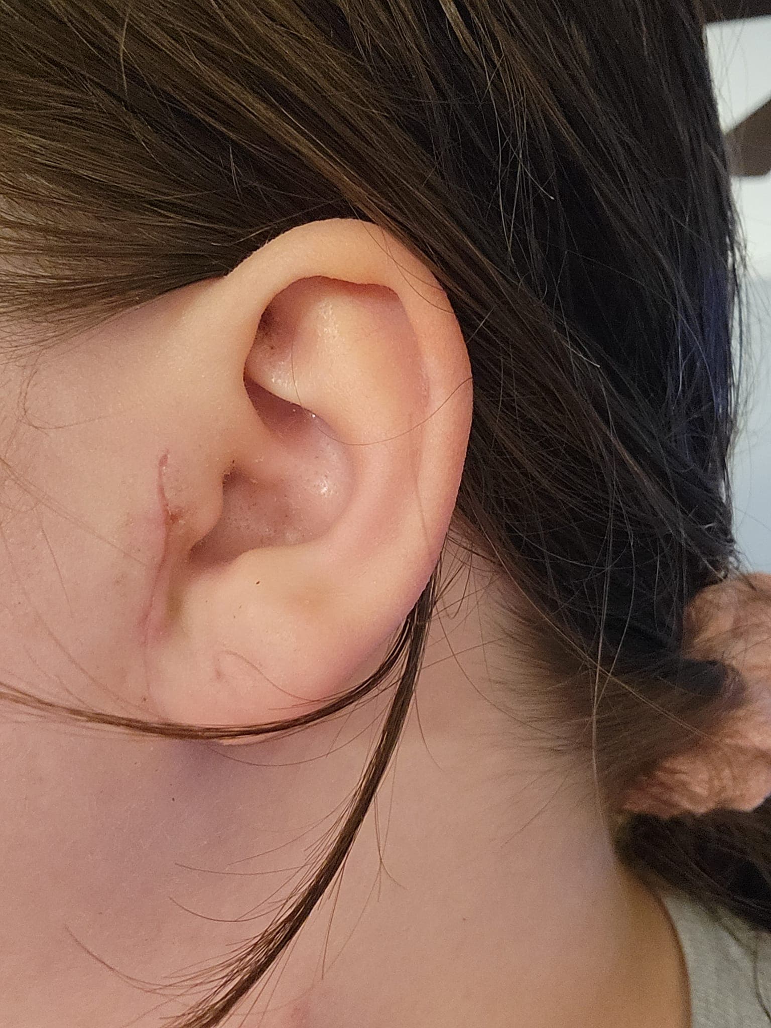
this was from the 5th!
this is from the 8th
this was on the 12th
this was yesterday!
This is my new home that my boyfriend and I just bought <3
For the first time in several months I’m starting to feel clear headed again. That foggy brain is going away!!
I’m ready to start studying for the actuarial exams again and I’m ready to finish my Economics degree!
Also: here are the medical notes from my surgery for anyone interested:
We designed bilareral preauricular incisions and bilateral 4 cm neck incision for access to remove the styloid process from the infratemporal fossa as the length of the process extended out into the neck.
We began on the right with design of the preauricular incision extended from the superior aspect of
the tragus down to just underneath the lobule of the ear bilaterally. This was marked out along the
skin crease and then injected with local anesthesia. We then incised through the skin and subcutaneous tissue and identify the cartilaginous ear canal. We then began dissection along the
perichondrium of the ear canal retract the parotid tissue anteriorly and dissecting down until we reached the bony cartilaginous junction of the ear canal. We further opened this plane from superior to inferior identified the styloid process originating off the skull base. After doing this, we then moved our dissection inferiorly and laterally and identify the main trunk of the facial nerve which was traced out towards the pes anserinus. At this point, we had identified the superior aspect of the styloid process deep within the infratemporal fossa. Periosteal elevator was used to dissected the soft tissue attachments around the styloid process down to the inferior aspect of the calcified tip.
The distal tip of the styloid process was fairly far inferior, and therefore for safety of access, we chose to access the lower neck incision next. The neck skin crease incision just below the submandibular gland was incised with 15 blade scalpel down through skin and subcutaneous tissue and through platysma. We then deepened our dissection along the posterior edge of the submandibular gland with sharp scissors and bipolar cautery. We retracted the posterior belly of digastric, and then dissected superior to this into the infratemporal fossa, identifying and preserving the hypoglossal nerve and the external carotid artery with its associated branches. With further dissection superiorly, we identified the distal tip of the calcified styloid process. We isolated this to prevent in severed the ligamentous attachment. We then used the periosteal elevator to elevate the other remaining soft tissue off of the distal half of the styloid process. Having done this, we then moved back to the preauricular incision.
A rongeur forceps was then used disarticulate the styloid process from the skull base, and then we retracted the process superiorly, using bipolar cautery to release/divide the ligaments and muscular attachments inferiorly. The process would not removed out through the preauricular incision, and
therefore we transition back to the neck where the process was grasped along its distal tip retracted inferiorly until we could release the remaining soft tissue attachments with sharp scissors and bipolar cautery. This allowed us to remove the 7 centimeter styloid process from the infratemporal fossa. There is no significant bleeding. There is a mild oozing at the skull base bone where the process was removed. This was controlled with bipolar cautery. We checked his facial nerve and hypoglossal nerve after completion procedure and they stimulated normally. The area was copiously irrigated sterile saline, and Valsalva confirmed good hemostasis. We then placed a #10 Blake drain into the neck and extended this up into the preauricular incision through the infratemporal fossa.
And then same thing for the left side and then they closed me up. Eek weird to read the medical version of my body being sliced LOL