I recently went to the doctor and had a CT scan of my head and neck, as well as a 3D X-ray of my head. My general practitioner said that it is not Eagle Syndrome. However, I still have all the symptoms: pain in the neck, pain in the facial region, pain around the Adam’s apple, pain when swallowing, and blurred vision in my left eye. These symptoms only improve when I do certain stretches or place my head in a specific position.
In addition, I am currently taking medications — Gabapentin and Amitriptyline at moderate doses — and my neurologist mentioned that I may need to increase the dosage because of the persistent pain.
At this point, I don’t know what else to say. Could any specialist please tell me if this could really be Eagle Syndrome?
@mjzago can you download radiantviewer to your computer (if it’s a windows computer) and then open your imaging on it and construct a 3D model? It’ll help us see the full styloids in one frame.
We’re not specialists/ doctors on here; we’re just people diagnosed with ES who have learnt a bit along the way… It’s not easy to see in some of your images, but the last one you definitely have one elongated styloid- you’ve ringed both sides in red, if the left side of the image is your styloid that would be the biggest styloid I’ve ever seen. I’m just wondering if it’s your jaw bone?
Did you get a report by the radiographer along with your imaging? Did they comment on your styloids? I’m not sure why your GP has dismissed your images, & given your symptoms I think that you should still pursue a diagnosis, so if you could get a referral to a doctor with experience, that would help. There are doctors on the list in Germany who do know about ES. If you need a referral from your GP to see another doctor, then perhaps you could print off a couple of the research papers which mention the common symptoms to argue your case? If you’re able to do the imaging that @TML suggests, then we might be able to give you more pointers…
@TML@Jules Thank you for the sugestions and explanations.
I’ve been dealing with symptoms for about 6 months now. At the beginning, I had a radiology report that said it was not Eagle Syndrome. However, I later took the images to my oral and maxillofacial surgeon, and he mentioned that the styloid process looked enlarged. Because of this, I’m now feeling unsure and a bit confused.
Thank you for the suggestion about the 3D software—I really appreciate it. I’m also attaching the images here for reference.
@mjzago styloids are typically considered elongated if over 3.0cm. Our measurements on 3D scans are typically slightly under the true length due to the nature of CT slices. I’d say your left is probably within normal limits but your right might be on the threshold.
What I’m more interested in after seeing your imaging is the differences in your greater horns of your hyoid bone. Your left greater horn is pretty straight, while your right one is curves upwards. It’s possible that this is where your swallowing/neck pain and issues are coming from and potential left eye symptoms.
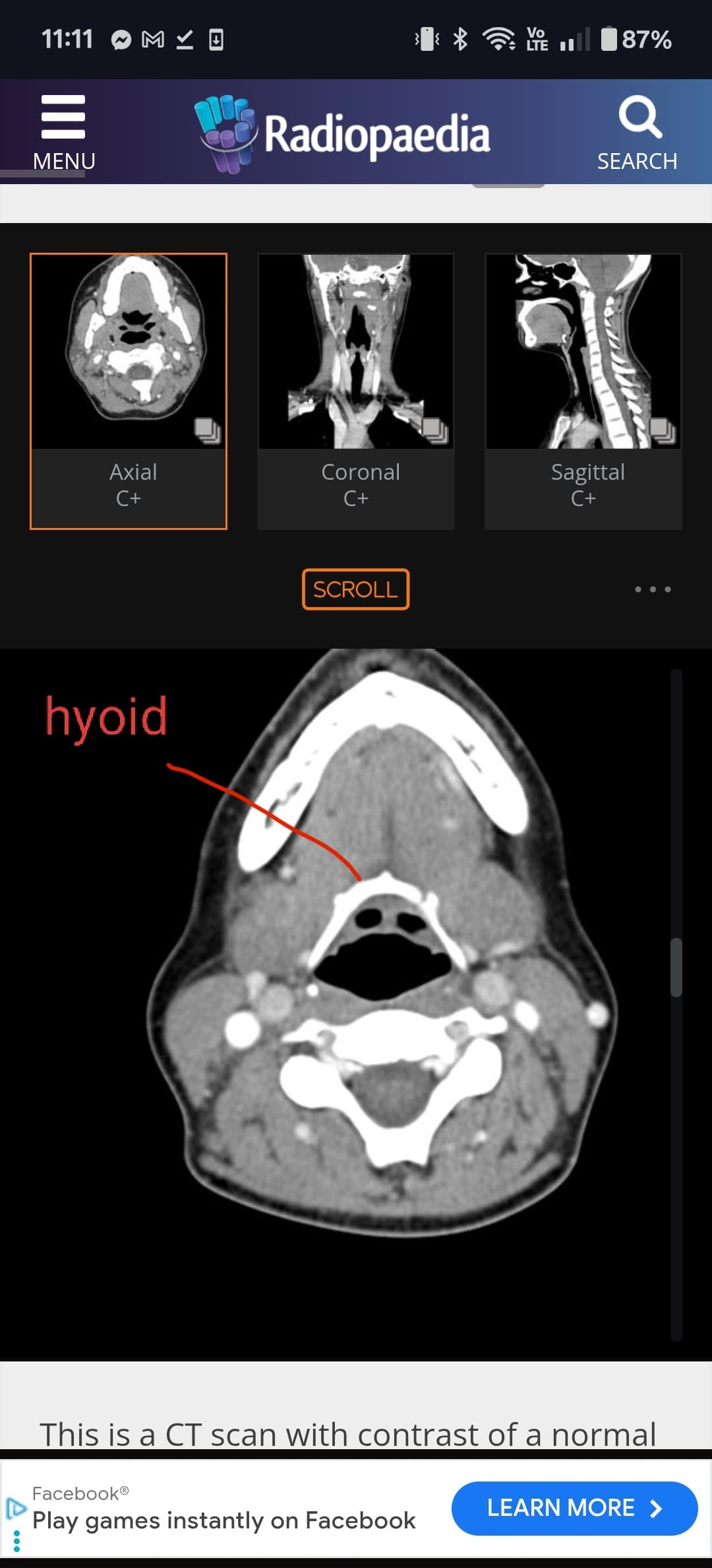
I think we should take a look a the greater horns in the axial view of your imaging to see if they are poking into your carotid arteries.
I’ve attached a couple of images showing what to look for. First locate the hyoid bone and then travel upwards until you get to the very tips of the greater horns:
@TML Thank you for the information. I suspected that something might be wrong with my hyoid bone. The pain and irritation are quite intense on that area. When I take a deep breath or swallow, the pain increases, and it often feels like something is stuck in my throat. I’m attaching the images as well. Thanks again for your help!
@TML I’ve been to ENTs in two different places — one ordered an MRI of the neck, and the other ordered a CT scan to check for possible Eagle syndrome. But neither of them really looked at the hyoid bone or the greater horn, I think.
I think there could be a couple things going on, and I personally think it’s more likely down at the level of the hyoid + thyroid cartilage and less so than the styloid(s).
I’m not 100% sure, but it appears that the left superior horn of the thyroid cartilage is pretty prominant. Not sure if this is normal limits, or if the ligament connecting the superior horn of the thyroid to the greater horn of the hyoid is calcified. If it is, it could be bumping into soft tissues and/or vascular structures when you swallow.
Your left greater horn of the hyoid seems quite long (maybe just looks this way since it’s less curved than your right greater horn). The tip is bumping the side of what looks to be a combination of your ICA, ECA and IJV. I really can’t tell though since your CT was without contrast. There is also a muscle (soft tissue) between your left greater horn and your vertebrae, which I believe is your anterior scalene. It’s normal for the scalene to be there, but I think less normal for a greater horn to be touching it. When you take a deep breath your anterior scalenes contract to elevate the first ribs, so I’m willing to guess that when you take a deep breath your greater horn digs into it more, and when you swallow the greater horn rubs against it (and maybe your vascular structures as well). Your right side seems less likely to be causing much problems, but really won’t know unless you get a CT with contrast. Hope this helps
There is a doctor list on the forum, but I’m not entirely sure if those doctors would be your best options since they specialize in ES and not hyoid/thyroid. That being said, I suspect that they would have some familiarity or know who to refer you to.
@TML thank you for the list! Would you mind to have a look on my full images?I can send you the Google drive folder. I’m desperate. I’m going on every single doctor in Germany. And getting miss diagnosed. Most say I need a psychosomatic clinic.
Your right styloid looks quite chunky, they’re not the longest of styloids but could still cause symptoms…but I agree with @TML that the right hyoid greater cornu looks quite alarming! The left side is quite long too, & your thyroid cartilage looks more obvious than it normally would in a CT image. It’s not something that we know much about, but we have seen a few members who’s thyroid cartilage looks more calcified than others- men’s cartilage does normally calcify, but it’s something you could look into…
@mjzago I took a look through your imaging. I have no idea how to look at MRI so I didn’t look at those files, and your maxillary CT didn’t provide much. I decided to do a full look through your head/neck CT. Unfortunately it lacks contrast so I can’t comment on vascular stuff.
See attached annotated imaging.
We already know your hyoid greater horns and/or superior horns of thyroid cartilage may be causing issues, but I also found small pockets of calcified stylohyoid ligament on each side which is around the area where you are experiencing pain. We couldn’t see them on the 3D models because they are small, but they are enough that they can cause pain. Every time you swallow these little calcification pockets are bouncing around so they could be causing symptoms.
@mjzago - Dr. Martini is the doctor most of our members from your country have gone to recently. You might want to start with him though I don’t know if he works on the hyoid bone, but it’s worth asking.
I was able to get an appointment with Dr. Martini. Yesterday I was reviewing the images again and I also found it quite strange how the structures of my hyoid look. The left side seems to have no curve, while the right side is much larger. I also think my thyroid cartilage looks unusual, though I’m not sure if that could be contributing to the pain. In any case, I’ll wait for the doctor’s evaluation.