First Post - Opinions Appreciated

Hello Everyone,
Greetings from the large island of Australia.

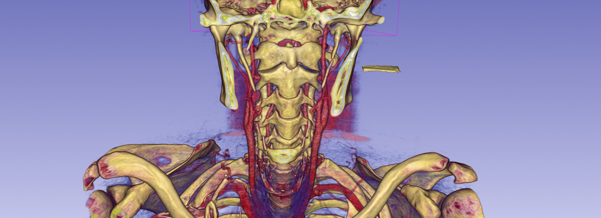
Right styloid process measures 4.6cm in length and left styloid process measures 4.4cm in length - “Mildly elongated styloid processes”

Perhaps my story might start In 2020 after a car crash, I would describe my symptoms at that time as severe whiplash, very sensitive neck and upper limb pain that never went away. If anything more symptoms appearing over time. Intermittent weeks and months of debilitation, originally thought to be something like CFS / Major Burnout.

Life is hard, no practitioner truly listens, people don’t understand. Luckily or tormentingly I am extremely stubborn and try to compensate as best I can, in between the occasional rotting/giving up periods.

My symptoms Include:

  • Being unable to look down, without rapidly worsening symptoms or it gets to that point anyway.
  • Vision issues - Blurry / double vision - it’s intermittent and variable in nature, I believe head position also influences it. Optometrist cant get my glasses right with the addition of prism and slight magnification, but I can read/test at 2020. Sometimes my vision wonders and I cant focus on anything.
    • I also experience this strange eye leaking (usually 1 eye - outer corner), which only happens after I lay partially reclined with my head back and turned after 10-15 mins, that happens when I have a dull headache, or after activity, often when i first wake up. This leaking is associated with a profound feeling of relief and brain fog elevating.
  • Hearing - I would describe as muffled/compressed, It makes music often un-enjoyable and i have difficulty understanding people sometimes. It also feels “full” - again i can somewhat reverse this with head position
    • Occasionally ill turn my head or look down with loud sudden tinnitus in one ear, again reveled by head position.
    • Often have some ringing/tinnitus that gets louder the worse i am, also pulsate tinnitus sometimes as well, often accompanied by a horrific headache.
  • Dizziness
  • Vertigo
  • Head and eye pressure
  • Positional sinus/ airway issues
  • Smell and taste
  • Weird nerve pain behind my ear (all round) - also temple
  • Jaw pain, teeth pain, scalp, neck
  • So overall symptoms like Occipital and Trigeminal neuralgia
  • Getting stuck in bed unable to wake/get up
  • Hyperadrenergic POTS - positional like symptoms
  • Sudden extreme nasua
  • Progressive difficulty swallowing - seems to correlate with how symptomatic I am, gets to the point where either it becomes to painful or I cant innovate a swallow, sometimes food gets suck for 10-15 mins (positional relief seems to help here). Smaller things seem to get stuck in throat without impacting/preventing swallow, I feel it release and go down, I think i also sometimes experience this but slightly different where its just the sensation.
    • I “choke” if i move my head to far back/ in the wrong position
    • I had a barium swallow test - which was “normal” - I would note that my Pyriform sinus stay filled between swallows, and I cant do a full big swallow in one go.
    • Talking seems to be a factor in making it worse
    • I have a “pointy bony protrusion” in the back of my throat near tonsil that pokes inward (can feel it with my finger) - ENT MISSED THIS! - I wonder if this is what others have described in the forum as the styloid shelf
    • Food and drink also goes up my nose quite regularly
  • Fatigue
  • Headaches / migraines
    • I have had these for weeks/ months at a time almost daily at points
  • Something seems to trigger Extreme anxiety / panic too, ill be feeling very unwell at the time usually, again usually reversed by position
  • Cognitive issues
    • Memory
    • Speech
    • Unable to access things i know i know
  • Thoracic / upper back and pain in both arms and hands
    • CRPS diagnosis for left hand/arm
    • Right hand is newer
    • Pain can be on any of the nerve distributions or move around
    • Outer 2 fingers on my hands occasionally have blood flow issue - they go blue
      • This again I think is positional, I think i have found some positions to reverse it, and triggered by say working at a desk
    • Arms above head, (I like to hold my head back and turned) - Both arms will tingle and then go completely numb - unable to move said arm

I have whinged to at least 10 GP’s, multiple physios and a few specialists (Neurologist (really really pissed me off and suggested FND), Sports doctor and ENT), all who all wouldn’t really listen, wouldn’t discuss the logical fallacies of their arguments, and said there was (sort of) nothing wrong with me.

I managed to get a 3D CT after finding the bony protrusion and presenting to a new GP with the dysphagia symptoms. The GP was willing to refer for CT with contrast/angiogram, however its very expensive and not covered by medicare unless a specialist refers it (frustrating).

  • The GP referred me to (with my input) Dr Ben Dixon at St Vincent’s hospital - they took a month to send me a letter so say no appointment time, waiting list.
  • Also referred me to Dr Michael Elliot (who seems to be the expert in the country) interstate in the hopes he might review over a phone appointment. They contacted me and said he reviewed it and I dont need to see him and I could see Dr Ben Dixon…
  • A 3rd referral to an OMFS - who have not gotten back to me and im questioning if that’s the best doctor to see anyway.

I rendered my CT scan, so ill provide a few images of that. Sure looks like there’s not enough room for the internal jugular/carotid. You’d think that’s enough for it to get taken seriously!

  • Also of note, is this what appears to be calcification between dens / C1??
  • I had an MRI that I am also pretty suspect on - I don’t believe the Nurologist personally reviewed it. (Anyone on here good at interpreting?)

Recently i also seen an Orthopedic surgeon, for an independent medical review, he actually listened and asked appropriate questions and tests (I didn’t get to/have time cover eagle syndrome type things). He seemed to think I had Thoracic Outlet Syndrome, but I am wondering if/ how much of that might be created by the Accessory cranial nerve?

Overall I am extremely frustrated at the medical system, Its very hard to convey the what and how I am so unwell and doctors don’t seem concerned as it doesn’t follow a particular pathology in 1 area or whatever. I often cant even communicate properly, but its dismissed as anxiety.

I probably forgot a few symptoms… (to many to remember)
Apologies if this is not formatted the best, using a computer/phone to type kinda sucks, and I am (probably) some form of dyslexic.

3 Likes

Hi, I recognised the mammothic tusks. Would just like to give my 2 cents.

Most important, you really need to get a contrast CT scan showing arteries and veins of your neck and brain especially seeing you have visual symptoms, head and eye pressure, scalp and neck pain, vertigo, brain fog, dizziness and tinnitus. Hopefully either the ENT or the Oral maxillofacial surgeon will be able to prescribe this scan given these symptoms. Surprised that Dr Elliott didn’t prescribe it.

The styloids certainly are causing your classic Eagle’s symptoms. I’ve annotated what appears to be the shadow of the internal jugular vein on the sagittal view of your CT scan. This is by no means diagnostic and needs to be verified with a contrast CT scan. In this neutral position it is more the transverse process of C1 that is creating the compression of your internal jugular veins. The styloids however would most likely come into play like a nutcracker when you look down and would explain your worsening symptoms and tinnitus in this position.

No doubt with the car accident you had 6 years ago this created alot of inflammation in the neck and tipped you from being able to compensate for any jugular compression to not being able to compensate and hence vascular symptoms. Take heed that a styloidectomy operation would create further inflammation in the neck potentially creating further compression of your internal jugular veins.

I would also ask Dr Ben Dixon what is his surgical approach as a summary on internet said he does a lot of operations via the intraoral route which is not the approach if you want to remove the styloids in there entirety.

4 Likes

Hello there,
Appreciate the 2 cents!

Yes I was quite surprised by the response from Dr Elliott/His office, I cant imagine looking at my CT and concluding that I didn’t need to. Though, the referral doesn’t list any suspected vascular type symptoms.

I appreciate the annotation,
Curious what might be the cause of the C1 positioning/ shape/ variance ? and judging by your comment suspect some of it might pre date 2020?

I assume take heed, in regards to inflammation is due to the C1 positioning, and resulting risk in recovery? Though, assuming the CT confirms the compression, as far as I am aware there is no alternative treatment?

Thank you for the reminder on the surgical approach!

1 Like

I agree with @Emerald 's comments & thanks for labelling the CT images!
It’s really frustrating as Prof Elliott does seem to be the most experienced doctor with vascular ES that we know of in Oz, and has declined to treat you- it looks as if there’s a fair amount of compression being caused by the C1 processes as well as styloids, so just removing the styloids might not be enough to help you. (But if that ends up being your only option it may well be worth a try to relieve your nerve pain and swallowing symptoms) It also sounds as if you do have high intracranial pressure, and that the symptoms are becoming worse from that- the leaking fluid from your eye sounds quite worrying, as this could be a CSF leak (rare to have this from your eye, but can happen- leaking from nose or ears is more common). Muffled hearing, headaches, dizziness, pulsatile tinnitus, visual symptoms, brain fog, etc are all vascular ES symptoms.
We are seeing members with more than one vascular compression syndrome, so TOS sounds like a possibility- proper testing would be needed to evaluate that…numbness, pain and/ or weakness in arms can be caused by a compressed spinal accessory nerve, and that is one of the nerves commonly affected by ES, but can’t say for sure which is the cause…
You could ask for a nerve pain medication to see if that settles the jaw, teeth & scalp pain, all common when the trigeminal or facial nerves are affected…
An OMFS might be able to diagnose ES for you, but you would really need a surgeon experienced with VES for the surgery, & doesn’t sound as if Dr Dixon would not be ideal as @Emerald said, if he only does intra-oral surgery…
It’s frustrating that a Neuro didn’t recognise the intracranial hypertension symptoms you have, we’re not very good at looking at MRIs on here I’m afraid…
I’m not really sure of what to suggest other than wait to see Dr Dixon or the OMFS doctor, whichever comes up first & try & get them to refer you for a CT with contrast, if you can’t afford to pay for one yourself? And even then it sounds like it’s going to be tough to get treatment, as if Prof Elliott won’t help I don’t know who else would treat this in your country…Others have resorted to getting an online appointment with Dr Aghayev in Turkey as he will review imaging, but not sure if he would still review without contrast images, and again there’s a cost (he does have a look for free & I think the cost to discuss your case with him is much lower than other doctors charge).
In the mean time, if you don’t already try sleeping propped up as this can help. I don’t know if your GP would be willing to prescribe a blood thinning medication like Diamox, Plavix, or you could ask about aspirin, or try supplements otherwise, there’s info here:
ES Information- Treatment: Pain Relief, Medications and Alternative Therapies - Welcome / Newbies Guide to Eagle Syndrome - Living with Eagle
There might be an Australian support group for intracranial hypertension you look at, they might have suggestions of doctors who treat this, and you might be able to get to see someone knowledgeable about IH who would actually look at your images & possible causes? If you’re on FB maybe have a look here:
IIH - Intracranial Hypertension Australia - Support Group | Facebook
Thinking of you, it’s such a difficult position to be in, and unfortunately we do understand on here, I wish that health care was better!

3 Likes

Is it common for a doctor like Prof Elliott to decline? I was refereed to the hospital, not privately, also the communication seemed odd.

On the Intracranial Hypertension significant changes in Barometric pressure knock me around too, elevators and sometimes stairs also. sudden urinary urgency issues that might relate also.

I also cant exercise with any intensity, even on say a ex bike, I get alot of head pressure, side of face goes numb, get very vague/stupid, cant get a good breath, even worse if someone tries to make my talk at the same time. Physio was also having me do some leg presses on the reformer that seemed to somehow give me head pressure! and when i get up my legs don’t work, plus feint

Come to think of it, I also forgot to explicitly mention, the occasional near blackouts/presyncope from positional changes

I have been quite suspicious of intracranial hypertension and intermittent leaks for quite a while.

As an indicative test, I used a glucose test strip on the fluid, once i got a small reading, the many other times it comes up as E3 (out of range to high) but it’s not exactly a reliable test. I should add, i would describe that my sinuses flush out at the same time, like it starts running down the back of my throat, tastes weird/sweet. The relief usually feels kinda euphoric.

The neurologist was very unhelpful, negligent even, didn’t even do any physical examination, tried to tell me it was all migraine and basically i am stupid for insisting that something is going on here and some kind of dynamic compression. Apparently if you do an MRI (without contrast) and EMG that rules out all neurological causes. And my lower back/ sciatic pain, that causes me difficulty walking and lifting my foot, didn’t warrant ticking the box to scan it too, and she would leave that “for the gp”… I think I am ranting at this point sorry.

In regards to TOS, the Practitioner seemed very familiar with it and i haven’t seen the report yet, I Indicated strongly on a number of specific tests (Addison’s, Surrender, and I forget), it was noted that my radial pulse drops completely in particular positions, I do get color change, coldness etc. I am not sure what more specific testing Is to be done though. I previously had a “normal” TOS ultrasound, IMO looked borderline and might be down to interpretation/expertise. I do think head position is a significant factor, and i do wonder about the accessory nerve causing partial palsy.

For me, it’s not so much the pain (although it sucks, but for me i think understanding why helps), it’s the disabling symptoms that worsen if i try to push through, or inadvertently trigger. and the constant compensation, just to half function. The cognitive difficulties are upsetting and embarrassing too. And the gaslighting and doubt from everyone.

And thank you, It’s been a very frustrating and isolating experience, with maybe some end in sight.
It is unfortunate to understand
and healthcare just truly baffles me!

4 Likes

@JustAGuy - If you were to get a CT scan w/ contrast that more clearly shows your IJV compression, perhaps you could get another opinion from Prof Elliot. Sometimes doctors just kind of wave their eyes over & dismiss imaging that’s not the type they prefer. If you provided him w/ imaging that definitively shows the level of IJV compression you have, you might get an invitation to visit him. Just speculating here, but it’s worth a try.

With IJV compression/IH, it’s very common for barometric pressure changes to cause symptoms to increase. Exercise increases blood flow & since the deoxygenated blood isn’t able to leave your brain via your IJVs at the rate freshly oxygenated blood enters through your internal carotids, your IH increases. The increased IH sounds to be giving you POTS (postural orthostatic tachycardia syndrome) symptoms i.e. feeling weak/faint when you change from sitting to standing or lying to sitting.

Sudden urinary urgency can be related to the vagus nerve being irritated. The vagus nerve lies w/in the carotid sheath w/ the IJV & internal carotid artery. In particular it snuggles next to the IJV right where the styloid/C1 compression occurs so when the IJV is crushed, so is the vagus nerve. The vagus is the largest of our cranial nerves & affects a significant number of bodily functions from skull base to pelvis. Thus when the IJV is decompressed, so is the vagus nerve & the symptoms it’s causing tend to subside over time just as the blood flow through the IJV normalizes over time.

Your left styloid is quite thick along w/ the calcification of your stylohyoid ligament is immensely long & your right one is long & quite curved bringing it close to your mandible. I have a hard time understanding how any doctor you’ve seen could have missed this in your imaging or been dismissive of it at least contributing to the symptoms you have. It’s downright scary that the evidence of CSF leaking you have from your eyes, nose & down your throat has also been dismissed.

Your cervical spine is very straight thus has lost its natural lordotic curve. That brings the styloids & cervical spine closer to nerves & vascular tissues in your neck. A straight cervical spine is referred to as military neck. This can be corrected by doing gentle PT exercises but it takes perseverance & time. With your potential CSF leak(s) & IJV compression, it might be best to wait on cervical spine correction until after your styloids/C1/IJV compression have been dealt with. If that continues to be a long time coming, you can explore the information in this post:

I’ve annotated a couple of your images, too. The thing I noticed is that you have decent space between your styloid & C1, however, the transverse processes (TPs) of your C1 vertebra are shaped differently than others I’ve seen. They’re slanted (which I’ve marked) & look to be flat vs more rounded. I’m wondering if it’s due to the slant that they are digging into your IJVs?

3 Likes

I think the best alternative open to you at this time without a CT scan with contrast is to get a private consultation with Geoffrey Parker an Interventional radiologist based in Sydney Australia. You could get him to revise all scans that you have had since your car accident. He has a special interest in head and neck radiology and CSF leaks.

I assume take heed, in regards to inflammation is due to the C1 positioning, and resulting risk in recovery? Though, assuming the CT confirms the compression, as far as I am aware there is no alternative treatment?

Yes, if you only get a styloidectomy done and the C1 positioning is causing significant compression of the internal jugular veins then you may have a very difficult surgical recovery, hence the importance of the CT scan with contrast. If the C1 is indeed causing significant compression of your internal jugulars veins then the alternative would be styloidectomy and a C1 shave (to open up the internal jugular) in the one operation.

5 Likes

Thank you, I really like the idea and Dr Geoffrey Parker sounds great!

I had been wanting someone to review it all actually, but wasn’t sure how to go about it/which specialty, I was wanting to seek a second opinion on the MRI in particular! The images weren’t annotated, and the report kinda glosses over, perhaps it was left to the Neurologist’s interpretation. (Ill put report bellow if interested/to give an idea)

Also appreciate the explanation, helped make a few things click!
I do also wonder about stability/CCI

I need to record all these details though so i don’t forget/ blank out when I go to these appointments eventually.

Report:

MR Cervical Spine:

Cord normal appearance, central canal patent, disc osteophyte bulging at most levels. No acute inflammation. Vertebral
body height maintained.
Foraminal stenoses - C6/7 mild bilaterally. Due to uncovertebral osteophytes. Facet OA is mild throughout.

MR Thoracic Spine:

Disc osteophyte bulging throughout, some of which contacts the ventral thoracic cord without significant compression or
compromise to the central canal. Vertebral body height maintained.
No foraminal compromise. No acute inflammation.
Conclusion:

  1. Low grade foraminal stenoses at C6/7 bilaterally.
  2. Disc osteophyte bulging at most levels of the thoracic spine producing slight indentations of the ventral cord
    without substantial central canal compromise.
  3. No demyelination of the cord or mass.

MR Brain:

No mass or oedema in the brain nor any significant white matter disease or atrophy for the age group. The DWI and SWI
are normal.
Paranasal sinuses and mastoid air cells are essentially clear with no upper airway or orbital mass detected. Flow voids
circle of Willis noted. Midline structures are normal.

2 Likes

@JustAGuy , it sounds like Dr Parker could be helpful if you could afford to see him privately. We’re not familiar with looking at MRI images, but there are changes with intracranial hypertension which can be seen on MRIs, so that would be something to ask about if you did get an appointment.
Otherwise I don’t know if you could get a referral with Dr Rao, he works with Prof Elliott doing the ES surgery- it might be a way to get your foot in the door with that team?
Dr. Prashanth Rao https://www.brainandspinesurgery.com.au
I don’t understand with the health care system how you can be dismissed either, but we see it so much on here- unfortunately I think since covid especially things have worsened, doctors are under so much pressure and often have to present cases to a panel to get their agreement as well before they can offer surgery. That’s certainly happening with the most well known ES surgeon in the UK where I am…
On your MRI report it does mention about stenosis C6/C7, so maybe that could be affecting your arms as well? The nerves for motor and sensory functions exit the spine here? Not saying it’s responsible for all the issues you have, but might be contributing?
Thanks for that info @Emerald , Dr Parker is on our list but we didn’t know how much expertise he has!

2 Likes

I do also wonder about stability/CCI

This is certainly a possibility and could explain the C1 positioning especially given that all these symptoms developed after a car accident. This is definitely a question to ask Dr Parker.

Just a point on the MRI, you don’t say when it was taken. Was it taken very soon after the car accident or later on after the development of your other symptoms as the MRI report states no signs of acute inflammation of the cervical spine ? Is there signs of chronic inflammation then? Another question for the radiologist.

Regardless of when the MRI was taken, you say that you have a very sensitive neck that never went away after the car accident. This could be due to damage to potentially preexisting collateral veins in the back of your neck that develop to compensate for internal jugular vein compression.

Often these collateral veins are tortuous anyway and a whiplash injury could certainly upset their capacity to compensate for internal jugular vein compression and damage them making them more tortuous than they already are; thus leading to cerebral venous congestion signs and a sensitve neck, a varicose vein type symptom (but in the neck).

This possible scenario however can only be excluded with a 3D rendering of a CT with contrast scan.

2 Likes

I read thru most of the comments but since you have POTS, do you have Ehlers Danlos -Hypermobility (hEDS)? Also have you checked out MCAS (Mast cell disorder) Alot of POTS patients, and now EDS are getting DX with MCAS. New theories about MCAS driving the inflammation in the body. BTW - Neurologists piss me off the most too! I just has a run in with my daughters POTS neurologist that doesn’t believe in IJV compression even thought I should him a picture of it on 3D. Grrrr.

I had whiplash and many of the same symptoms as you. Hearing loss documented in right ear over 10 years. I has TOS and several docs wont touch me because of my hEDS. I was just encouraged by vascular surgeon to get further evaluation. My fingers go numb and its from TOS.

As for CCI docs - he is in Barcelona: https://drgilete.com

Your in the right place. Put those gaslighting docs behind you. Its all about finding the right doc.

2 Likes

Sorry about the delay in responding to everyone! Haven’t been well, lacked the cognitive capacity of late.

@Jules - Are those changes with Intracranial hypertension typically picked up on standard reporting?

Yes, I do think spinal stenosis is a possible root cause for some of it, although this has been dismissed as doesn’t correlate. MRI’s are supposedly tricky due to “incidental findings”. I’ll throw a slice in of C6/7 and my “normal for my age (28) MRI” “Symptoms not neurological” and what I might call canal stenosis/narrowing which is apparently also not a problem… and I shouldn’t concern myself with what that looks like with flexion. (Can someone tell me if I am being a silly patient here?)

I do get some relief by pushing in somewhere around C6-T2 that often feels like it budges out. Profound relief from holding my head up (behind my ear) and back tucking in or curving my neck back, usually rotate to the left slightly, but if I rotate to far to the left I think I compress the facial nerve (lower part of face/ear goes numb and then hurts), I feel like there’s also a blood vessel involved here near my ear/cheek. An aggressive chin tuck that pulls in the thyroid area and hold also helps.

I do agree with the TOS diagnosis though, “Bilateral clinical signs indicative for thoracic outlet syndrome with positive “surrender”, Roos and Adson’s tests affecting all fingers and a decline of the radial artery and appearance of tachycardia during the provocation tests”.

@Emerald regarding the timeline of MRI’s, the report I listed was from mid-late 2025 after being rear ended in another accident early 2025, the CT scan was a few months after that. Thanks for the tip regarding chronic inflammation. The initial accident was mid-late 2020, left wrist MRI 3 weeks later, Cervical and Brachial plexus MRI 6 months post, 7 months post left wrist arthroscopy and debridement of the triangular fibrocartilage complex (TFCC) and removal of a large dorsal wrist ganglion, CRPS post surgery.

Interesting regarding potential collateral veins, hopefully we see soon. To me it’s absolutely baffling that Venus inflow and outflow is not checked, I don’t understand why that’s a pathology that’s completely ignored, I know so some extent they can see upstream changes, but it’s not really sufficient.

I have noticed that my mid back, lower back/leg/foot/bladder issues seem to correlate with aggravating neck positions and especially when I am experiencing eye leaking, I think I had a lot of thoughts around this but I have forgotten (If I don’t write it down I forget… and then good luck collating that into something meaningful). I actually presented to the GP with both eyes that would not stop, doc seemed to think viral conjunctivitis, I could barely communicate and basically said concerned about CSF leak, to which naturally he responded oh there’s no way, you’d be very unwell etc.

Maybe I was wanting to ask about an increase in visual disturbances like seeing starts on lights (day and night), vss (screens), blur, confusion, difficulty communicating, fatigue etc - a bunch of these things appeared together for about a week. Above my left eye often looks puffy/droopy in the morning after getting up.

Edit: also maybe the doc was right about the conjunctivitis, days later and they are a little gunky. I also wonder about some kind of forced lacrimation due to nerve involvement, but that doesn’t explain why it feels so relieving/ good when it does leak, and also the positional nature.

1 Like

I am not sure about hEDS, I have considered the possibility, but it’s likely difficult to peruse. I don’t quite qualify as hyper mobile, but i think you don’t have to? MCAS also considered as I feel like I get weird allergies/reactions, but again another rabbit hole and unsure how much of it is explained by Eagles etc.

Sorry to hear about your frustrating experiences also! I don’t understand why it’s like this, and this arrogant oppositional attitude to evidence and common sense. Their ego cant handle it or something.

2 Likes

There are changes which can be seen on an MRI with intracranial hypertension, yes, but I don’t know how severe it has to be/ how symptomatic you would be before the changes show (empty sella where the pituitary gland is, and a swollen optic nerve sheath for example), or otherwise whether it’s one of those things that radiologists won’t look for or ignore unless specifically asked to?

No, not a silly patient, so much is dismissed as within normal boundaries etc, like we’re all the same and it’s just another example of the medical professions’ one size fits all approach! If it causes symptoms then you can’t dismiss it as normal! Given your medical history too, this should be taken into account…
I get that numbness sometimes rotating my head too far as well! Just be careful experimenting with different head positions to try to alleviate symptoms, you might inadvertently make them worse! :hugs:

2 Likes

I’m not good at seeing things in MRIs so can’t comment on the images you posted, however, your radiology report noted several larger than normal lymph nodes. That can be indicative of an occult Lymes Disease or other infection. Have you been checked for Lymes or anything else?

Have you been treated for conjunctivitis? If not, it seems odd that your doctor would suggest it then not provide you with the means to treat it.

That’s an interesting observation which does sort of lead to eye leaking possibly being CSF leak related though bladder issues would have more to do w/ your vagus nerve the other issues would be connected to irritated spinal nerves which could make sense in light of the disc bulge comments in your MRI report.

1 Like