Imaging/symptom input? Theories?

I’m about stumped for answers at this point so any input/opinions would be appreciated, my imagination is open. I had a Venogram was “negative” with right head rotation. Symptom wise I have about everything in the book (stroke/TIA-like symptoms, face pain and swelling, objective pulsatile tinnitus, myelopathy, cognitive symptoms, feel arterial pulse in teeth on left side, swelling in throat, stridor with exercise, photophobia, personality change, constant nausea, you name it).

I see an MS specialist next week (don’t think I have it but at least an academic neurologist can decide where to send me).

3D reconstruction shows internal jugular veins both compressed by C1 (~1yr ago)
Left - Feel face and neck pulling when rotating head to right


Right side

Neck CT with contrast more recent

Size of IVJs superior to C1

Size of IJVs (pink) at level of C1 with calcified stylohyoid ligaments (cyan)

Dilated collateral veins of pterygoid venous plexus (green above) (left side of throat feels and looks like it’s caving in, and tongue swelling on that side also)

Veins all contributing to the jugular vein below level of C1 giving appearance the vein is “large” as one ENT put it. I suspect the catheter for the venogram was not high enough and reads as normal but most of the blood is coming from elsewhere and not the jugular vein. Both sides have similar appearance on this more recent CT than the 3D from a year ago.

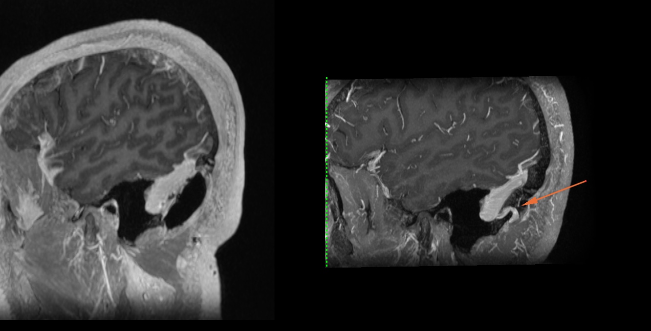
Left side collateral vessels on angiogram which I firmly believe to be causing face and eye symptoms

This all started with face pain ~7 years ago, and a large emissary vein draining from the back of the skull on the left side was sacrificed for a craniotomy (right before, left after). An already compromised venous system now missing one of its routes. A lot of my Eagle symptoms started right after the craniotomy. Diagnosis was unknown but couldn’t handle the facial pain any longer and there was a visibly large vein compressing it so I took my chances. Helped for a while but slowly gotten worse over time.

Unfortunately I think I also may have Bowhunter syndrome on the Right side (osteophyte compressing R vertebral artery) after reviewing my CT (only explains a few of my symptoms and not left face issues) but will have to get in line for another angiogram or maybe Doppler ultrasound

I’ve probably seen 10+ specialists between ENT/neurologists/neurosurgeons who usually want to send me to a headache clinic (also didn’t help).

Thanks!

@Rad1 - Very thorough discussion & imaging to support the possible causes of your symptoms. Unfortunately, I don’t have enough skill looking at CT images to give any sort of an opinion. I can usually recognize the styloid processes & hyoid bone (but even then have been wrong :scream: :crazy_face:) so I’ll leave further discussion to the likes of @vdm, @Kooldude & @coldbear & others who are better versed in reading & commenting on these. I expect you’ll get some helpful input.

1 Like

@Rad1 Just a word of encouragement. You are doing a great job in advocating for yourself and also see that you have a better knowledge of reading these images. When Thomas Edison was inventing the light bulb, he went through a series of failures ( or may I say porotypes that did not work) before he can get to the right one and he was famously quoted as saying

“I have not failed 10,000 times. I have not failed once. I have succeeded in proving that those 10,000 ways will not work. When I have eliminated the ways that will not work, I will find the way that will work".

All your efforts in seeking answers whether seeing multiple doctors, learning how to research and read images will pay off one day and give you the answer you are looking for.

Every imaging needs to be interpreted with symptoms and I feel Jugular vein compression alone does not explain the myriad symptoms you have but definitely might account for some. I do see IJV narrowing & compression as you said along with dilated veins in different places as you rightly pointed. I have a number of questions for you to better guide my suspicion/theory here.

  1. When they did the Catheter Angiogram, did they rule out any Dural Arteriovenous Fistulas (DAVFs) such as Cavernous Fistula. These can cause objective pulsatile tinnitus with a whole host of symptoms in the facial area along with cognitive symptoms. I am assuming they did but sometimes, depending whoever is reading it, they might miss it.

  2. What symptoms started after craniotomy and what symptoms preceded it ( I know facial pain preceded it but would like to establish a good hypothesis to pursue as the surgery can also be confounding factor)

  3. Do you have short of breath/breathlessness and blueish lips with facial/neck swelling

  4. Do you have the contrast CT scan of the head and neck uploaded somewhere we can take a quick look (Not radiologists here but we might point to our suspicion and you can confirm it with competent radiologists to either establish our suspicions or dismiss it) I have a few suspicions of my own but would like both CT and MRA of neck & head if you had one.

3 Likes

@Rad1 First a disclaimer. I am not a doctor nor a radiologist and all my assertions here need to be established with proper imaging and verified by a trained radiologist or doctors. Secondly, assuming that the recent Catheter Angiogram ruled out DAVFs such as cavernous DAVF and other vascular malformations, I think the limited images you shared show what appears to be Jugular Vein Reflux. That is usually indicated in imaging as hyperintensities on the Jugular Vein. The brightness shows in-flow instead of outflow of blood (as pointed by the large cyan arrow below images). Normally Jugular Vein carries blood out of the head and towards the heart but in cases where there is compression or obstruction in the central veins such as left brachiocephalic vein/Vena cava, the blood refluxes back to the Jugular vein and back to the head area causing facial pain, breathlessness sensation, neck/face swelling, tachycardia, temperature changes and blueish lips due to deoxygenated blood backing up to the veins of the head and neck. This correlates with your symptoms of facial pain and the engorged/dilated veins of the face such as facial vein/Superior Ophthalmic Vein that was causing pain prior to the craniotomy. It is mainly caused by left brachiocephalic vein compression between the Aorta and the Sternum so if you have chest CT scan, we can establish that is the case. There was one of our fellows here called @DogLover who suffered this type of compression and we talked about her case here (Dilated Collaterals Cause Progressive Weakness on All Limbs as result of Bilateral Jugular Vein Occlusion - Interesting IJV Bypass Treatment - #37 by DogLover) if you want to read her case more. That link has it all.

So this is the suspicion I have but in order to establish it, we need chest contrast CT at the level of the Aorta to see if that is the case. I do not think CT of neck and head go all the way to that level. Also keep in mind that sometimes, Cavernous DAVF or cDAVF as it is known can mimic Jugular Vein Reflux from BC compression as this article (MRI and MR Angiography Findings to Differentiate Jugular Venous Reflux From Cavernous Dural Arteriovenous Fistula : American Journal of Roentgenology : Vol. 202, No. 4 (AJR)) detailed how to differentiate the two.

Big Cyan Arrows pointing what I believe to be hyperintensities in the Jugular Vein potentially caused by retrograde blood flow.


2 Likes