@kel34ban if you can send the DICOM files somehow, happy to take a look if i can work it out.
1 Like
Not sure if you can private message your email on here as I can email you the files i have.
Thank you
Hi
It will not send the file via email ![]() but thank you
but thank you
@kel34ban - you should be able to upload your dicom file in a private message to @LimeZest. Files that are too big for normal email seem to load ok there.
2 Likes
I will try this now. Thank you
3 Likes
@kel34ban your email did come through - received it at 6 am this morning but it went to spam. Will look at your scans (of course not a doctor) but can at least post some images here for people to comment.
2 Likes
Thank you. It said to big to send each way I tried to send it so I assumed you would not get it.
When you have had your appointment can you tag me so I can see how it went please. My brain is non existent these days and I may miss the chat ![]()
If it my brain function ever comes back I think I’ll retrain as a neurosurgeon lol
2 Likes
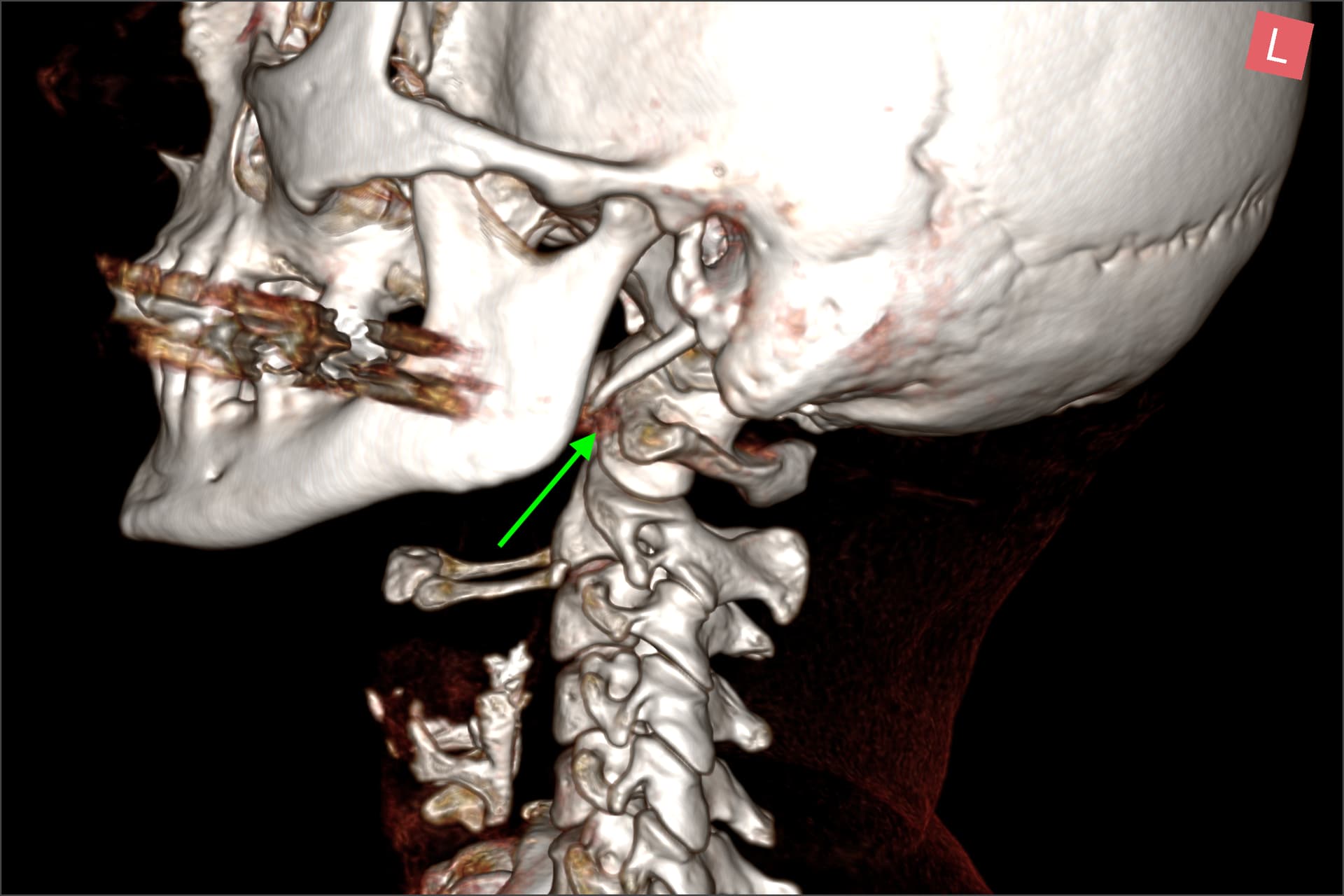
@kel34ban i’ve uploaded your CTV images here with annotations of what i think is what
Have put some 3D images so you can see styloids and also the jugular veins.
Picture of right styloid

Left styloid
Frontal view

Frontal view with veins

Left IJV with some compression - note i think an artefact (i.e not really there but present in scan) at IJV level coming from teeth (maybe metal filling?) but kind of shows compression

Can see compression from C1 a bit better here

I dont know if this is just me but i think the big vein running down spine looks bigger on right (dont know for sure but looks like it could be venous plexus which is typically dominant when upright)

Right IJV picture

This is axial image, so imagine a slice through your neck horizontally showing styloids and round bits being the IJVs

Another axial image a few slices down showing left IJV compression (looks like its on right side but image is reversed)

Sagittal view of R IJV

Sagittal vie of L IJV showing compression from C1

Again not a doctor so dont take anything i say here as medical information. I also note that there seems to be calcification around thyroid perhaps? Or is that normal thyroid cartilage?
Perhaps others have some ideas of what is going on here.
2 Likes
Thank you for this. I think Dr T did not want to help as he thinks I have EDS! I don’t think physio can help with this so I’ll not waste more money and energy.
Thank you @LimeZest for sorting the scans!
The C1 process seems to be pushing the IJV, but doesn’t look to me as if that’s the only compression as the jugular veins are distended from lower down, below where the scan ends?
1 Like
no problem @Jules very happy to help. let me know if there is a better view you think may help. I did read a paper that suggested people with TOS are also likely due have compression higher up. But @kel34ban i think maybe best to just get the blood patch done and see how you feel after. Even if the results are temporary maybe it will give you a better idea of what symptoms are coming from where.
Hope you are managing ok!
2 Likes
Thank you both. I suspected compression lower down as I get fluid and bulging veins when I lean forward.
I’m going to have one more private consultation with a vascular consultant whose interest is TOS and then definitely no more. I’m too weak and I really do give up😩
I definitely feel like I have more than just a csf leak going on as it feels very vascular. My blood patch is on the 15th February and I’m scared because deep down I know something else is very wrong.
Who knows, miracles might happen and all my symptoms could be csf leak related. I know that is not the case as I can feel it but any relief is better than none.
2 Likes
And what is sad is that the consultant never even mentioned that. I get that it is not his area but he should have been honest and said that the issue may be further down.
We are all just left to piece the puzzle together and it’s so difficult when you are unwell.
2 Likes
This thing you mention before about having to lie flat to resolve the feeling, thats super consistent with a CSF leak. I really hope the blood patch at least gives you some relief temporarily.
3 Likes

This also seems really similar. Even down to the distended vein you have on your neck, and the strange knotted vasculature you show.
1 Like
That could be me lol
I’m guessing there is no fix to all these issues and certainly not in the UK ![]()
My vein pulses massively on the left side of my neck.
Where did you find the video?
Thank you
2 Likes
I also noticed that your cervical spine is ramrod straight, & your styloids are coming off your skull base straighter toward your cervical spine than we usually see. They may only appear that way due to your head position when getting scanned but normally they point more downward. The styloids are also fairly thick & pointed.
Doing some gentle neck work to work on restoring your cervical curve might be helpful in reducing symptoms. A simple exercise to start on this is to lie on your back w/ knees bent & gently press the back of your head into the surface you’re lying on for a slow count of 5 then relax for a slow count of 5 & repeat 5-10 times. You can do this several times a day IF it doesn’t cause pain or stir up symptoms. A firm surface (carpeted floor, yoga mat) is better than a soft surface like a bed or couch.
1 Like
Thank you for all the info
1 Like
Another member @Warrick who is in the UK has been studying these videos. So i have watched a couple and the most recent one (the one i posted) was put on youtube yesterday or so. I just found quite a lot of parallels in your story, CSF leak, distended veins. I did find it curious that the one on the outside of your neck was clearly not your IJV, it sat far too on the surface. So it seems its a collateral vein that has swollen to compensate for compression elsewhere.
Again i think best thing to do is get the blood patch, see how you feel and then if (which i hope not) the leak happens again go from there.
2 Likes
Hi @kel34ban
Sorry to hear your ordeal here. I think it is hard to conclude anything from 3D images alone but I see something that might be behind your CSF leak. Both of your transverse sinuses are narrowed (stenosed). This can induce Intracranial hypertension which can lead to CSF leak as the pressure goes up in the spine and brain dura tear can occur. Not a doctor but that is what I can see with the limited images you posted without studying the whole CTV. There could be other stuff going on but I will need to see the CTV to confirm it.
I think this what you need.
1 - you need to book Catheter angiogram/venogram to measure the pressure gradient in the transverse sinus.
2 - Address the transverse sinus stenosis by stenting
3 - Get a blood patch to seal the leak.
Images below show bilateral transverse sinus stenosis ( yellow arrows)


4 Likes