New here - my story

Hi everyone,

Thank you for welcoming me into this group. I am new. I have been in hell since 2023, but before that have had bizarre and bad symptoms since around 2016, but really bad since 2018: severe migraines and pain shooting into the left side of my face (one of the worst symptoms), neck pain, ears blocked and ringing in my ears, sinuses blocked and pressure, dizziness and vertigo, severe neck cracking, brain fog, vision problems (the other worst symptom) on the left side, pain around the ear, jaw pain and bite problems (teeth don’t touch at all in back suddenly for no reason), occasional numbness in the back of my head, trouble talking and fatigue talking (bad symptom), tongue fatigue and problems, throat issues, painful spot behind left upper molar area, and more. It’s been a hellscape. I do not know what is going on but my sister read about Eagle Syndrome, and then she and I both uploaded my X-rays into AI and it said they were elongated and prominent. If it’s not this, it’s my cervical spine, but my cervical spine MRI only shows “mild degenerative changes.” I am 57 years old and female. Most people my age would have mild degenerative changes. I have been to several doctors for the neck issue and most have said my cervical spine could not be responsible. But, one new one I saw recently said my spine IS causing my “headaches” but he did not say what part of my spine is responsible or how it is causing all of this. He just said “the pain is coming from your neck.” He wants to put Botox into my SCM muscles and if that does not work, do facet joint injections at C5, C6, and C7. I am not necessarily opposed to this, but my question is: Why? What evidence is this being based upon? The treatment seems to be just a shot in the dark. How long do I go on taking all kind of meds (been there, done that, to no effect) and letting all kind of docs do all kinds of scary procedures on me without knowing what is wrong?

To date I have had: many, many meds, Botox for migraine, C3-C4 facet ablation, nerve root injections at C5 and C6 , and PT. I have had cervical and brain MRIs (brain completely normal, thank God). PT with dry needling has helped more than anything . I have been sent to pain management mental health groups and CBT therapy for anxiety contributing to my pain levels. I mean, sure, anxiety probably is, but I am in pain so yes, I am anxious. I have been anxious before many times in my life and it never created pain. I feel like something is really wrong in my head and neck and I am not being listened to. I feel like I am living on a different planet. Maybe it is my neck or maybe it’s Eagle Syndrome, or maybe it is something else, I do not know.

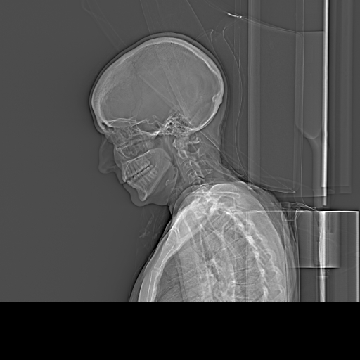
Anyway I have two lateral X-rays taken in 2023 and 2024 when I went to the ER with severe pain and they ruled out stroke. Is anyone available to read them?

Thank you to all of you - and I offer my sympathy to all of you who are suffering.

2 Likes

Hi @Quiju01 - Welcome to our forum! Many of your symptoms sound like they could be related to ES w/ internal jugular vein (IJV) compression while other are the result of nerve compression or irritation most likely by your elongated styloids.

Migraines, neck pain, blocked/ringing ears, dizziness/vertigo, brain fog, vision problems, numbness on back of head are all symptoms of a vascular issue.

Sinus blockage/pressure, facial pain, neck pain, pain around the ear, jaw pain, facial pain, vocal fatigue, tongue fatigue throat issues, pain behind your upper molar, & anxiety are all related to nerves that can be negatively affected by effected by elongated styloids.

The following post is a bit long but it will help you see why we’d guess your symptoms are predominantly being caused by your styloids affecting nerves & vascular tissues in your neck thus the doctor who said your “spine” is responsible is correct but only if he’s referring to the upper part of your cervical spine (C1) & styloid processes.

IJV compression coinciding w/ elongated styloids most often occurs between the styloid(s) & C1 vertebra up near the skull base. When the IJV(s) is/are not fully open, the rate of blood flow out of the brain via IJVs is reduced whereas the blood flow into the brain via the internal carotid arteries (ICAs) remains normal so a situation of intracranial hypertension (IH) occurs which causes the migraine level headaches & other symptoms you mentioned. Additionally, the vagus nerve passes between the styloids & C1 alongside the IJV so when the IJV gets compressed, so does the vagus nerve which is responsible for managing anxiety, heart rate, blood pressure, vocalization, breathing, swallowing, to some degree, & stomach/intestinal functions among other things. Thus, a number of the symptoms you mentioned above could be related to vagus irritation or compression.

We’d be happy for you to post your lateral x-ray images, but we won’t be able to give you any insight regarding vascular compression until you’ve had a CT scan with contrast. It’s best to get one w/ & w/o so all aspects of what’s going on with your styloids can be seen clearly. You want the imaging to be done between your skull base & hyoid bone.

2 Likes

I forgot to add that I somehow managed to get an appointment with an ENT at dr Donald Annino’s office in Boston on 11/25. Dr Annino is one of the Eagle providers on the provider page. I talked the guy into it who answered the phone.

2 Likes

So you aren’t seeing Dr. Annino himself?

No I’m not seeing Dr Annino. I’m seeing a Dr Prince. He is a head and neck surgeon who is on Dr Annino’s team.

1 Like

Thank you. I did have a CT angiogram on both of those occasions when I went to the ER. So I have the scans but I can’t manage to get them out of the patient system (what appears to be the 3D image). I can only export the lateral x-ray. When I have my appt on 11/25, Dr Prince at Dr Annino’s office will have access to them in the system.

2 Likes

Dr. Prince hasn’t been mentioned on or forum so I hope he knows about ES. It seems like even in a team of doctors where one is an ES specialist, the other barely know anything about it. That’s not me being negative, it’s just that if you prepare for that & then Dr. Prince actually has good ES knowledge, it will be a pleasant surprise.

Regarding your imaging, if you can see the 3D images, but just can’t upload them, try taking screen shots & posting those here. It’s best to remove any personal information from the images before posting them on our forum.

1 Like

Well I will let you all know what happens, if he knows or not and if he is receptive or not to what I have to say. I don’t think you’re being negative, just realistic. I get it - I’ve been through the mill with these doctors.

2 Likes

Fyi

Having a quick look at your imaging, it looks like you do have an elongated styloid process (although it’s not that clear), but what stands out to me is your hyoid bone processes- even without enlarging the image you can see they look abnormally curved upwards, with little hooks on the end! It could be that the stylo-hyoid ligaments have calcified from the hyoid end, or the processes themselves are elongated. So it could be that you have both ES & HBS. So if you can get access to your other images &/ or new images it’ll be interesting to see the hyoid on those. If Dr Prince is receptive to ES, then it might be worth asking him about the hyoid too.
Also looking at the image, I think your neck has lost it’s natural curve, aka military neck. We see this quite a bit, it can make vascular symptoms worse potentially or it could be that this is your body’s way of trying to make more space for blood vessels. There’s info & exercises which might help in this discussion:
List of my favourite resources on YouTube to learn anatomy - General - Living with Eagle
I hope that he is helpful!

1 Like

thank you so much for taking a look at my X-ray & for your thoughts on the hyoid, etc. I will most definitely speak to Dr. Prince about all of it. He will be able to access my CT angiogram which I believe has the 3D reconstruction. As far as the neck goes, I had 2 MRIs of the cervical spine. In this image, which is a lateral X-ray from my CT angiogram, my head is flexed forward (laying on a pillow or something you can sort of see in the image). My head is pretty angled down and forward. The spine would not have the lordosis in that position as it would have in a normal position. My MRI assessed this as well. My entire cervical MRI was fairly unimpressive in my opinion, which is sort of why I am so tired of hearing from (some) doctors that all of this mayhem is coming from my neck (!).

1 Like

@Quiju01 - I’ve annotated your x-ray so you can better see what @Jules is talking about. Your styloid is quite long & curvy, & the greater horn of your hyoid bone is as she described. You also have fairly significant forward head posture (which causes loss of the lordotic curve) & your shoulders are rolled forward. Those 2 things are correctible w/ PT but take time to fix. It’s best to deal with the styloid & hyoid bone situations before working on your posture too much though, if it’s not uncomfy, you can try doing some of the exercises in the link Jules sent you.

Thank you for the annotations! See my explanation above about the neck. I was lying down for that CT scan and was propped up on something. Thankfully I do not stand like this in real life :grin: . I have actually been told I stand up really straight and look like I have good posture just FYI. Also I am in PT.

2 Likes

Got it! Your post must have immediately preceded mine in landing on the forum, & I didn’t see it once mine was up. Curious that they put your head on a pillow for an x-ray as that could be making your hyoid situation look worse than it might be & the position of your styloid worse, too.

I do have to tell you that even among our members who have great posture, their cervical lordosis is gone. That’s not something that’s necessarily visible from the outside.

2 Likes

Good afternoon @Quiju01

I have not posted for a while so hi to @Jules and @Isaiah_40_31 too.

Your problems and symptoms are much like mine and I await c1/2 fusion (yes still waiting). Could your tests include mri? CT with contrast? Make sure they look at and report the c0-c2 area as I’ve known many reports that fail to even mention this critical area. Often the symptoms can overlap several areas and my diagnosis of ES is overshadow by AAI. The other parts of the c spine get degenerative changes sure, but the very top where so many structures are affected do very badly. Symptoms are severe for me now so get the ball rolling as the journey is long and there are many rabbit holes open for us to dive into

Feel free to message me if you wish?

All the best Xx

Ps my MP has got involved now due to long waits

1 Like

@Mcwelly - It’s good to see you back, but I’m sorry you’re awaiting a fusion now & wish for you that it’s tremendously helpful in resolving your worst symptoms & hopefully others as well. It sounds like you’ve made some diagnosis progress since you last posted so that’s a huge plus, but your journey has been tremendously long. I’m really glad your MP is now advocating for you. I hope that expedites your procedure significantly. :hugs:

2 Likes

Good your MP is involved, hope they can get you pushed forward! :hugs:

2 Likes