Hi VDM,
Yes, the catheter venogram was done at supine position and I agree the fluid dynamics are affected by position and heart rate in the case of the vascular system along with many other physiological stuff such as whether you are awake or sleeping since that affects the heart rate. So standing/sitting will be different from supine position, similarly, running vs stationary.
It turns out though that the Jugular Veins tend to do all the draining exclusively in supine position as opposed to standing or sitting where they are partially helped by the collateral veins along the spine. So if you see collateral filling while supine position, it can only mean that your Jugulars are somewhat impaired and blood is rerouted to collateral veins and since being upright naturally collapses the jugular vein, an impaired jugular drainage is even more impaired while standing or sitting. So doing the test in supine position might more accurately assess your Jugular drainage than doing it in upright is my understanding of it.
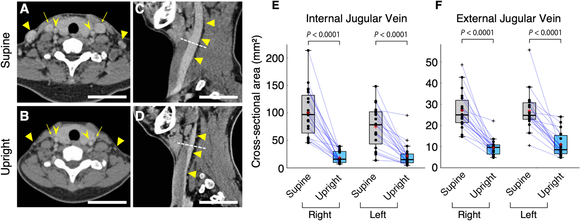
I have included a figures from a study comparing the jugular vein in upright position versus supine below.
Figure 1

Neck veins are collapsed in an upright body position. ( A , B ) Axial computed tomography (CT) section from the recirculation phase. Arrows, triangles, and arrowheads show the internal jugular veins (IJVs), external jugular veins (EJVs), and internal carotid arteries, respectively. Although the internal carotid arteries hardly change in different postures, the IJVs and EJVs are significantly collapsed in an upright posture. Scale bars: 50 mm. ( C , D ) Sagittal CT section from the same phase. The IJVs (arrowheads) are significantly collapsed. The dashed white line indicates the level of area measurement. ( E , F ) The cross-sectional area of the IJVs ( E , n = 20, two-sided paired t -test, right: P < 0.0001; left: P < 0.0001) and EJVs ( F , n = 19, two-sided paired t -test, right: P < 0.0001; left: P < 0.0001). In the box plots, the central mark indicates the median, the red cross indicates the mean, and the bottom and top edges of the box indicate the 25th and 75th percentiles, respectively. Whiskers extend to the maximum and minimum values within 1.5 interquartile ranges below the first quartile or above the third quartile, and black crosses indicate outliers. Increases and decreases from a supine position to an upright position are colored in red and blue, respectiv
Figure 4

Summary of the positional changes in craniocervical venous structure between supine and upright posture. In the cervical region, the internal jugular vein (IJV) significantly collapses. In the craniocervical junction, the IJV shrinks in an upright posture; in contrast, the anterior condylar vein (ACV), anterior condylar confluence (ACC), and vertebral venous system—including the suboccipital cavernous sinus (SOCS), vertebral artery venous plexus (VAVP), and anterior internal vertebral venous plexus (AIVVP), as described in the figure—were enlarged in an upright posture. The following three venous routes become more prominent in the upright position: (1) the ACV, originating from the ACC and draining into the SOCS, VAVP, and AIVVP; (2) the LCV, originating from the ACC and draining into the SOCS, VAVP, and AIVVP; (3) the PCV, originating from the ACC or jugular bulb and draining into the SOCS, as represented by numbers in the figure. As opposed to those venous structures, the pterygoid plexus (PP), located anteriorly, does not undergo consistent changes depending on posture. In the intracranial space, the venous structure undergoes almost no change between postures. The vertebral venous plexus (VVP) at the cervical level, which could not be evaluated in this study, is shown as a dotted line.