Question for those with military/straight neck (loss of cervical lordosis)

Hi,

Just a question/observation from my side: do you also get some relief from symptoms, esp. jugular vein compression symptoms, while lying on your back and holding your hands under neck/just above the neck, like in this picture?

UPDATE: that’s a serious question, no joke, despite the picture (couldn’t find a better one quickly to fully explain what I meant).

2 Likes

Hi VDM,

I tried the technique last night to see if I get a relief but my finding are mixed. I felt bit better for short period but it was not sustained for long. Don’t know if I am doing it the right way though.

1 Like

True, it would very unlikely give any long-lasting effects, but merely a way to give those stiff muscles some short-lasting relief from tension and bring the “normal” curves into the spine.

But if it works temporarily, I’d guess in that case you might get some benefit of a good physio by trying to:

  • release tight upper trapezius muscles
  • release tight scalenes
  • release tight levator scapulae muscles
  • release tight pec minor and pec major muscles
  • possibly release all the knots in rhomboids and lat.dorsi muscles
  • release hamstring muscles
  • release rectus abdominis
  • strengthen everything, and “awake” the postural muscles in the lower back (wouldn’t be surprised if it hurts too right now), as they are probably overtaken by action muscles right now.

I’d really suggest to pay a visit to a good physio, it might help you tremendously while waiting for any next steps in Eagle’s treatment.

Based on my own experience, I even suspect my styloid processes became thick (they were much thinner on panoramic dental xrays taken in 2014) to stabilize the head and prevent it from being pulled too much backwards. For the last 3-4 months or so, I’ve been working on myself with this theory in mind, and now see some results - getting spontaneous relief in neck, shoulders and lower back (esp. around the SI joint). It’s crazily slow process though.

The pics below might give more context into this.


3 Likes

I do not mind getting a good physio but since my jugular vein is compressed and I do not know if there is a good physio-therapist that won’t make things worse by messing with my neck muscles & bones.

For example, when I was doing the dynamic catheter angiogram/venogram, they found when I turn my head to right, they do not see a collateral filling via condylar vein which was translated as a temporary relief of styloid & C1 compression but on head turned to the left and on a neutral position, the compression does not get relieved hence the prominent appearance of collateral venous filling. So provocative movements, while they could potentially relieve my symptoms, they also have the potential to make things worse. so I am undecided on this at this point. May be after surgery to make my neck bit more flexible.

BTW, I put some of the images of head movement during the Catheter Angiogram along with the report below.

Head tilted to left - Collateral filling can be seen.

Head tilted to right - No Collateral filling can be seen.

Head in a neutral position - Collateral filling can be seen.

Report finding:

Diagnostic catheter was advanced over the wire under roadmap navigation to selectively catheterize the right common
carotid artery, left vertebral artery left common carotid artery. Venous phase angiograms were obtained from each
selective catheterization including visualization of brachiocephalic/jugular system in the chest, neck as well as intracranial
dural venous sinuses. Venous manometry was performed with the catheter in left common carotid artery. Additionally,
provocative testing was performed with head turned to the right and to the left respectively and the venous outflow was
assessed.
The following observations are made:

  • On the right common carotid artery injection and left vertebral injection, the left transverse sinus is the dominant sinus
    but there is also nondominant flow into the right transverse sinus. Right transverse sinus is not hypoplastic but only
    nondominant. Bilateral prominent condylar veins are seen, left more than the right.
  • The left common carotid artery angiogram shows that the left hemisphere is draining almost exclusively through the left
    transverse sinus.
  • There is compression of internal jugular vein at the level of C1 transverse process-styloid process bilaterally.
  • On provocative testing with head turned to the right and left respectively, an interesting observation is made: When head
    is turned to the right, the left condylar vein is no longer opacified. With the head in neutral position or turned to the left, the
    left condylar vein is prominently seen. This suggests that with head turned to the right, there is improvement of the venous
    outflow, by relief of the compression at the level of styloid process-C1 transverse process with a better downstream flow
    towards the heart with consequent reduction of venous reflux into the left condylar vein. This may explain his left-sided
    pulsatile tinnitus.
  • There is no evidence of dural arteriovenous fistula.

There were no complications during the procedure.
The catheters were removed. Hemostasis at right common femoral vein access was achieved with manual compression.
Left common femoral artery was closed using Angio-Seal device after confirmation with a groin angiogram.
Patient was transferred to surgical daycare with stable vital signs and at neurological baseline.

IMPRESSION:
Eagle syndrome, with prominent venous reflux into left condylar vein - this is the most likely cause of his left-sided
pulsatile tinnitus. This venous reflux improves with head turned to the right. See detailed explanation above.
Dominant left transverse sinus.

1 Like

I think holding off on physio would be wise for now too…

2 Likes

I agree w/ Jules because a part of PT can be massage or mobilization of the injured body part plus exercises to help strengthen muscles or relax tight muscles. All of those things could potentially further stimulate your symptoms instead of reducing them.

2 Likes

Well, it’s always a hit-or-miss with physiotherapists, as I had quite a few of them in the last 10 years or so. Just looking retrospectively, I wish I had one really good physiotherapist as soon as I became aware of styloid process problems. “Good” - I mean, they work with you rather on you, and could basically give you a good overview of how human’s skeletomuscular system is designed to work, monitor your motion/movement/posture, give advice what to do and (even more important) what not do.

Until I found enough sources and materials explaining body’s muscular system in simple layman terms, flowcharts and pictures like the above, or youtube videos that show them in 3D in action, I was just navigating among the individual trees without seeing how big the forest is. And the more I Iearn, the more I understand how limited my understanding still is. I guess, a good physio would have helped me to find out these things much earlier.

For example, just recently it “clicked” with me that for very long time my superficial abdominal muscles (including ABS) were desperately and subconsciously compensating for weak back muscles.

It might sound total nonsense, as according to the individual-muscle-analysis and typical description (e.g. Rectus abdominis muscle - Wikipedia), you would find that antagonist muscle is Erector spinae, and they pull in “opposite” directions. So first thought is that if ABS needs to work hard to keep the body bent, that means it is doing that to counteract strong/stiff Erector spinae. But… Not that simple.

How? Well, the ABS would stiffen, become short and rigid, almost solid, to just support me leaning forward as the back muscles are not capable of this by pulling/holding the spine from the back. Tight ABS plus diaphragm creates pressure inside the abdominal area, and in that way the whole belly/waist area becomes like a barrel holding the ribcage, neck and head on top. Result - the body feels strong and solid, but that strength soon turns into pain in the chest and shoulders, after sitting like ten minutes, and breathing is also really difficult. *** Breathing. Needs. Effort***. Because breathing happens by using chest and ribs rather than diaphragm. Plus sitting like that is extremely uncomfortable if the chair does not have a good back support. Why? Not all muscles are made from the same type of fibre, and that’s why some muscles are better at long, light load, while others better withstand short lasting but explosive loads. The trick is that muscle composition also can slightly adapt to the needs, and eventually those “action” muscles might become too much “postural”-like… (https://academic.oup.com/ptj/article/81/11/1810/2857618)
So, muscles can literally somehow adapt to perform the function that they are not optimal at, both functionally and biologically, and the result is also non-optimal body performance or even mis-performance.

After this discovery and re-learning how to simply sit using the muscles “designed” for that task, one month later I am able to sit on a flat bench without any back support and not holding myself by elbows for about 20 mins at a time without any significant pain in the chest, breath quite easily while sitting, and feel much less tension. Only after re-learning all this I understood how messed up my skeletomuscular system was (and still is). And you hear this from a person who just a few years ago used to casually run 10-20 km a few times a week, occasionally road-cycle distances between 50-150 km, or do 50 push-ups at once without thinking twice, and thought quite high about his own physical abilities and strength.

The biggest problem, in my opinion, was that I used to spend eight or more hours at the desk, holding my ABS tight, and then do quite hard exercising sessions involving the same muscles again, and then lie flat in bed sleeping. As a result, my deep postural muscles apparently became quite weak, as they were barely exposed to any real physical loads, while ABS never had proper chance to relax and recover their flexibility. Healthy muscles must be strong when engaged and elastic when relaxed, not stiff and rigid disguising as strong.

Muscle imbalance can be devastating, that’s why I’d still suggest to get a good physiotherapist (not chiropractor adjustments, not massage or accupuncture sessions, but really good physiotherapist who could identify imbalances in the whole body and help to gradually bring the tension away off the impacted areas).

And based on the fact that muscles “adapt”, reverting it back is a slow process, doesn’t happen overnight, and shouldn’t even be tried for the same gradual muscle adaptation reasons.

But certainly, it’s a personal choice, and I am not a doctor to give universal guidelines. Just talking about what I wish I knew and did in the past, and now paying the price :slight_smile:

2 Likes

@KoolDude The diagnostics were impressive, I wish I could get something like that in the place where I live now…
Certainly, in this case it might be better to avoid any physio activities overloading the neck.
But on the other hand, was the venogram performed in a supine position? The fluid dynamics might be different while sitting/sitting with the head down/standing/running/walking/looking up etc.

4 Likes

vdm,

There has been much discussion about military neck aka forward head posture aka upper-crossed syndrome, but what you just discussed is called “lower-crossed syndrome” & goes hand in hand w/ upper-crossed syndrome. There are two doctors Jules & Janda who did a lot of research about these two syndromes & may have even named them. I learned about them in my orthopedic Pilates training course. Based on what you said above, you have watched YouTube videos w/ exercises that can help reverse both syndromes - upper & lower-crossed. As you noted, the process is slow & takes perseverance. Retraining our muscles to do the jobs they’re intended to do can be quite the challenge when they’ve been called on to do jobs outside of their “job description” for a long time. This again points to how amazingly our bodies are designed in order to thrive & survive, however, sometimes the thrive part gets lost when the body gets into survive mode.

3 Likes

Hi VDM,

Yes, the catheter venogram was done at supine position and I agree the fluid dynamics are affected by position and heart rate in the case of the vascular system along with many other physiological stuff such as whether you are awake or sleeping since that affects the heart rate. So standing/sitting will be different from supine position, similarly, running vs stationary.

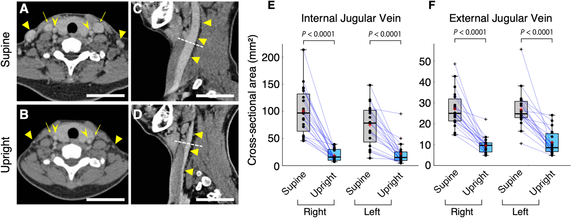
It turns out though that the Jugular Veins tend to do all the draining exclusively in supine position as opposed to standing or sitting where they are partially helped by the collateral veins along the spine. So if you see collateral filling while supine position, it can only mean that your Jugulars are somewhat impaired and blood is rerouted to collateral veins and since being upright naturally collapses the jugular vein, an impaired jugular drainage is even more impaired while standing or sitting. So doing the test in supine position might more accurately assess your Jugular drainage than doing it in upright is my understanding of it.

I have included a figures from a study comparing the jugular vein in upright position versus supine below.

Figure 1

From: Posture-induced changes in the vessels of the head and neck: evaluation using conventional supine CT and upright CT

Figure 1

Neck veins are collapsed in an upright body position. ( A , B ) Axial computed tomography (CT) section from the recirculation phase. Arrows, triangles, and arrowheads show the internal jugular veins (IJVs), external jugular veins (EJVs), and internal carotid arteries, respectively. Although the internal carotid arteries hardly change in different postures, the IJVs and EJVs are significantly collapsed in an upright posture. Scale bars: 50 mm. ( C , D ) Sagittal CT section from the same phase. The IJVs (arrowheads) are significantly collapsed. The dashed white line indicates the level of area measurement. ( E , F ) The cross-sectional area of the IJVs ( E , n = 20, two-sided paired t -test, right: P < 0.0001; left: P < 0.0001) and EJVs ( F , n = 19, two-sided paired t -test, right: P < 0.0001; left: P < 0.0001). In the box plots, the central mark indicates the median, the red cross indicates the mean, and the bottom and top edges of the box indicate the 25th and 75th percentiles, respectively. Whiskers extend to the maximum and minimum values within 1.5 interquartile ranges below the first quartile or above the third quartile, and black crosses indicate outliers. Increases and decreases from a supine position to an upright position are colored in red and blue, respectiv

Figure 4

From: Posture-induced changes in the vessels of the head and neck: evaluation using conventional supine CT and upright CT

Figure 4

Summary of the positional changes in craniocervical venous structure between supine and upright posture. In the cervical region, the internal jugular vein (IJV) significantly collapses. In the craniocervical junction, the IJV shrinks in an upright posture; in contrast, the anterior condylar vein (ACV), anterior condylar confluence (ACC), and vertebral venous system—including the suboccipital cavernous sinus (SOCS), vertebral artery venous plexus (VAVP), and anterior internal vertebral venous plexus (AIVVP), as described in the figure—were enlarged in an upright posture. The following three venous routes become more prominent in the upright position: (1) the ACV, originating from the ACC and draining into the SOCS, VAVP, and AIVVP; (2) the LCV, originating from the ACC and draining into the SOCS, VAVP, and AIVVP; (3) the PCV, originating from the ACC or jugular bulb and draining into the SOCS, as represented by numbers in the figure. As opposed to those venous structures, the pterygoid plexus (PP), located anteriorly, does not undergo consistent changes depending on posture. In the intracranial space, the venous structure undergoes almost no change between postures. The vertebral venous plexus (VVP) at the cervical level, which could not be evaluated in this study, is shown as a dotted line.

4 Likes

@KoolDude that’s really interesting study!
Could it mean that healthy people with good collateral venous flow have no problem when standing upright, and if it is restricted (e.g. due to the military neck or other reasons), the IJVs try to take some load off, and that’s why people get better after the surgery?.. i.e. because they start getting the venous blood flow, even through the route which usually is not the primary route when standing up?.. :thinking:

Okay, that raises the question: how much leeway and mobility does a healthy atlantooccipital joint have?
[Cervical Spine Functional Anatomy and the Biomechanics of Injury Due to Compressive Loading - PMC] says

Normal flexion to hyperextension at the atlanto-occipital joint ranges from approximately 15° to 20°.15,16 Rotation and lateral flexion between the occiput and atlas are not possible due to the depth of the atlantal sockets, in which the occipital condyles rest. Rotation to one side causes the contralateral occipital condyle to contact the anterior wall of its atlantal socket and the ipsilateral condyle to contact the posterior wall of its respective atlantal socket.15 Similarly, lateral flexion requires the contralateral occipital condyle to lift out of its socket, a movement that is restrained by the tight atlanto-occipital joint capsule.15

So it is quite possible that after a whiplash injury, or chronic neck muscle spasm/tension, the area where the collateral venous flow is supposed to happen is… squashed, and restricting the blood flow? And similarly, while supine, the occipital bone does not properly shift forward on top of the c1 “rails” and that prevents the styloids from moving away from the c1 and giving enough space to open up the IJVs?.. :thinking::thinking::thinking:

(On the lighter side: page 9,

Nightingale et al24,25,27 assessed the dynamic responses of the cervical spine to axial loading using high-speed video and cadaver specimens mounted (inverted) to a drop-track apparatus.

I already see cadaver rights activists coming to our chat…)

Here’s what I found: Venous Compression in Cerebral Venous System: Light and Shadows (VCS - venous compression syndrome)

If the first vertebra or another cervical vertebra has a rotation, a lateral or anterior dislocation or abnormal angle, we could have a direct effect of internal jugular or vertebral compression syndrome [4]. VCS is multifactorial, in fact there are at least three structures involved in its genesis. The bone apparatus (cervical vertebrae), the muscular apparatus (sternocleidomastoid, omohyoid, sternum thyroid and scalene) and the fascial system (superficial, medium and deep fascia)
[…]
A dislocation of the first vertebra (C1-Atlas) or more distal vertebras such as C3, C4 or other even more distal, can determine a VCS. VCS can affect the vertebral veins at various levels with a circle of compensation via the intra-vertebral veins or other veins vicarious such as the cervical ones
[…]
The decompression treatments are possible and can be non-invasive and invasive. The currently used are: 1. adjustment of the first cervical vertebra, 2. adjustment of all cervical vertebrae, 3. postural gymnastics, 4. resection omohyoid muscle [7]. The decompression treatments to be developed in the future are 1. Decompressive fasciotomy 2. Resection of the scalene muscle 3. The re-alignment of the cervical spine with exo-prosthesis or arthrodesis.

Alright, so it seems, finally we found some more credible source than my speculations saying that vascular-venous Eagle’s syndrome and military neck MIGHT be related.

Other resources:

6 Likes

Interesting information & definitely some good deeper research on your parts @KoolDude & @vdm. Thank you for sharing the information that you’re finding. The way it’s presented is pretty technical & likely a bit above some of our forum member’s ability to understand it (including me, but I get the gist of it). If you could possibly summarize your findings in layman’s terms that would be tremendously helpful for others on our forum. Thank you so much!!

3 Likes

In layman’s terms, it would be something like this:

  • there are multiple vein systems draining blood out of the brain, one major of them is Internal Jugular Veins (IJV), and let’s put the others together under one name “collateral system”. Some of the collateral system veins go through the same “canals” in the spine as vertebrobasilar arteries (transverse foramen - Vertebrae- transverse foramen)
  • in healthy people, IJV is dominating while lying down, the collateral system dominating when standing/sitting
  • the collateral system might be constricted by neck misalignments, including C1, C2, neck rotation or others (presumably military neck too)
  • the IJV among other reasons might be constricted by stiff neck muscles, (scalene, omohyoid…), also due to the styloid process pressing against the C1 vertebrae, and possibly by neck misalignments too (if the misaligned part of the neck starts compressing the vein)
  • as long as one of the systems is functioning properly, especially the collateral system, there is a good chance there is little trouble with blood flow or pressure in the head
  • if both systems get in trouble, surgery to decompress IJV system might be a relatively easy way to let the blood circulate again, especially when lying down (because surgery involving transverse foramenae, I guess, is much more problematic due to the nature of vertebrae dynamics, but that’s to be confirmed)
  • unfortunately, neck/spine misalignments potentially can push both systems out of their optimal performance, and those misalignments like military neck might be caused by many reasons, from whiplash or bad posture to cervical disc degradation.
  • depending on the individual body, surgically opening up IJVs might not be sufficient, as they might still remain constricted when not lying down, and in that case stenting might help (?)

That’s probably the easiest way to sum it up.

I speculate that gradually restoring neck’s shape and mobility might help with vascular issues, given it’s not the nerve-related variant of Eagle’s, or maybe even put the styloid processes further away from the affected areas. Definitely not something to be done using high velocity and sheer force tractions done by chiropractors.

6 Likes

WOW! You did a phenomenal summary, @vdm! Thank you so much for taking your time to do that. This information should be tremendously helpful & informative to those who have IJV flow issues.

I really appreciate you! :hugs::heart:

4 Likes

@vdm There is reason why I avoided to answer the question when you raised it here. The simple answer is I really do not know and can only speculate what it means. Based on this research (Venous collapse regulates intracranial pressure in upright body positions), the collapse of the IJVs in upright position might be a natural way of regulating the intercranial pressure (ICP) since hydrostatic pressure (pressure on the blood due to gravity) is different when upright compared to when lying down. So our bodies adapt to the gravitational pull of the fluids such as blood when we are upright so the overall pressure of the fluids in the brain does not get negatively affected. Of course, the story is different when you have an impaired IJVs or any other disease that affects CFS & intracranial pressure of the brain. I really do not know what the effect of the posture would be in that case. According to @Jules & @Isaiah_40_31, we know that sleeping on an incline such as wedge pillow does help with symptoms since we sleep on supine position, the impaired IJVs are expected to do all the draining and obviously can’t do it efficiently potentially causing more symptoms in sleep vs when awake. So sleeping on your head elevated should drain bit better since the collaterals come to their rescue. It is also important to note that upright position does not appear to collapse the collateral veins and brain sinuses such as transverse sinus, straight sinues, sigmoid …etc. There is another interesting limited study (Internal jugular vein blood flow in the upright position during external compression and increased central venous pressure: an ultrasound study in healthy volunteers | SpringerLink) conducted on upright position with compressed IJVs to study the blood flow and drainage. It only found that drainage (flow rate) was not affected as you would think but intercranial venous volumes increased. So without going deep into the physics of fluid dynamics, it is hard to answer without doing a proper, controlled study which takes into consideration of all the pressures, forces and postures.

@KoolDude that’s really interesting study!
Could it mean that healthy people with good collateral venous flow have no problem when standing upright, and if it is restricted (e.g. due to the military neck or other reasons), the IJVs try to take some load off, and that’s why people get better after the surgery?.. i.e. because they start getting the venous blood flow, even through the route which usually is not the primary route when standing up?.. :thinking:

3 Likes

I did not do well in physics class when in college. I enjoyed it immensely, but as w/ chemistry, it all ended up being a bit more than my brain could wrap itself around. I’m making this confession because I really appreciate it when you & @vdm & others put things in “layman’s terms”. I can understand more complex concepts that way and bet I’m not alone on this forum.

I continue to really appreciate all who have put time into researching symptoms & their causes at a deeper level. It is truly the icing on the cake for our forum to be able to educate & not just encourage & inform at a basic level.

:blush:

3 Likes
3 Likes

I’ve watched Bob & Brad before in other YouTube PT videos. I really like their approach to PT in the videos I’ve seen. Thank you for sharing this one @vdm!

1 Like

Some simple exercises for tight upper neck muscles

3 Likes

Interesting question. My wife frequently sees me raising my arm above my head on my more symptomatic side while I am sleeping - straight up and back. It likely relieves some pressure from something being compressed.

2 Likes