I promise I’ll come back and share my full story soon, but I wanted to get this post out now in hopes of getting some advice before my ENT appointment.
I’ve had symptoms for quite a while, but about 18 months ago they started to debilitating. . About a year ago, I convinced my neurologist to order a CT Soft Tissue Neck with Contrast even though he thought I was completely fine and didn’t need it. While I was drafting “my story” tonight, I remembered that scan and went back to review it.
The radiology report doesn’t mention anything about my styloids or possible stylohyoid ligament calcification, most likely because that wasn’t what was requested.
Would you recommend asking my doctor to have the radiologist re-read the scan with those concerns in mind? Based on my symptoms, I also want to know if there’s compression of the arteries or jugular vein. Or should I wait and ask my ENT to reorder the correct imaging? My goal is to have a clear diagnosis before reaching out to surgeons, but I’m a little unsure of the best next step.
I also tried converting the images into 3D but realized it’s almost 2:30 a.m. and I have work tomorrow, so I’ll pick that back up later.
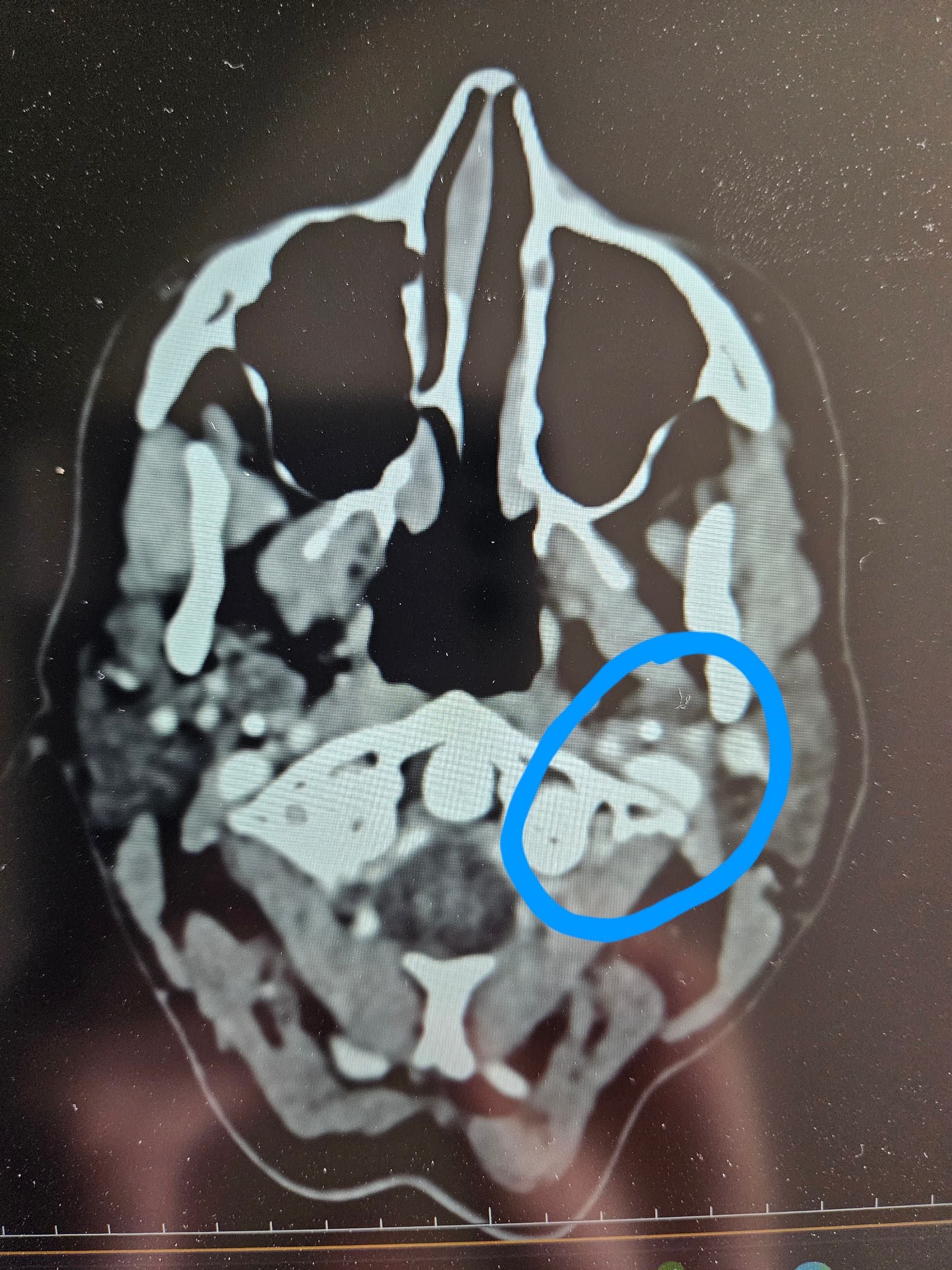
Any advice on how you would move forward would be greatly appreciated. Here are some of my images in the meantime.
Hello @Drea welcome to the forum. Just looking at your images you have uploaded, the measurements you have taken are of your jaw bone. I have attached my images for you as a guide to what you are looking for. If you can find these on your CT then I am sure other members will be able to tell you if they think anything could be causing you problems.
As @Rosie says, those aren’t the styloids; I’m afraid I can’t label your images for you, but on your second one you can see the styloid process which looks quite thick, & it does look as if it could be compressing your IJV, although it’s not the clearest of images. It also looks as if there could be a collateral vein at the back of your head, which can indicate compression & the body compensates by using & enlarging other veins…On the 3rd one you can see clearly what looks to be a calcified stylo-hyoid ligament on your left side, it looks quite long so may well be causing symptoms! Obviously we’re not doctors on here, but can give you pointers, & hopefully others will be able to chip in too…
It’s possible if your symptoms have worsened since you got the CT done that things have changed, & as some of the imaging isn’t the clearest you could ask for it to be done again, but then that’s more radiation which isn’t ideal, the CT you had done was the correct scan, so you could just see if you can get this re-read…
I woke up right at 8 a.m. to call the ENT’s office and saw your messages. I have to admit, they gave me a good laugh—which I really needed. Of course, I had the wrong images, lol!
I was able to get an appointment today at 3 p.m. and will be requesting a re-read. You’re absolutely right—I don’t want to be exposed to more radiation. Over the past 18 months, I’ve already had six CTs/MRIs trying to figure this out.
Good you’ve got an appointment, well done, & hopefully they’ll be able to look back over the images again… 6 CTs is alot ! MRIs don’t use radiation, so that’s not a problem… Let us know how you get on
@Isaiah_40_31 i think what you have labeled as calcified stylohyoid ligament is actually the greater horn of the hyoid. Not on the right trajectory to be attached to styloid. Worth seeing if it’s digging into anything!
I saw the doctor yesterday, and she agreed that my scans should be reviewed for Eagle Syndrome. She mentioned that my styloids looked only slightly longer than 3 cm and didn’t think the stylohyoid ligament was calcified. She was kind but also admitted this isn’t her area of expertise.
Last night, I went back through my images and had trouble finding the styloids. It seems like my lower jaw bone is blocking them. On some of the images, the styloids look broken up into little spots, as if they’re partially visible—if that makes sense.
I’m wondering if it would be best to repeat the CT, since it’s been a year and my symptoms have gotten much worse. I am at the point where I have had food come back up and out my nose while choking, I struggle to breathe, am on BP medicine (have never had BP plms), lose my voice every day, feel like something is stuck in my throat, feel like I am going to pass out, I know something is tight or pinched off because I can feel it on the left side of my neck, my face tingles on the left side, it goes down to my collarbone and my arm tingles….just so tired of advocating and fighting doctors….
@TML - Thx for the correction & I agree. I’ll edit my annotation. I was thinking the same thing last night & meant to put that it was either one but was tired & didn’t follow through.
Getting an updated CT w/ contrast would be most helpful toward getting a proper diagnosis, @Drea. With the symptoms you mentioned above, it sounds like your internal carotid artery may be partially compressed or is being irritated by your styloid, calcified stylohyoid ligament or elongation of the greater horn of your hyoid. If you notice that certain head positions cause your symptoms to flare, ask your doctor to request the scan be done in that/those position(s) as well as neutral & also request that some 3D images be provided.
It’s not uncommon for the stylohyoid ligament to calcify in little sections which is what your’s looks like it’s done, however, as you noted, your jaw is in the way so you can’t see the whole thing, Your next CT scan should cover the area between skull base & hyoid bone or even further down than the hyoid. You can ask for your doctor to have it evaluated for styloid length, thickness, curve, stylohyoid calcification & length, & whether there’s any sign of IJV or carotid compression.
@Drea if the request hasn’t already been submitted, I suggest you ask for a neurologist re-read specifically. Neurologists seem to provide the better reads when compared to general radiologists (at least in my experience)!
Update: I’ve now shared my full story in a new thread here: My Full Story Thank you to everyone who responded before — your advice really helped me decide to move forward with a new CT.